HGF R, also known as Met (from N-methyl-N’-nitro-N-nitrosoguanidine induced), is a glycosylated receptor tyrosine kinase that plays a central role in epithelial morphogenesis and cancer development. HGF R is synthesized as a single chain precursor which undergoes cotranslational proteolytic cleavage. This generates a mature HGF R that is a disulfide-linked dimer composed of a 50 kDa extracellular alpha chain and a 145 kDa transmembrane beta chain (1, 2). The extracellular domain (ECD) contains a seven bladed beta -propeller sema domain, a cysteine-rich PSI/MRS, and four Ig-like E-set domains, while the cytoplasmic region includes the tyrosine kinase domain (3, 4). Proteolysis and alternate splicing generate additional forms of human HGF R which either lack of the kinase domain, consist of secreted extracellular domains, or are deficient in proteolytic separation of the alpha and beta chains (5-7). The sema domain, which is formed by both the alpha and beta chains of HGF R, mediates both ligand binding and receptor dimerization (3, 8). Ligand-induced tyrosine phosphorylation in the cytoplasmic region activates the kinase domain and provides docking sites for multiple SH2-containing molecules (9, 10). HGF stimulation induces HGF R downregulation via internalization and proteasome-dependent degradation (11). In the absence of ligand, HGF R forms non-covalent complexes with a variety of membrane proteins including CD44v6, CD151, EGF R, Fas, Integrin alpha 6/ beta 4, Plexins B1, 2, 3, and MSP R/Ron (12-19). Ligation of one complex component triggers activation of the other, followed by cooperative signaling effects (12-19). Formation of some of these heteromeric complexes is a requirement for epithelial cell morphogenesis and tumor cell invasion (12, 16, 17). Paracrine induction of epithelial cell scattering and branching tubulogenesis results from the stimulation of HGF R on undifferentiated epithelium by HGF released from neighboring mesenchymal cells (20). Genetic polymorphisms, chromosomal translocation, over-expression, and additional splicing and proteolytic cleavage of HGF R have been described in a wide range of cancers (1). Within the ECD, human HGF R shares 86-88% amino acid sequence identity with canine, mouse, and rat HGF R.
Human HGFR/c-MET Antibody (95106)
R&D Systems | Catalog # MAB3582

Key Product Details
Species Reactivity
Validated:
Cited:
Applications
Validated:
Cited:
Label
Antibody Source
Product Specifications
Immunogen
Glu25-Thr932
Accession # P08581
Specificity
Clonality
Host
Isotype
Scientific Data Images for Human HGFR/c-MET Antibody (95106)
Detection of HGF R/c‑MET in MDA‑MB‑231 Human Cell Line by Flow Cytometry.
MDA-MB-231 human breast cancer cell line was stained with Human HGF R/c-MET Monoclonal Antibody (Catalog # MAB3582, filled histogram) or isotype control antibody (Catalog # MAB002, open histogram), followed by Allophycocyanin-conjugated Anti-Mouse IgG F(ab')2Secondary Antibody (Catalog # F0101B).
Detection of HGFR/c-MET by Western Blot
Generation of CARs and analysis of IL-2 secretion by c-Met CAR Jurkat cell. (A) Schematic representation of c-Met CAR and FITC CAR construct. (B) c-Met gene (pCMV3-Met) was transduced by electroporation into K562, and Western blot was performed. (C) After transducing the mock CD8sp-c-Met CAR, CSF2Rsp-c-Met CAR, or FITC CAR construct into Jurkat, expressions of CAR were confirmed through Western blot. (D) The mock CD8sp-c-Met CAR, CSF2Rsp-c-Met CAR, or FITC CAR Jurkat were co-cultured with K562 or c-Met-K562 at an E:T ratio of 15:1. After overnight incubation, supernatants were collected and ELISA was performed to measure IL-2 level. (The whole western blots figure see Figure S1). Image collected and cropped by CiteAb from the following open publication (https://pubmed.ncbi.nlm.nih.gov/34830894), licensed under a CC-BY license. Not internally tested by R&D Systems.
Detection of HGFR/c-MET by Western Blot
c-Met CAR KHYG-1 specifically lyses the c-Met positive GC cells. (A) Western blot with the cell lysates of mock CD8sp-c-Met, CSF2Rsp-c-Met, or FITC KHYG-1 cells to see the CAR expression (B) The mock CD8sp-c-Met CAR, CSF2Rsp-c-Met CAR, or FITC CAR KHYG-1 were co-incubated with MKN-45, SNU-5, SNU-1, and SNU-484 at E:T ratio of 5:1 or 10:1 for 5 h. The cytotoxicity of CAR KHYG-1 was measured by the Bright-Glo (luciferase) assay system. (The whole western blots figure see Figure S1). Image collected and cropped by CiteAb from the following open publication (https://pubmed.ncbi.nlm.nih.gov/34830894), licensed under a CC-BY license. Not internally tested by R&D Systems.Applications for Human HGFR/c-MET Antibody (95106)
CyTOF-ready
Flow Cytometry
Sample: MDA‑MB‑231 human breast cancer cell line
Reviewed Applications
Read 4 reviews rated 4 using MAB3582 in the following applications:
Flow Cytometry Panel Builder
Bio-Techne Knows Flow Cytometry
Save time and reduce costly mistakes by quickly finding compatible reagents using the Panel Builder Tool.
Advanced Features
- Spectra Viewer - Custom analysis of spectra from multiple fluorochromes
- Spillover Popups - Visualize the spectra of individual fluorochromes
- Antigen Density Selector - Match fluorochrome brightness with antigen density

Formulation, Preparation, and Storage
Purification
Reconstitution
Reconstitute at 0.5 mg/mL in sterile PBS. For liquid material, refer to CoA for concentration.
Formulation
Shipping
Stability & Storage
- 12 months from date of receipt, -20 to -70 °C as supplied.
- 1 month, 2 to 8 °C under sterile conditions after reconstitution.
- 6 months, -20 to -70 °C under sterile conditions after reconstitution.
Calculators
Background: HGFR/c-MET
References
- Birchmeier, C. et al. (2003) Nat. Rev. Mol. Cell Biol. 4:915.
- Corso, S. et al. (2005) Trends Mol. Med. 11:284.
- Gherardi, E. et al. (2003) Proc. Natl. Acad. Sci. USA 100:12039.
- Park, M. et al. (1987) Proc. Natl. Acad. Sci. USA 84:6379.
- Crepaldi, T. et al. (1994) J. Biol. Chem. 269:1750.
- Prat, M. et al. (1991) Mol. Cell. Biol. 12:5954.
- Rodrigues, G.A. et al. (1991) Mol. Cell. Biol. 11:2962.
- Kong-Beltran, M. et al. (2004) Cancer Cell 6:75.
- Naldini, L. et al. (1991) Mol. Cell. Biol. 11:1793.
- Ponzetto, C. et al. (1994) Cell 77:261.
- Jeffers, M. et al. (1997) Mol. Cell. Biol. 17:799.
- Orian-Rousseau, V. et al. (2002) Genes Dev. 16:3074.
- Klosek, S.K. et al. (2005) Biochem. Biophys. Res. Commun. 336:408.
- Jo, M. et al. (2000) J. Biol. Chem. 275:8806.
- Wang, X. et al. (2002) Mol. Cell 9:411.
- Trusolino, L. et al. (2001) Cell 107:643.
- Giordano, S. et al. (2002) Nat. Cell Biol. 4:720.
- Conrotto, P. et al. (2004) Oncogene 23:5131.
- Follenzi, A. et al. (2000) Oncogene 19:3041.
- Sonnenberg, E. et al. (1993) J. Cell Biol. 123:223.
Long Name
Alternate Names
Gene Symbol
UniProt
Additional HGFR/c-MET Products
Product Documents for Human HGFR/c-MET Antibody (95106)
Certificate of Analysis
To download a Certificate of Analysis, please enter a lot or batch number in the search box below.
Note: Certificate of Analysis not available for kit components.
Product Specific Notices for Human HGFR/c-MET Antibody (95106)
For research use only
Citations for Human HGFR/c-MET Antibody (95106)
 Powered by Bioz
Customer Reviews for Human HGFR/c-MET Antibody (95106) (4)
Have you used Human HGFR/c-MET Antibody (95106)?
Submit a review and receive an Amazon gift card!
$25/€18/£15/$25CAN/¥2500 Yen for a review with an image
$10/€7/£6/$10CAN/¥1110 Yen for a review without an image
Submit a review
Customer Images

-
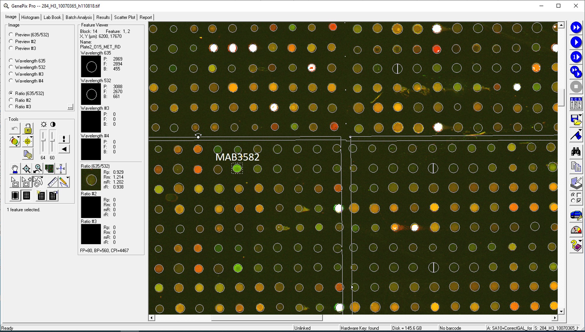
Application: MicroarraysSample Tested: EDTA PlasmaSpecies: HumanVerified Customer | Posted 01/14/2021

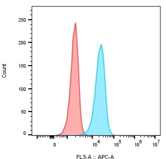
-
Application: Flow CytometrySample Tested: A-431 epidermoid carcinoma cell lineSpecies: HumanVerified Customer | Posted 12/23/2020Detection of human HGFR/c-MET on epidermoid carcinoma cell line A-431. A-431 cells were treated with 100 nM of the human HGFR/c-MET antibody (catalog # MAB3582) or mouse IgG1 isotype control, followed by a secondary antibody goat a-mouse IgG Fc APC. Isotype control (RED), human HGFR/c-MET antibody (BLUE).
-
Application: MicroarraySample Tested: EDTA PlasmaSpecies: HumanVerified Customer | Posted 02/08/2020Antibody was printed on custom arrays and incubated with fluorescently labeled human EDTA plasma
-
Application: MicroarraysSample Tested: EDTA PlasmaSpecies: HumanVerified Customer | Posted 11/14/2018
There are no reviews that match your criteria.
Protocols
Find general support by application which include: protocols, troubleshooting, illustrated assays, videos and webinars.
- 7-Amino Actinomycin D (7-AAD) Cell Viability Flow Cytometry Protocol
- Extracellular Membrane Flow Cytometry Protocol
- Flow Cytometry Protocol for Cell Surface Markers
- Flow Cytometry Protocol for Staining Membrane Associated Proteins
- Flow Cytometry Staining Protocols
- Flow Cytometry Troubleshooting Guide
- Intracellular Flow Cytometry Protocol Using Alcohol (Methanol)
- Intracellular Flow Cytometry Protocol Using Detergents
- Intracellular Nuclear Staining Flow Cytometry Protocol Using Detergents
- Intracellular Staining Flow Cytometry Protocol Using Alcohol Permeabilization
- Intracellular Staining Flow Cytometry Protocol Using Detergents to Permeabilize Cells
- Propidium Iodide Cell Viability Flow Cytometry Protocol
- Protocol for Liperfluo
- Protocol for the Characterization of Human Th22 Cells
- Protocol for the Characterization of Human Th9 Cells
- Protocol: Annexin V and PI Staining by Flow Cytometry
- Protocol: Annexin V and PI Staining for Apoptosis by Flow Cytometry
- Troubleshooting Guide: Fluorokine Flow Cytometry Kits
- View all Protocols, Troubleshooting, Illustrated assays and Webinars
FAQs for Human HGFR/c-MET Antibody (95106)
-
Q: Does Human HGFR/c-MET Antibody, Catalog # MAB3582, bind to the alpha chain or beta chain of HGFR/c-MET?
A: The immunogen for MAB3582 (Recombinant human HGF R/cMET, aa Glu25-Thr932) spans both the alpha and beta chain regions of Human HGFR and we do not epitope map our antibodies. However, staining in flow cytometry detects a surface epitope. Since only the alpha chain is extracellular in location and the beta chain is transmembrane in location, the antibody likley binds the alpha chain of this protein.


