LAMP1 (Lysosome-associated membrane protein-1; also CD107a) is a 100‑130 kDa member of the LAMP family of glycoproteins. It is expressed in lysosomal and plasma membranes of macrophages, NK and T-cells, and with LAMP2, is essential for the formation of phagolysosomes. On the cell surface, it also presents carbohydrates to selectins. Mature human LAMP1 is a 389 amino acid (aa) type I transmembrane glycoprotein. It contains a 354 aa luminal/extracellular domain (ECD) (aa 28‑381) and a 12 aa cytoplasmic tail (aa 405‑416). The ECD has two large looping regions (aa 28‑193 and 227‑381) plus multiple N- and O-linked glycosylation sites. There is one potential splice variant that shows a 26 aa substitution in the signal sequence. Over aa 28‑380, human LAMP1 shares 64% aa identity with mouse LAMP1.
Human LAMP-1/CD107a Lumenal Domain Antibody
R&D Systems | Catalog # AF4800

Key Product Details
Species Reactivity
Validated:
Human
Cited:
Human, Mouse, Rat, Primate - Chlorocebus aethiops (African Green Monkey)
Applications
Validated:
Western Blot, Immunocytochemistry
Cited:
Western Blot, Immunocytochemistry, Microarray
Label
Unconjugated
Antibody Source
Polyclonal Sheep IgG
Loading...
Product Specifications
Immunogen
Mouse myeloma cell line NS0-derived recombinant human LAMP1/CD107a
Ala28-Asn380
Accession # P11279
Ala28-Asn380
Accession # P11279
Specificity
Detects human LAMP1/CD107a Lumenal Domain in direct ELISAs and Western blots. In direct ELISAs, less than 1% cross-reactivity with recombinant mouse LAMP1 is observed.
Clonality
Polyclonal
Host
Sheep
Isotype
IgG
Scientific Data Images for Human LAMP-1/CD107a Lumenal Domain Antibody
Detection of Human LAMP‑1/CD107a by Western Blot.
Western blot shows lysates of THP-1 human acute monocytic leukemia cell line and HL-60 human acute promyelocytic leukemia cell line. PVDF membrane was probed with 1 µg/mL of Sheep Anti-Human LAMP-1/CD107a Lumenal Domain Antigen Affinity-purified Polyclonal Antibody (Catalog # AF4800) followed by HRP-conjugated Anti-Sheep IgG Secondary Antibody (Catalog # HAF016). A specific band was detected for LAMP-1/CD107a at approximately 120 kDa (as indicated). This experiment was conducted under reducing conditions and using Immunoblot Buffer Group 1.
LAMP1/CD107a in THP-1 Human Cell Line.
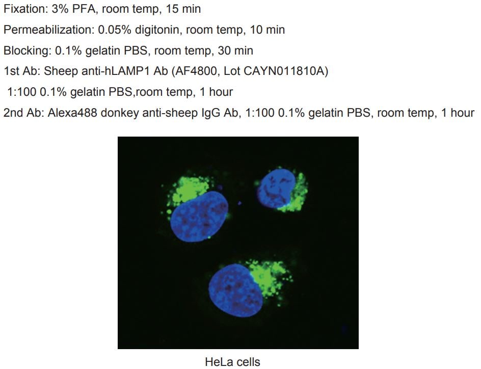
LAMP1/CD107a was detected in immersion fixed THP-1 human acute monocytic leukemia cell line using 10 µg/mL Sheep Anti-Human LAMP1/CD107a Lumenal Domain Antigen Affinity-purified Polyclonal Antibody (Catalog # AF4800) for 3 hours at room temperature. Cells were stained with the NorthernLights™ 557-conjugated Anti-Sheep IgG Secondary Antibody (red; Catalog # NL010) and counterstained with DAPI (blue). View our protocol for Fluorescent ICC Staining of Cells on Coverslips.Applications for Human LAMP-1/CD107a Lumenal Domain Antibody
Application
Recommended Usage
Immunocytochemistry
5-15 µg/mL
Sample: Immersion fixed THP-1 human acute monocytic leukemia cell line
Sample: Immersion fixed THP-1 human acute monocytic leukemia cell line
Western Blot
1 µg/mL
Sample: THP‑1 human acute monocytic leukemia cell line and HL‑60 human acute promyelocytic leukemia cell line
Sample: THP‑1 human acute monocytic leukemia cell line and HL‑60 human acute promyelocytic leukemia cell line
Reviewed Applications
Read 2 reviews rated 5 using AF4800 in the following applications:
Formulation, Preparation, and Storage
Purification
Antigen Affinity-purified
Reconstitution
Reconstitute at 0.2 mg/mL in sterile PBS. For liquid material, refer to CoA for concentration.
Loading...
Formulation
Lyophilized from a 0.2 μm filtered solution in PBS with Trehalose. *Small pack size (SP) is supplied either lyophilized or as a 0.2 µm filtered solution in PBS.
Shipping
Lyophilized product is shipped at ambient temperature. Liquid small pack size (-SP) is shipped with polar packs. Upon receipt, store immediately at the temperature recommended below.
Stability & Storage
Use a manual defrost freezer and avoid repeated freeze-thaw cycles.
- 12 months from date of receipt, -20 to -70 °C as supplied.
- 1 month, 2 to 8 °C under sterile conditions after reconstitution.
- 6 months, -20 to -70 °C under sterile conditions after reconstitution.
Calculators
Background: LAMP-1/CD107a
Long Name
Lysosome-associated Membrane Glycoprotein 1
Alternate Names
CD107a, LAMP1
Gene Symbol
LAMP1
UniProt
Additional LAMP-1/CD107a Products
Product Documents for Human LAMP-1/CD107a Lumenal Domain Antibody
Certificate of Analysis
To download a Certificate of Analysis, please enter a lot or batch number in the search box below.
Note: Certificate of Analysis not available for kit components.
Product Specific Notices for Human LAMP-1/CD107a Lumenal Domain Antibody
For research use only
Citations for Human LAMP-1/CD107a Lumenal Domain Antibody
 Powered by Bioz
Customer Reviews for Human LAMP-1/CD107a Lumenal Domain Antibody (2)
5 out of 5
2 Customer Ratings
Have you used Human LAMP-1/CD107a Lumenal Domain Antibody?
Submit a review and receive an Amazon gift card!
$25/€18/£15/$25CAN/¥2500 Yen for a review with an image
$10/€7/£6/$10CAN/¥1110 Yen for a review without an image
Submit a review
Customer Images
Showing
1
-
2 of
2 reviews
Showing All
Filter By:
-
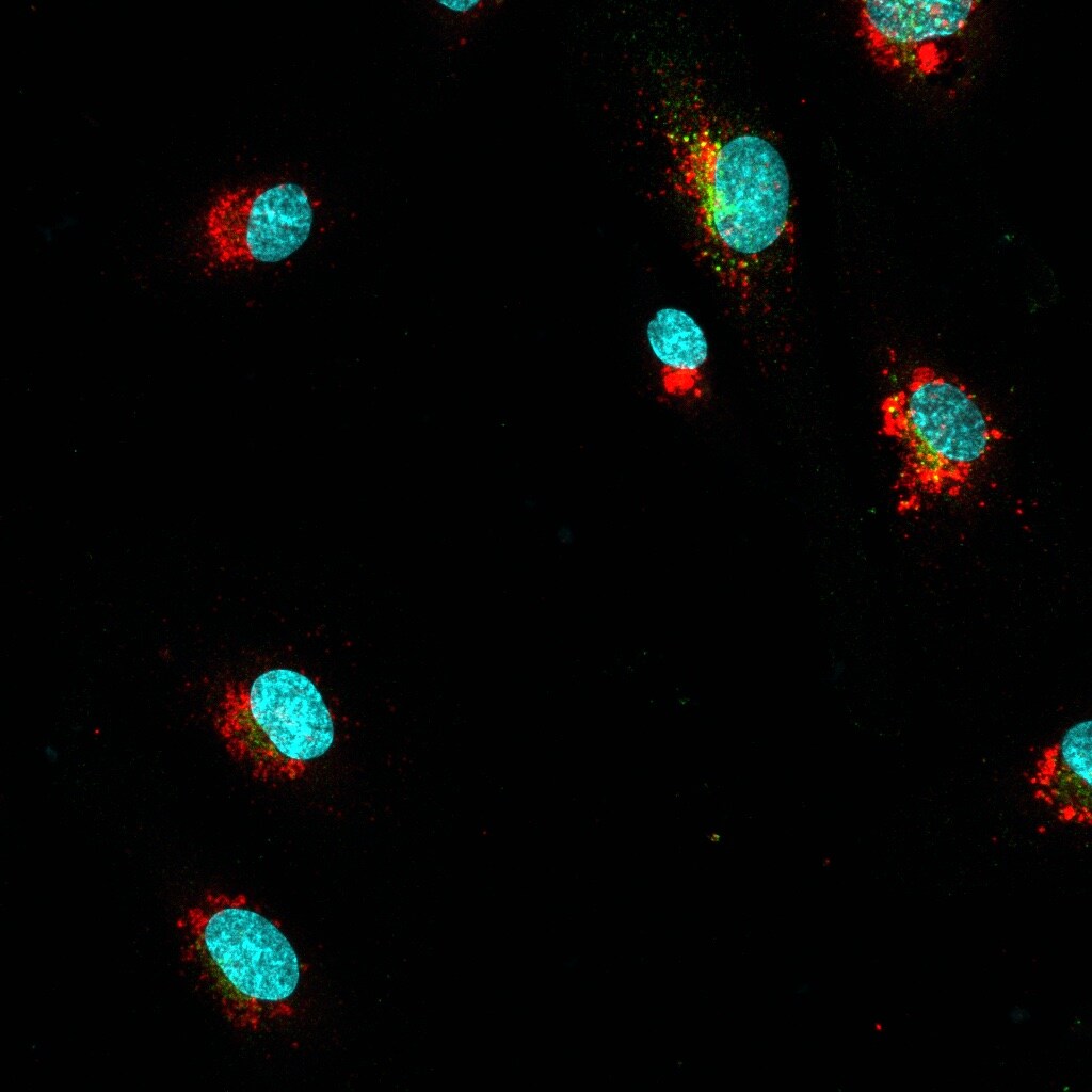
Application: Immunocytochemistry/ImmunofluorescenceSample Tested: HUVEC human umbilical vein endothelial cellsSpecies: HumanVerified Customer | Posted 08/04/2021LAMP1 (red), VEGFR2 (green), Nucleus (blue)

-
Application: ImmunocytochemistrySample Tested: hela cellSpecies: HumanVerified Customer | Posted 10/19/2018
There are no reviews that match your criteria.
Protocols
Find general support by application which include: protocols, troubleshooting, illustrated assays, videos and webinars.
- Appropriate Fixation of IHC/ICC Samples
- Cellular Response to Hypoxia Protocols
- ClariTSA™ Fluorophore Kits
- Detection & Visualization of Antibody Binding
- ICC Cell Smear Protocol for Suspension Cells
- ICC Immunocytochemistry Protocol Videos
- ICC for Adherent Cells
- Immunocytochemistry (ICC) Protocol
- Immunocytochemistry Troubleshooting
- Immunofluorescence of Organoids Embedded in Cultrex Basement Membrane Extract
- Immunohistochemistry (IHC) and Immunocytochemistry (ICC) Protocols
- Preparing Samples for IHC/ICC Experiments
- Preventing Non-Specific Staining (Non-Specific Binding)
- Primary Antibody Selection & Optimization
- Protocol for VisUCyte™ HRP Polymer Detection Reagent
- Protocol for the Fluorescent ICC Staining of Cell Smears - Graphic
- Protocol for the Fluorescent ICC Staining of Cultured Cells on Coverslips - Graphic
- Protocol for the Preparation and Fluorescent ICC Staining of Cells on Coverslips
- Protocol for the Preparation and Fluorescent ICC Staining of Non-adherent Cells
- Protocol for the Preparation and Fluorescent ICC Staining of Stem Cells on Coverslips
- Protocol for the Preparation of a Cell Smear for Non-adherent Cell ICC - Graphic
- R&D Systems Quality Control Western Blot Protocol
- TUNEL and Active Caspase-3 Detection by IHC/ICC Protocol
- The Importance of IHC/ICC Controls
- Troubleshooting Guide: Western Blot Figures
- Western Blot Conditions
- Western Blot Protocol
- Western Blot Protocol for Cell Lysates
- Western Blot Troubleshooting
- Western Blot Troubleshooting Guide
- View all Protocols, Troubleshooting, Illustrated assays and Webinars
Loading...
Associated Pathways
