Human Microtubule-associated Protein (MAP) Light Chain 3 (LC3) A is a121 amino acid (aa) protein with a predicted molecular weight of 14 kDa. It is a member of the LC3 subfamily of Autophagy-related 8 (Atg8) proteins (1). The LC3 subfamily also includes LC3B andLC3C. LC3 exhibits 100% aa sequence identity with its mouse and rat orthologs, and is orthologous to the yeast autophagy-related protein Atg8. Atg8 family members show structural similarity with Ubiquitin, but lack aa sequence similarity. LC3 was originally described as part is part of a complex that includes heavy and light chains comprising the MAP1 family of microtubule regulatory proteins (3). However, LC3 has gained attention for MAP1-independent functions in autophagy. LC3 utilizes a ubiquitin-like conjugation system that includes E1-, E2-, and E3-like enzymes to covalently attach phosphatidylethanolamine (PE) to its C-terminus, incorporating it into the phagophore membrane during the early stages of autophagasome formation (4). Recruitment of LC3 to the phagophore may promote membrane elongation (4,5). It may also be involved in cargo recruitment to autophagosomes (1). LC3 is often used as a marker of autophagy.

Key Product Details
Validated by
Biological Validation
Species Reactivity
Validated:
Human
Cited:
Human
Applications
Validated:
Immunohistochemistry, Western Blot
Cited:
Immunocytochemistry
Label
Unconjugated
Antibody Source
Monoclonal Rat IgG2B Clone # 877005
Loading...
Product Specifications
Immunogen
E. coli-derived recombinant human LC3A
Accession # Q9H492
Accession # Q9H492
Specificity
Detects human LC3A in direct ELISAs.
Clonality
Monoclonal
Host
Rat
Isotype
IgG2B
Scientific Data Images for Human LC3A Antibody (877005)
Detection of Human, Mouse, and Rat LC3A by Western Blot.
Western blot shows lysates of human brain tissue, NIH-3T3 mouse embryonic fibroblast cell line, and PC-12 rat adrenal pheochromocytoma cell line untreated (-) or treated (+) with 50 µM Chloroquine for 18 hours. PVDF membrane was probed with 2 µg/mL of Rat Anti-Human LC3A Antibody (Catalog # MAB8558) followed by HRP-conjugated Anti-Rat IgG Secondary Antibody (Catalog # HAF005). Specific bands were detected for LC3A at approximately 14 and 16 kDa (as indicated). This experiment was conducted under reducing conditions and using Immunoblot Buffer Group 1.
LC3A in Human Brain Cortex Tissue.
LC3A was detected in immersion fixed paraffin-embedded sections of human brain cortex tissue using Rat Anti-Human LC3A Monoclonal Antibody (Catalog # MAB8558) at 15 µg/mL overnight at 4 °C. Tissue was stained using the Anti-Rat HRP-DAB Cell & Tissue Staining Kit (brown; Catalog # CTS017) and counterstained with hematoxylin (blue). Specific staining was localized to neurons. View our protocol for Chromogenic IHC Staining of Paraffin-embedded Tissue Sections.
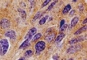
LC3A in Human Brain.
LC3A was detected in immersion fixed paraffin-embedded sections of human brain (cortex) using Rat Anti-Human LC3A Monoclonal Antibody (Catalog # MAB8558) at 1.7 µg/mL overnight at 4 °C. Tissue was stained using the Anti-Rat HRP-DAB Cell & Tissue Staining Kit (brown; Catalog # CTS017) and counterstained with hematoxylin (blue). Specific staining was localized to cytoplasm. View our protocol for Chromogenic IHC Staining of Paraffin-embedded Tissue Sections.Applications for Human LC3A Antibody (877005)
Application
Recommended Usage
Immunohistochemistry
8-25 µg/mL
Sample: Immersion fixed paraffin-embedded sections of human brain cortex tissue
Sample: Immersion fixed paraffin-embedded sections of human brain cortex tissue
Western Blot
2 µg/mL
Sample: Human brain tissue (untreated), NIH‑3T3 mouse embryonic fibroblast cell line and PC‑12 rat adrenal pheochromocytoma cell line treated with Chloroquine
Sample: Human brain tissue (untreated), NIH‑3T3 mouse embryonic fibroblast cell line and PC‑12 rat adrenal pheochromocytoma cell line treated with Chloroquine
Reviewed Applications
Read 2 reviews rated 5 using MAB8558 in the following applications:
Formulation, Preparation, and Storage
Purification
Protein A or G purified from hybridoma culture supernatant
Reconstitution
Reconstitute at 0.5 mg/mL in sterile PBS. For liquid material, refer to CoA for concentration.
Loading...
Formulation
Lyophilized from a 0.2 μm filtered solution in PBS with Trehalose. *Small pack size (SP) is supplied either lyophilized or as a 0.2 µm filtered solution in PBS.
Shipping
Lyophilized product is shipped at ambient temperature. Liquid small pack size (-SP) is shipped with polar packs. Upon receipt, store immediately at the temperature recommended below.
Stability & Storage
Use a manual defrost freezer and avoid repeated freeze-thaw cycles.
- 12 months from date of receipt, -20 to -70 °C as supplied.
- 1 month, 2 to 8 °C under sterile conditions after reconstitution.
- 6 months, -20 to -70 °C under sterile conditions after reconstitution.
Calculators
Background: LC3A
References
- Shpilka, T. et al. (2011) Genome Biol. 12:226.
- He, H. et al. (2003) J. Biol. Chem. 278:29278.
- Kuznetsov, S.A. & V.I. Gelfand (1987) FEBS Let. 212:145.
- Weidberg, H. et al. (2011) Ann Rev. Biochem. 80:125.
- Weidberg, H. et al. (2010) EMBO J. 29:1792.
Long Name
Microtubule-associated Protein 1 Light Chain 3 alpha
Alternate Names
Apg8, APG8a, Apg8p3, ATG8E, LC3, MAP1ALC3, MAP1LC3A, MLP3A
Gene Symbol
MAP1LC3A
UniProt
Additional LC3A Products
Product Documents for Human LC3A Antibody (877005)
Certificate of Analysis
To download a Certificate of Analysis, please enter a lot or batch number in the search box below.
Note: Certificate of Analysis not available for kit components.
Product Specific Notices for Human LC3A Antibody (877005)
For research use only
Related Research Areas
Citations for Human LC3A Antibody (877005)
 Powered by Bioz
Customer Reviews for Human LC3A Antibody (877005) (2)
5 out of 5
2 Customer Ratings
Have you used Human LC3A Antibody (877005)?
Submit a review and receive an Amazon gift card!
$25/€18/£15/$25CAN/¥2500 Yen for a review with an image
$10/€7/£6/$10CAN/¥1110 Yen for a review without an image
Submit a review
Customer Images
Showing
1
-
2 of
2 reviews
Showing All
Filter By:
-
Application: ImmunohistochemistrySample Tested: gastric cancerSpecies: HumanVerified Customer | Posted 10/29/2021

-
Application: ImmunohistochemistrySample Tested: Cancer cell lysatesSpecies: HumanVerified Customer | Posted 08/30/2017
There are no reviews that match your criteria.
Protocols
Find general support by application which include: protocols, troubleshooting, illustrated assays, videos and webinars.
- Antigen Retrieval Protocol (PIER)
- Antigen Retrieval for Frozen Sections Protocol
- Appropriate Fixation of IHC/ICC Samples
- Cellular Response to Hypoxia Protocols
- Chromogenic IHC Staining of Formalin-Fixed Paraffin-Embedded (FFPE) Tissue Protocol
- Chromogenic Immunohistochemistry Staining of Frozen Tissue
- ClariTSA™ Fluorophore Kits
- Detection & Visualization of Antibody Binding
- Fluorescent IHC Staining of Frozen Tissue Protocol
- Graphic Protocol for Heat-induced Epitope Retrieval
- Graphic Protocol for the Preparation and Fluorescent IHC Staining of Frozen Tissue Sections
- Graphic Protocol for the Preparation and Fluorescent IHC Staining of Paraffin-embedded Tissue Sections
- Graphic Protocol for the Preparation of Gelatin-coated Slides for Histological Tissue Sections
- IHC Sample Preparation (Frozen sections vs Paraffin)
- Immunofluorescent IHC Staining of Formalin-Fixed Paraffin-Embedded (FFPE) Tissue Protocol
- Immunohistochemistry (IHC) and Immunocytochemistry (ICC) Protocols
- Immunohistochemistry Frozen Troubleshooting
- Immunohistochemistry Paraffin Troubleshooting
- Preparing Samples for IHC/ICC Experiments
- Preventing Non-Specific Staining (Non-Specific Binding)
- Primary Antibody Selection & Optimization
- Protocol for Heat-Induced Epitope Retrieval (HIER)
- Protocol for Making a 4% Formaldehyde Solution in PBS
- Protocol for VisUCyte™ HRP Polymer Detection Reagent
- Protocol for the Preparation & Fixation of Cells on Coverslips
- Protocol for the Preparation and Chromogenic IHC Staining of Frozen Tissue Sections
- Protocol for the Preparation and Chromogenic IHC Staining of Frozen Tissue Sections - Graphic
- Protocol for the Preparation and Chromogenic IHC Staining of Paraffin-embedded Tissue Sections
- Protocol for the Preparation and Chromogenic IHC Staining of Paraffin-embedded Tissue Sections - Graphic
- Protocol for the Preparation and Fluorescent IHC Staining of Frozen Tissue Sections
- Protocol for the Preparation and Fluorescent IHC Staining of Paraffin-embedded Tissue Sections
- Protocol for the Preparation of Gelatin-coated Slides for Histological Tissue Sections
- R&D Systems Quality Control Western Blot Protocol
- TUNEL and Active Caspase-3 Detection by IHC/ICC Protocol
- The Importance of IHC/ICC Controls
- Troubleshooting Guide: Immunohistochemistry
- Troubleshooting Guide: Western Blot Figures
- Western Blot Conditions
- Western Blot Protocol
- Western Blot Protocol for Cell Lysates
- Western Blot Troubleshooting
- Western Blot Troubleshooting Guide
- View all Protocols, Troubleshooting, Illustrated assays and Webinars
Loading...