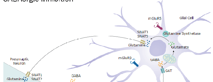
Metabotropic glutamate receptor 3 (mGluR3) is a 90-100 kDa, 7-transmembrane glycoprotein that belongs to group II of the C-family of G-protein coupled receptors. It is a presynaptic receptor expressed on both neurons and glia, whose activation reduces adenylate cyclase activity. Mature human mGluR3 is 857 amino acids in length and contains a 554 amino acid (aa) N-terminal extracellular domain (ECD) (aa 23-576). The ECD binds glutamate and forms homodimers. There is one alternative splice form that is soluble, 515 aa in length and shows a 96 aa substitution for aa 442-879. Over aa 25-507, human mGluR3 shares 97% aa sequence identity with mouse and rat mGluR3 and 67% aa sequence identity with hGluR2.

Key Product Details
Species Reactivity
Human
Applications
Immunohistochemistry, Western Blot
Label
Unconjugated
Antibody Source
Polyclonal Sheep IgG
Loading...
Product Specifications
Immunogen
Chinese hamster ovary cell line CHO-derived recombinant human mGluR3
Asp25-Ser507
Accession # Q14832
Asp25-Ser507
Accession # Q14832
Specificity
Detects human mGluR3 in direct ELISAs and Western blots. In direct ELISAs and Western blots, less than 10% cross-reactivity with recombinant human (rh) mGluR2, rhmGluR4, rhmGluR8 is observed, and less than 1% cross-reactivity with rhmGluR1, rhmGluR5, and rhmGluR7 is observed.
Clonality
Polyclonal
Host
Sheep
Isotype
IgG
Scientific Data Images for Human mGluR3 Antibody
mGluR3 in Human Brain.
mGluR3 was detected in immersion fixed paraffin-embedded sections of human brain (caudate nucleus) using Sheep Anti-Human mGluR3 Antigen Affinity-purified Polyclonal Antibody (Catalog # AF4668) at 10 µg/mL overnight at 4 °C. Tissue was stained using the Anti-Sheep HRP-DAB Cell & Tissue Staining Kit (brown; Catalog # CTS019) and counterstained with hematoxylin (blue). Specific labeling was localized to the plasma membrane and cytoplasm of neurons. View our protocol for Chromogenic IHC Staining of Paraffin-embedded Tissue Sections.Applications for Human mGluR3 Antibody
Application
Recommended Usage
Immunohistochemistry
5-15 µg/mL
Sample: Immersion fixed paraffin-embedded sections of human brain (caudate putamen) subjected to Antigen Retrieval Reagent-Basic (Catalog # CTS013)
Sample: Immersion fixed paraffin-embedded sections of human brain (caudate putamen) subjected to Antigen Retrieval Reagent-Basic (Catalog # CTS013)
Western Blot
0.1 µg/mL
Sample: Recombinant Human mGluR3
Sample: Recombinant Human mGluR3
Formulation, Preparation, and Storage
Purification
Antigen Affinity-purified
Reconstitution
Reconstitute at 0.2 mg/mL in sterile PBS. For liquid material, refer to CoA for concentration.
Loading...
Formulation
Lyophilized from a 0.2 μm filtered solution in PBS with Trehalose. *Small pack size (SP) is supplied either lyophilized or as a 0.2 µm filtered solution in PBS.
Shipping
Lyophilized product is shipped at ambient temperature. Liquid small pack size (-SP) is shipped with polar packs. Upon receipt, store immediately at the temperature recommended below.
Stability & Storage
Use a manual defrost freezer and avoid repeated freeze-thaw cycles.
- 12 months from date of receipt, -20 to -70 °C as supplied.
- 1 month, 2 to 8 °C under sterile conditions after reconstitution.
- 6 months, -20 to -70 °C under sterile conditions after reconstitution.
Calculators
Background: mGluR3
Long Name
Metabotropic Glutamate Receptor 3
Alternate Names
GPRC1C, GRM3
Gene Symbol
GRM3
UniProt
Additional mGluR3 Products
Product Documents for Human mGluR3 Antibody
Certificate of Analysis
To download a Certificate of Analysis, please enter a lot or batch number in the search box below.
Note: Certificate of Analysis not available for kit components.
Product Specific Notices for Human mGluR3 Antibody
For research use only
Related Research Areas
Customer Reviews for Human mGluR3 Antibody
There are currently no reviews for this product. Be the first to review Human mGluR3 Antibody and earn rewards!
Have you used Human mGluR3 Antibody?
Submit a review and receive an Amazon gift card!
$25/€18/£15/$25CAN/¥2500 Yen for a review with an image
$10/€7/£6/$10CAN/¥1110 Yen for a review without an image
Submit a review
Protocols
Find general support by application which include: protocols, troubleshooting, illustrated assays, videos and webinars.
- Antigen Retrieval Protocol (PIER)
- Antigen Retrieval for Frozen Sections Protocol
- Appropriate Fixation of IHC/ICC Samples
- Cellular Response to Hypoxia Protocols
- Chromogenic IHC Staining of Formalin-Fixed Paraffin-Embedded (FFPE) Tissue Protocol
- Chromogenic Immunohistochemistry Staining of Frozen Tissue
- ClariTSA™ Fluorophore Kits
- Detection & Visualization of Antibody Binding
- Fluorescent IHC Staining of Frozen Tissue Protocol
- Graphic Protocol for Heat-induced Epitope Retrieval
- Graphic Protocol for the Preparation and Fluorescent IHC Staining of Frozen Tissue Sections
- Graphic Protocol for the Preparation and Fluorescent IHC Staining of Paraffin-embedded Tissue Sections
- Graphic Protocol for the Preparation of Gelatin-coated Slides for Histological Tissue Sections
- IHC Sample Preparation (Frozen sections vs Paraffin)
- Immunofluorescent IHC Staining of Formalin-Fixed Paraffin-Embedded (FFPE) Tissue Protocol
- Immunohistochemistry (IHC) and Immunocytochemistry (ICC) Protocols
- Immunohistochemistry Frozen Troubleshooting
- Immunohistochemistry Paraffin Troubleshooting
- Preparing Samples for IHC/ICC Experiments
- Preventing Non-Specific Staining (Non-Specific Binding)
- Primary Antibody Selection & Optimization
- Protocol for Heat-Induced Epitope Retrieval (HIER)
- Protocol for Making a 4% Formaldehyde Solution in PBS
- Protocol for VisUCyte™ HRP Polymer Detection Reagent
- Protocol for the Preparation & Fixation of Cells on Coverslips
- Protocol for the Preparation and Chromogenic IHC Staining of Frozen Tissue Sections
- Protocol for the Preparation and Chromogenic IHC Staining of Frozen Tissue Sections - Graphic
- Protocol for the Preparation and Chromogenic IHC Staining of Paraffin-embedded Tissue Sections
- Protocol for the Preparation and Chromogenic IHC Staining of Paraffin-embedded Tissue Sections - Graphic
- Protocol for the Preparation and Fluorescent IHC Staining of Frozen Tissue Sections
- Protocol for the Preparation and Fluorescent IHC Staining of Paraffin-embedded Tissue Sections
- Protocol for the Preparation of Gelatin-coated Slides for Histological Tissue Sections
- R&D Systems Quality Control Western Blot Protocol
- TUNEL and Active Caspase-3 Detection by IHC/ICC Protocol
- The Importance of IHC/ICC Controls
- Troubleshooting Guide: Immunohistochemistry
- Troubleshooting Guide: Western Blot Figures
- Western Blot Conditions
- Western Blot Protocol
- Western Blot Protocol for Cell Lysates
- Western Blot Troubleshooting
- Western Blot Troubleshooting Guide
- View all Protocols, Troubleshooting, Illustrated assays and Webinars
FAQs for Human mGluR3 Antibody
Showing
1
-
1 of
1 FAQ
Showing All
-
Q: What was the antigen retrieval method for this product?
A: We used basic pH antigen retrieval (citrate buffer) with primary antibody incubation overnight at 4 degrees C at 10ug/mL.
Loading...
Associated Pathways