
Key Product Details
Species Reactivity
Validated:
Human, Mouse
Cited:
Human, Mouse
Applications
Validated:
Immunohistochemistry, Western Blot, Flow Cytometry, Immunocytochemistry, CyTOF-ready
Cited:
Immunohistochemistry-Frozen, Western Blot
Label
Unconjugated
Antibody Source
Monoclonal Rat IgG2A Clone # 772906
Loading...
Product Specifications
Immunogen
Mouse myeloma cell line NS0-derived recombinant mouse Desmocollin-1
Arg135-Lys691
Accession # P55849
Arg135-Lys691
Accession # P55849
Specificity
Detects mouse Desmocollin-1 in ELISAs and Western blots. In direct ELISAs, 100% cross-reactivity with
recombinant human Desmocollin-1 is observed, and no cross-reactivity with
recombinant mouse Desmocollin-2 or -3 is observed.
Clonality
Monoclonal
Host
Rat
Isotype
IgG2A
Scientific Data Images for Desmocollin-1 Antibody (772906)
Detection of Human and Mouse Desmocollin‑1 by Western Blot.
Western blot shows lysates of SK-Mel-28 human malignant melanoma cell line and B16-F1 mouse melanoma cell line. PVDF membrane was probed with 0.1 µg/mL of Rat Anti-Human/Mouse Desmocollin-1 Monoclonal Antibody (Catalog # MAB7367) followed by HRP-conjugated Anti-Rat IgG Secondary Antibody (Catalog # HAF005). A specific band was detected for Desmocollin-1 at approximately 98 kDa (as indicated). This experiment was conducted under reducing conditions and using Immunoblot Buffer Group 1.
Detection of Desmocollin-1 in B16‑F1 Mouse Cell Line by Flow Cytometry.
B16-F1 mouse melanoma cell line was stained with Rat Anti-Human/Mouse Desmocollin-1 Monoclonal Antibody (Catalog # MAB7367, filled histogram) or isotype control antibody (Catalog # MAB006, open histogram), followed by Allophycocyanin-conjugated Anti-Rat IgG Secondary Antibody (Catalog # F0113).
Detection of Desmocollin‑1 in A549 Human Cell Line by Flow Cytometry.
A549 human lung carcinoma cell line was stained with Rat Anti-Human/Mouse Desmocollin-1 Monoclonal Antibody (Catalog # MAB7367, filled histogram) or isotype control antibody (Catalog # MAB006, open histogram), followed by Allophycocyanin-conjugated Anti-Rat IgG Secondary Antibody (Catalog # F0113).
Desmocollin‑1 in A549 Human Cell Line.
Desmocollin-1 was detected in immersion fixed A549 human lung carcinoma cell line using Rat Anti-Human/Mouse Desmocollin-1 Monoclonal Antibody (Catalog # MAB7367) at 10 µg/mL for 3 hours at room temperature. Cells were stained using the NorthernLights™ 557-conjugated Anti-Rat IgG Secondary Antibody (red; Catalog # NL013) and counterstained with DAPI (blue). Specific staining was localized to cytoplasm and nuclei. View our protocol for Fluorescent ICC Staining of Cells on Coverslips.
Desmocollin‑1 in Mouse Skin.
Desmocollin-1 was detected in perfusion fixed frozen sections of mouse skin using Rat Anti-Human/Mouse Desmocollin-1 Monoclonal Antibody (Catalog # MAB7367) at 1.7 µg/mL overnight at 4 °C. Tissue was stained using the Anti-Rat HRP-DAB Cell & Tissue Staining Kit (brown; Catalog # CTS017) and counterstained with hematoxylin (blue). Specific staining was localized to hair root sheath. View our protocol for Chromogenic IHC Staining of Frozen Tissue Sections.Applications for Desmocollin-1 Antibody (772906)
Application
Recommended Usage
CyTOF-ready
Ready to be labeled using established conjugation methods. No BSA or other carrier proteins that could interfere with conjugation.
Flow Cytometry
0.25 µg/106 cells
Sample: B16‑F1 mouse melanoma cell line and A549 human lung carcinoma cell line
Sample: B16‑F1 mouse melanoma cell line and A549 human lung carcinoma cell line
Immunocytochemistry
8-25 µg/mL
Sample: Immersion fixed A549 human lung carcinoma cell line
Sample: Immersion fixed A549 human lung carcinoma cell line
Immunohistochemistry
1-25 µg/mL
Sample: Perfusion fixed frozen sections of mouse skin
Sample: Perfusion fixed frozen sections of mouse skin
Western Blot
0.1 µg/mL
Sample: SK‑Mel‑28 human malignant melanoma cell line and B16‑F1 mouse melanoma cell line
Sample: SK‑Mel‑28 human malignant melanoma cell line and B16‑F1 mouse melanoma cell line
Reviewed Applications
Read 2 reviews rated 5 using MAB7367 in the following applications:
Flow Cytometry Panel Builder
Bio-Techne Knows Flow Cytometry
Save time and reduce costly mistakes by quickly finding compatible reagents using the Panel Builder Tool.
Advanced Features
- Spectra Viewer - Custom analysis of spectra from multiple fluorochromes
- Spillover Popups - Visualize the spectra of individual fluorochromes
- Antigen Density Selector - Match fluorochrome brightness with antigen density

Formulation, Preparation, and Storage
Purification
Protein A or G purified from hybridoma culture supernatant
Reconstitution
Sterile PBS to a final concentration of 0.5 mg/mL. For liquid material, refer to CoA for concentration.
Loading...
Formulation
Lyophilized from a 0.2 μm filtered solution in PBS with Trehalose. *Small pack size (SP) is supplied either lyophilized or as a 0.2 µm filtered solution in PBS.
Shipping
Lyophilized product is shipped at ambient temperature. Liquid small pack size (-SP) is shipped with polar packs. Upon receipt, store immediately at the temperature recommended below.
Stability & Storage
Use a manual defrost freezer and avoid repeated freeze-thaw cycles.
- 12 months from date of receipt, -20 to -70 °C as supplied.
- 1 month, 2 to 8 °C under sterile conditions after reconstitution.
- 6 months, -20 to -70 °C under sterile conditions after reconstitution.
Calculators
Background: Desmocollin-1
DSC‑1 expression is induced by DSG-1. It serves as a component of desmosomes, forming a linkage that unites adjacent cells with cytoplasmic intermediate filaments. In particular, homodimeric DSC-1 may form heterotypic interactions with DSG-1 in-trans, and bind to the cytoskeleton intracellularly via plakophilin-1. Mature mouse DSC-1 is a 760 amino acid (aa) type I transmembrane glycoprotein (aa 135-691). The mature molecule contains a 557 aa extracellular region with five cadherin domains (aa 135-682), and a 172 aa cytoplasmic domain. There is one splice variant that shows an 11 aa substitution for aa 822-886. Over aa 135-691, mouse DSC‑1 shares 82% aa sequence identity with human DSC-1.
Alternate Names
CDHF1, Desmocollin1, DG2/DG3, DSC1
Gene Symbol
DSC1
UniProt
Additional Desmocollin-1 Products
Product Documents for Desmocollin-1 Antibody (772906)
Certificate of Analysis
To download a Certificate of Analysis, please enter a lot or batch number in the search box below.
Note: Certificate of Analysis not available for kit components.
Product Specific Notices for Desmocollin-1 Antibody (772906)
For research use only
Related Research Areas
Citations for Desmocollin-1 Antibody (772906)
 Powered by Bioz
Customer Reviews for Desmocollin-1 Antibody (772906) (2)
5 out of 5
2 Customer Ratings
Have you used Desmocollin-1 Antibody (772906)?
Submit a review and receive an Amazon gift card!
$25/€18/£15/$25CAN/¥2500 Yen for a review with an image
$10/€7/£6/$10CAN/¥1110 Yen for a review without an image
Submit a review
Customer Images
Showing
1
-
2 of
2 reviews
Showing All
Filter By:
-
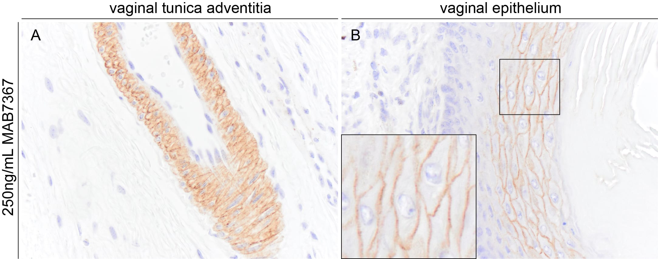
Application: Immunohistochemistry-ParaffinSample Tested: VaginaSpecies: MouseVerified Customer | Posted 04/18/2025Representative images demonstrating DSC 1 immunoreactivity in FFPE sections of mouse vagina. MAB7367 was used at 125 ng per mL and was left on tissue sections for 30min at room temperature.Requires proteinase induced epitope retrieval with 5 min proteinase K at room temperature

-
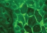
Application: Immunocytochemistry/ImmunofluorescenceSample Tested: Oral squamous cell carcinomaSpecies: HumanVerified Customer | Posted 11/04/2021
There are no reviews that match your criteria.
Protocols
Find general support by application which include: protocols, troubleshooting, illustrated assays, videos and webinars.
- 7-Amino Actinomycin D (7-AAD) Cell Viability Flow Cytometry Protocol
- Antigen Retrieval Protocol (PIER)
- Antigen Retrieval for Frozen Sections Protocol
- Appropriate Fixation of IHC/ICC Samples
- Cellular Response to Hypoxia Protocols
- Chromogenic IHC Staining of Formalin-Fixed Paraffin-Embedded (FFPE) Tissue Protocol
- Chromogenic Immunohistochemistry Staining of Frozen Tissue
- ClariTSA™ Fluorophore Kits
- Detection & Visualization of Antibody Binding
- Extracellular Membrane Flow Cytometry Protocol
- Flow Cytometry Protocol for Cell Surface Markers
- Flow Cytometry Protocol for Staining Membrane Associated Proteins
- Flow Cytometry Staining Protocols
- Flow Cytometry Troubleshooting Guide
- Fluorescent IHC Staining of Frozen Tissue Protocol
- Graphic Protocol for Heat-induced Epitope Retrieval
- Graphic Protocol for the Preparation and Fluorescent IHC Staining of Frozen Tissue Sections
- Graphic Protocol for the Preparation and Fluorescent IHC Staining of Paraffin-embedded Tissue Sections
- Graphic Protocol for the Preparation of Gelatin-coated Slides for Histological Tissue Sections
- ICC Cell Smear Protocol for Suspension Cells
- ICC Immunocytochemistry Protocol Videos
- ICC for Adherent Cells
- IHC Sample Preparation (Frozen sections vs Paraffin)
- Immunocytochemistry (ICC) Protocol
- Immunocytochemistry Troubleshooting
- Immunofluorescence of Organoids Embedded in Cultrex Basement Membrane Extract
- Immunofluorescent IHC Staining of Formalin-Fixed Paraffin-Embedded (FFPE) Tissue Protocol
- Immunohistochemistry (IHC) and Immunocytochemistry (ICC) Protocols
- Immunohistochemistry Frozen Troubleshooting
- Immunohistochemistry Paraffin Troubleshooting
- Intracellular Flow Cytometry Protocol Using Alcohol (Methanol)
- Intracellular Flow Cytometry Protocol Using Detergents
- Intracellular Nuclear Staining Flow Cytometry Protocol Using Detergents
- Intracellular Staining Flow Cytometry Protocol Using Alcohol Permeabilization
- Intracellular Staining Flow Cytometry Protocol Using Detergents to Permeabilize Cells
- Preparing Samples for IHC/ICC Experiments
- Preventing Non-Specific Staining (Non-Specific Binding)
- Primary Antibody Selection & Optimization
- Propidium Iodide Cell Viability Flow Cytometry Protocol
- Protocol for Heat-Induced Epitope Retrieval (HIER)
- Protocol for Liperfluo
- Protocol for Making a 4% Formaldehyde Solution in PBS
- Protocol for VisUCyte™ HRP Polymer Detection Reagent
- Protocol for the Characterization of Human Th22 Cells
- Protocol for the Characterization of Human Th9 Cells
- Protocol for the Fluorescent ICC Staining of Cell Smears - Graphic
- Protocol for the Fluorescent ICC Staining of Cultured Cells on Coverslips - Graphic
- Protocol for the Preparation & Fixation of Cells on Coverslips
- Protocol for the Preparation and Chromogenic IHC Staining of Frozen Tissue Sections
- Protocol for the Preparation and Chromogenic IHC Staining of Frozen Tissue Sections - Graphic
- Protocol for the Preparation and Chromogenic IHC Staining of Paraffin-embedded Tissue Sections
- Protocol for the Preparation and Chromogenic IHC Staining of Paraffin-embedded Tissue Sections - Graphic
- Protocol for the Preparation and Fluorescent ICC Staining of Cells on Coverslips
- Protocol for the Preparation and Fluorescent ICC Staining of Non-adherent Cells
- Protocol for the Preparation and Fluorescent ICC Staining of Stem Cells on Coverslips
- Protocol for the Preparation and Fluorescent IHC Staining of Frozen Tissue Sections
- Protocol for the Preparation and Fluorescent IHC Staining of Paraffin-embedded Tissue Sections
- Protocol for the Preparation of Gelatin-coated Slides for Histological Tissue Sections
- Protocol for the Preparation of a Cell Smear for Non-adherent Cell ICC - Graphic
- Protocol: Annexin V and PI Staining by Flow Cytometry
- Protocol: Annexin V and PI Staining for Apoptosis by Flow Cytometry
- R&D Systems Quality Control Western Blot Protocol
- TUNEL and Active Caspase-3 Detection by IHC/ICC Protocol
- The Importance of IHC/ICC Controls
- Troubleshooting Guide: Fluorokine Flow Cytometry Kits
- Troubleshooting Guide: Immunohistochemistry
- Troubleshooting Guide: Western Blot Figures
- Western Blot Conditions
- Western Blot Protocol
- Western Blot Protocol for Cell Lysates
- Western Blot Troubleshooting
- Western Blot Troubleshooting Guide
- View all Protocols, Troubleshooting, Illustrated assays and Webinars
Loading...