
FANCD2 Antibody (1290C)
R&D Systems | Catalog # MAB93691

Scientific images may be resized, cropped, or adjusted for brightness or contrast for presentation purposes. These updates are limited to presentation and do not affect the underlying scientific interpretation of the data. Where available, additional source data may be provided upon request.
Key Product Details
Validated by
Species Reactivity
Validated:
Cited:
Applications
Validated:
Cited:
Label
Antibody Source
Product Specifications
Immunogen
Glu11-Glu230
Accession # Q9BXW9
Specificity
Clonality
Host
Isotype
Scientific Data Images for FANCD2 Antibody (1290C)
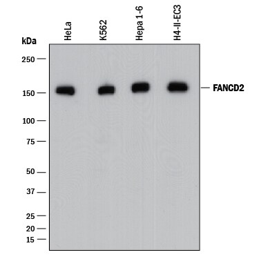
Detection of Human, Mouse, and Rat FANCD2 by Western Blot.
Western blot shows lysates of HeLa human cervical epithelial carcinoma cell line, K562 human chronic myelogenous leukemia cell line, Hepa 1-6 mouse hepatoma cell line, and H4-II-E-C3 rat hepatoma cell line. PVDF membrane was probed with 0.5 µg/mL of Rabbit Anti-Human/Mouse/Rat FANCD2 Monoclonal Antibody (Catalog # MAB93691) followed by HRP-conjugated Anti-Rabbit IgG Secondary Antibody (Catalog # HAF008). A specific band was detected for FANCD2 at approximately 150 kDa (as indicated). This experiment was conducted under reducing conditions and using Immunoblot Buffer Group 1.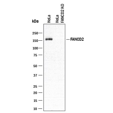
Western Blot Shows Human FANCD2 Specificity by Using Knockout Cell Line.
Western blot shows lysates of HeLa human cervical epithelial carcinoma parental cell line and FANCD2 knockout HeLa cell line (KO). PVDF membrane was probed with 0.5 µg/mL of Rabbit Anti-Human/Mouse/Rat FANCD2 Monoclonal Antibody (Catalog # MAB93691) followed by HRP-conjugated Anti-Rabbit IgG Secondary Antibody (Catalog # HAF008). A specific band was detected for FANCD2 at approximately 150 kDa (as indicated) in the parental HeLa cell line, but is not detectable in knockout HeLa cell line. This experiment was conducted under reducing conditions and using Immunoblot Buffer Group 1.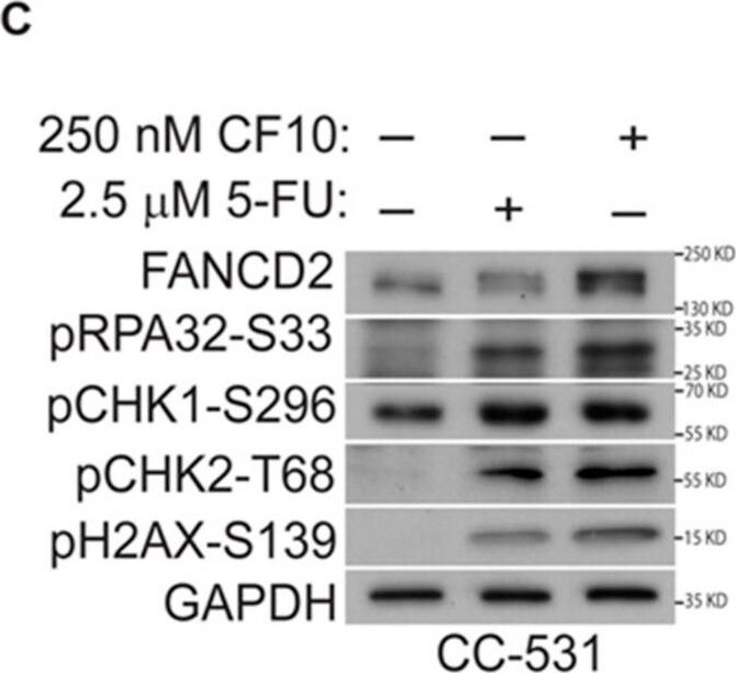
Detection of FANCD2 by Western Blot
CF10 activates the ATR/Chk1 pathway, consistent with replication stress, and causes DNA DSBs in CC531 cells. Western blots from CC531 cell lysates: (A) CF10 dose–response graphs demonstrate that a 250 nM dose is sufficient for ATR/Chk1 pathway activation and DNA DSBs; (B) equimolar 5-FU is not effective in ATR/Chk1 activation and DNA DSBs; and (C) a 10-fold greater 5-FU dose is relatively less effective than 250 nM CF10. * indicates mono-ubiquitnated FANCD2. Image collected and cropped by CiteAb from the following open publication (https://www.mdpi.com/2072-6694/16/7/1360), licensed under a CC-BY license. Not internally tested by R&D Systems.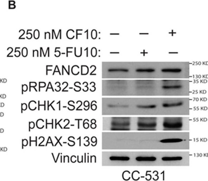
Detection of FANCD2 by Western Blot
CF10 activates the ATR/Chk1 pathway, consistent with replication stress, and causes DNA DSBs in CC531 cells. Western blots from CC531 cell lysates: (A) CF10 dose–response graphs demonstrate that a 250 nM dose is sufficient for ATR/Chk1 pathway activation and DNA DSBs; (B) equimolar 5-FU is not effective in ATR/Chk1 activation and DNA DSBs; and (C) a 10-fold greater 5-FU dose is relatively less effective than 250 nM CF10. * indicates mono-ubiquitnated FANCD2. Image collected and cropped by CiteAb from the following open publication (https://www.mdpi.com/2072-6694/16/7/1360), licensed under a CC-BY license. Not internally tested by R&D Systems.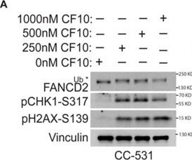
Detection of FANCD2 by Western Blot
CF10 activates the ATR/Chk1 pathway, consistent with replication stress, and causes DNA DSBs in CC531 cells. Western blots from CC531 cell lysates: (A) CF10 dose–response graphs demonstrate that a 250 nM dose is sufficient for ATR/Chk1 pathway activation and DNA DSBs; (B) equimolar 5-FU is not effective in ATR/Chk1 activation and DNA DSBs; and (C) a 10-fold greater 5-FU dose is relatively less effective than 250 nM CF10. * indicates mono-ubiquitnated FANCD2. Image collected and cropped by CiteAb from the following open publication (https://www.mdpi.com/2072-6694/16/7/1360), licensed under a CC-BY license. Not internally tested by R&D Systems.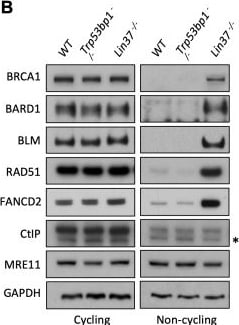
Detection of afpdau30tbxqqdhcvrla.jpg by Western Blot
LIN37 suppresses HR protein expression in non-cycling cells.(A) Gene ontology (GO) analysis of genes upregulated in non-cycling Lig4−/−:Lin37−/− abl pre-B cells. Enriched GO terms with the 20 lowest p-values (on the right of each bar) are shown. (B) Western blot analysis of indicated proteins in cycling or non-cycling WT, Trp53bp1−/−, and Lin37−/− abl pre-B cells. HR, homologous recombination; WT, wild type. Image collected and cropped by CiteAb from the following open publication (https://pubmed.ncbi.nlm.nih.gov/34477552), licensed under a CC-BY license. Not internally tested by R&D Systems.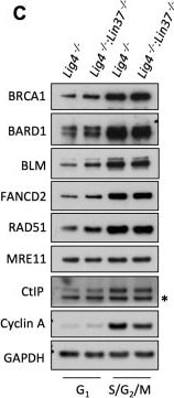
Detection of beax31b3yqaeebr9muas.jpg by Western Blot
LIN37 function in DNA end protection is restricted to G0.(A, B) Western blot analysis of indicated proteins in cycling and non-cycling abl pre-B cells (A) or MCF10A cells (B). (C, D) Western blot analysis of indicated proteins in cycling G1 or S/G2/M abl pre-B cells (C) or MCF10A cells (D), isolated by flow cytometric cell sorting based on the PIP-FUCCI reporter. Representative of two independent experiments. Asterisk indicates non-specific recognizing bands. (E, F) Flow cytometric analysis of chromatin-bound RPA and gamma H2AX before and after IR treatment of G1-phase Lig4−/−, Lig4−/−:Trp53bp1−/− and Lig4−/−:Lin37−/− abl pre-B cells (E) or WT, Trp53bp1−/−, and Lin37−/− MCF10A cells (F). Representative of three experiments. IR, ionizing radiation; WT, wild type.Figure 7—source data 1.RNA-Seq result and GO analysis in cycling G1Lig4−/− and Lig4−/−:Lin37−/− abl pre-B cells.GO, gene ontology; RNA-Seq, RNA sequencing.Figure 7—source data 2.GO analysis of genes upregulated in non-cycling G0 and/or cycling G1Lig4−/−:Lin37−/− abl pre-B cells.GO, gene ontology.RNA-Seq result and GO analysis in cycling G1Lig4−/− and Lig4−/−:Lin37−/− abl pre-B cells.GO, gene ontology; RNA-Seq, RNA sequencing.GO analysis of genes upregulated in non-cycling G0 and/or cycling G1Lig4−/−:Lin37−/− abl pre-B cells.GO, gene ontology.Identification of G1-phase cells in proliferating cells.(A, B) Flow cytometric analysis of EdU incorporation and DNA content (7-AAD) of cycling Lig4−/−, Lig4−/−:Trp53bp1−/−, and Lig4−/−:Lin37−/− abl pre-B cells (A) or cycling WT, Trp53bp1−/−, and Lin37−/− MCF10A cells (B). Cells were treated (+EdU) or not treated (−EdU) with EdU and analyses were carried out before or after IR. The percentage of G1-phase cells is shown. IR, ionizing radiation; WT, wild type. Image collected and cropped by CiteAb from the following open publication (https://pubmed.ncbi.nlm.nih.gov/34477552), licensed under a CC-BY license. Not internally tested by R&D Systems.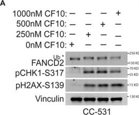
Detection of bsj8zokabieoi4xhbttv.jpg by Western Blot
CF10 activates the ATR/Chk1 pathway, consistent with replication stress, and causes DNA DSBs in CC531 cells. Western blots from CC531 cell lysates: (A) CF10 dose–response graphs demonstrate that a 250 nM dose is sufficient for ATR/Chk1 pathway activation and DNA DSBs; (B) equimolar 5-FU is not effective in ATR/Chk1 activation and DNA DSBs; and (C) a 10-fold greater 5-FU dose is relatively less effective than 250 nM CF10. * indicates mono-ubiquitnated FANCD2. Image collected and cropped by CiteAb from the following open publication (https://www.mdpi.com/2072-6694/16/7/1360), licensed under a CC-BY license. Not internally tested by R&D Systems.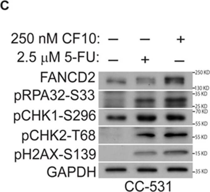
Detection of c7qkvvj0koq9cvooruff.jpg by Western Blot
CF10 activates the ATR/Chk1 pathway, consistent with replication stress, and causes DNA DSBs in CC531 cells. Western blots from CC531 cell lysates: (A) CF10 dose–response graphs demonstrate that a 250 nM dose is sufficient for ATR/Chk1 pathway activation and DNA DSBs; (B) equimolar 5-FU is not effective in ATR/Chk1 activation and DNA DSBs; and (C) a 10-fold greater 5-FU dose is relatively less effective than 250 nM CF10. * indicates mono-ubiquitnated FANCD2. Image collected and cropped by CiteAb from the following open publication (https://www.mdpi.com/2072-6694/16/7/1360), licensed under a CC-BY license. Not internally tested by R&D Systems.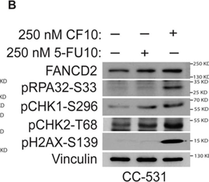
Detection of gx3yg3gne4afqelpe86c.jpg by Western Blot
CF10 activates the ATR/Chk1 pathway, consistent with replication stress, and causes DNA DSBs in CC531 cells. Western blots from CC531 cell lysates: (A) CF10 dose–response graphs demonstrate that a 250 nM dose is sufficient for ATR/Chk1 pathway activation and DNA DSBs; (B) equimolar 5-FU is not effective in ATR/Chk1 activation and DNA DSBs; and (C) a 10-fold greater 5-FU dose is relatively less effective than 250 nM CF10. * indicates mono-ubiquitnated FANCD2. Image collected and cropped by CiteAb from the following open publication (https://www.mdpi.com/2072-6694/16/7/1360), licensed under a CC-BY license. Not internally tested by R&D Systems.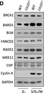
Detection of FANCD2 by Western Blot
LIN37 function in DNA end protection is restricted to G0.(A, B) Western blot analysis of indicated proteins in cycling and non-cycling abl pre-B cells (A) or MCF10A cells (B). (C, D) Western blot analysis of indicated proteins in cycling G1 or S/G2/M abl pre-B cells (C) or MCF10A cells (D), isolated by flow cytometric cell sorting based on the PIP-FUCCI reporter. Representative of two independent experiments. Asterisk indicates non-specific recognizing bands. (E, F) Flow cytometric analysis of chromatin-bound RPA and gamma H2AX before and after IR treatment of G1-phase Lig4−/−, Lig4−/−:Trp53bp1−/− and Lig4−/−:Lin37−/− abl pre-B cells (E) or WT, Trp53bp1−/−, and Lin37−/− MCF10A cells (F). Representative of three experiments. IR, ionizing radiation; WT, wild type.Figure 7—source data 1.RNA-Seq result and GO analysis in cycling G1Lig4−/− and Lig4−/−:Lin37−/− abl pre-B cells.GO, gene ontology; RNA-Seq, RNA sequencing.Figure 7—source data 2.GO analysis of genes upregulated in non-cycling G0 and/or cycling G1Lig4−/−:Lin37−/− abl pre-B cells.GO, gene ontology.RNA-Seq result and GO analysis in cycling G1Lig4−/− and Lig4−/−:Lin37−/− abl pre-B cells.GO, gene ontology; RNA-Seq, RNA sequencing.GO analysis of genes upregulated in non-cycling G0 and/or cycling G1Lig4−/−:Lin37−/− abl pre-B cells.GO, gene ontology.Identification of G1-phase cells in proliferating cells.(A, B) Flow cytometric analysis of EdU incorporation and DNA content (7-AAD) of cycling Lig4−/−, Lig4−/−:Trp53bp1−/−, and Lig4−/−:Lin37−/− abl pre-B cells (A) or cycling WT, Trp53bp1−/−, and Lin37−/− MCF10A cells (B). Cells were treated (+EdU) or not treated (−EdU) with EdU and analyses were carried out before or after IR. The percentage of G1-phase cells is shown. IR, ionizing radiation; WT, wild type. Image collected and cropped by CiteAb from the following open publication (https://pubmed.ncbi.nlm.nih.gov/34477552), licensed under a CC-BY license. Not internally tested by R&D Systems.Applications for FANCD2 Antibody (1290C)
Knockout Validated
Western Blot
Sample: HeLa human cervical epithelial carcinoma cell line, K562 human chronic myelogenous leukemia cell line, Hepa 1‑6 mouse hepatoma cell line, and H4‑II‑E‑C3 rat hepatoma cell line
Formulation, Preparation, and Storage
Purification
Reconstitution
Reconstitute at 0.5 mg/mL in sterile PBS. For liquid material, refer to CoA for concentration.
Formulation
Shipping
Stability & Storage
- 12 months from date of receipt, -20 to -70 °C as supplied.
- 1 month, 2 to 8 °C under sterile conditions after reconstitution.
- 6 months, -20 to -70 °C under sterile conditions after reconstitution.
Calculators
Background: FANCD2
Long Name
Alternate Names
Gene Symbol
UniProt
Additional FANCD2 Products
Product Documents for FANCD2 Antibody (1290C)
Certificate of Analysis
To download a Certificate of Analysis, please enter a lot or batch number in the search box below.
Note: Certificate of Analysis not available for kit components.
Product Specific Notices for FANCD2 Antibody (1290C)
For research use only
Citations for FANCD2 Antibody (1290C)
 Powered by Bioz
Customer Reviews for FANCD2 Antibody (1290C)
There are currently no reviews for this product. Be the first to review FANCD2 Antibody (1290C) and earn rewards!
Have you used FANCD2 Antibody (1290C)?
Submit a review and receive an Amazon gift card!
$25/€18/£15/$25CAN/¥2500 for a review with an image
$10/€7/£6/$10CAN/¥1110 for a review without an image
Submit a review
Protocols
Find general support by application which include: protocols, troubleshooting, illustrated assays, videos and webinars.
- Cellular Response to Hypoxia Protocols
- R&D Systems Quality Control Western Blot Protocol
- Troubleshooting Guide: Western Blot Figures
- Western Blot Conditions
- Western Blot Protocol
- Western Blot Protocol for Cell Lysates
- Western Blot Troubleshooting
- Western Blot Troubleshooting Guide
- View all Protocols, Troubleshooting, Illustrated assays and Webinars