
Key Product Details
Species Reactivity
Validated:
Human, Mouse, Rat
Cited:
Mouse
Applications
Validated:
Immunohistochemistry, Western Blot, Simple Western
Cited:
Immunohistochemistry
Label
Unconjugated
Antibody Source
Polyclonal Sheep IgG
Loading...
Product Specifications
Immunogen
E. coli-derived recombinant human PKC iota
Ile455-Val596
Accession # P41743
Ile455-Val596
Accession # P41743
Specificity
Detects human, mouse and rat PKC iota / lambda / zeta in Western blots.
Clonality
Polyclonal
Host
Sheep
Isotype
IgG
Scientific Data Images for PKC iota/lambda/zeta Antibody
Detection of Human, Mouse, and Rat PKC iota / lambda / zeta by Western Blot.
Western blot shows lysates of A431 human epithelial carcinoma cell line, DU145 human prostate carcinoma cell line, Balb/3T3 mouse embryonic fibroblast cell line, and PC-12 rat adrenal pheochromocytoma cell line. PVDF membrane was probed with 1 µg/mL Sheep Anti-Human/Mouse/Rat PKC iota/lambda/zeta Cross-reactive Antigen Affinity-purified Polyclonal Antibody (Catalog # AF4465) followed by HRP-conjugated Anti-Sheep IgG Secondary Antibody (Catalog # HAF016). For additional reference, recombinant human PKC iota, PKC zeta, and PKC alpha (2 ng/lane) were included. Specific bands were detected at approximately 22 kDa for recombinant PKC iota/zeta and ~80 kDa for natural PKC iota/zeta (as indicated). This experiment was conducted under reducing conditions and using Immunoblot Buffer Group 1.
PKC iota / lambda / zeta in Human Pancreas.
PKC iota/lambda/zeta was detected in immersion fixed paraffin-embedded sections of human pancreas using Sheep Anti-Human/Mouse/Rat PKC iota/lambda/zeta Cross-reactive Antigen Affinity-purified Polyclonal Antibody (Catalog # AF4465) at 15 µg/mL overnight at 4 °C. Tissue was stained using the Anti-Sheep HRP-DAB Cell & Tissue Staining Kit (brown; Catalog # CTS019) and counterstained with hematoxylin (blue). View our protocol for Chromogenic IHC Staining of Paraffin-embedded Tissue Sections.This application has not been tested in mouse or rat samples.
Detection of Human PKC iota / lambda / zeta by Simple WesternTM.
Simple Western shows lysates of A431 human epithelial carcinoma cell line, loaded at 0.5 mg/ml. A specific band was detected for PKC iota / lambda / zeta at approximately 80 kDa (as indicated) using 10 µg/mL of Sheep Anti-Human/Mouse/Rat PKC iota / lambda / zeta Antigen Affinity-purified Polyclonal Antibody (Catalog # AF4465). This experiment was conducted under reducing conditions and using the 12-230kDa separation system.Applications for PKC iota/lambda/zeta Antibody
Application
Recommended Usage
Immunohistochemistry
5-15 µg/mL
Sample: Immersion fixed paraffin-embedded sections of human pancreas
Sample: Immersion fixed paraffin-embedded sections of human pancreas
Simple Western
10 µg/mL
Sample: A431 human epithelial carcinoma cell line
Sample: A431 human epithelial carcinoma cell line
Western Blot
1 µg/mL
Sample: A431 human epithelial carcinoma cell line, DU145 human prostate carcinoma cell line, Balb/3T3 mouse embryonic fibroblast cell line, and PC-12 rat adrenal pheochromocytoma cell line
Sample: A431 human epithelial carcinoma cell line, DU145 human prostate carcinoma cell line, Balb/3T3 mouse embryonic fibroblast cell line, and PC-12 rat adrenal pheochromocytoma cell line
Reviewed Applications
Read 1 review rated 4 using AF4465 in the following applications:
Formulation, Preparation, and Storage
Purification
Antigen Affinity-purified
Reconstitution
Reconstitute at 0.2 mg/mL in sterile PBS. For liquid material, refer to CoA for concentration.
Loading...
Formulation
Lyophilized from a 0.2 μm filtered solution in PBS with Trehalose. See Certificate of Analysis for details.
*Small pack size (-SP) is supplied either lyophilized or as a 0.2 µm filtered solution in PBS.
*Small pack size (-SP) is supplied either lyophilized or as a 0.2 µm filtered solution in PBS.
Shipping
Lyophilized product is shipped at ambient temperature. Liquid small pack size (-SP) is shipped with polar packs. Upon receipt, store immediately at the temperature recommended below.
Stability & Storage
Use a manual defrost freezer and avoid repeated freeze-thaw cycles.
- 12 months from date of receipt, -20 to -70 °C as supplied.
- 1 month, 2 to 8 °C under sterile conditions after reconstitution.
- 6 months, -20 to -70 °C under sterile conditions after reconstitution.
Calculators
Background: PKC iota/lambda/zeta
NF‑ kappa B activation. Furthermore, insulin-stimulated atypical PKC activation has been directly implicated in the translocation of GLUT4 and glucose uptake in adipocytes.
Long Name
Protein Kinase C iota/lambda/zeta
UniProt
Additional PKC iota/lambda/zeta Products
Product Documents for PKC iota/lambda/zeta Antibody
Certificate of Analysis
To download a Certificate of Analysis, please enter a lot or batch number in the search box below.
Note: Certificate of Analysis not available for kit components.
Product Specific Notices for PKC iota/lambda/zeta Antibody
For research use only
Related Research Areas
Citations for PKC iota/lambda/zeta Antibody
 Powered by Bioz
Customer Reviews for PKC iota/lambda/zeta Antibody (1)
4 out of 5
1 Customer Rating
Have you used PKC iota/lambda/zeta Antibody?
Submit a review and receive an Amazon gift card!
$25/€18/£15/$25CAN/¥2500 Yen for a review with an image
$10/€7/£6/$10CAN/¥1110 Yen for a review without an image
Submit a review
Customer Images
Showing
1
-
1 of
1 review
Showing All
Filter By:
-
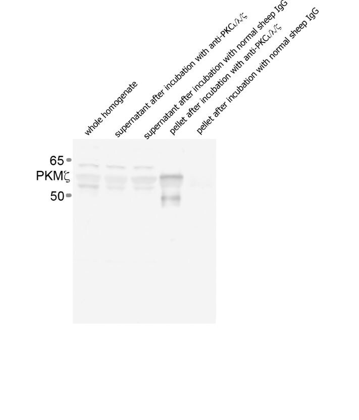
Application: ImmunoprecipitationSample Tested: HippocampusSpecies: MouseVerified Customer | Posted 07/31/2015

There are no reviews that match your criteria.
Protocols
Find general support by application which include: protocols, troubleshooting, illustrated assays, videos and webinars.
- Antigen Retrieval Protocol (PIER)
- Antigen Retrieval for Frozen Sections Protocol
- Appropriate Fixation of IHC/ICC Samples
- Cellular Response to Hypoxia Protocols
- Chromogenic IHC Staining of Formalin-Fixed Paraffin-Embedded (FFPE) Tissue Protocol
- Chromogenic Immunohistochemistry Staining of Frozen Tissue
- ClariTSA™ Fluorophore Kits
- Detection & Visualization of Antibody Binding
- Fluorescent IHC Staining of Frozen Tissue Protocol
- Graphic Protocol for Heat-induced Epitope Retrieval
- Graphic Protocol for the Preparation and Fluorescent IHC Staining of Frozen Tissue Sections
- Graphic Protocol for the Preparation and Fluorescent IHC Staining of Paraffin-embedded Tissue Sections
- Graphic Protocol for the Preparation of Gelatin-coated Slides for Histological Tissue Sections
- IHC Sample Preparation (Frozen sections vs Paraffin)
- Immunofluorescent IHC Staining of Formalin-Fixed Paraffin-Embedded (FFPE) Tissue Protocol
- Immunohistochemistry (IHC) and Immunocytochemistry (ICC) Protocols
- Immunohistochemistry Frozen Troubleshooting
- Immunohistochemistry Paraffin Troubleshooting
- Preparing Samples for IHC/ICC Experiments
- Preventing Non-Specific Staining (Non-Specific Binding)
- Primary Antibody Selection & Optimization
- Protocol for Heat-Induced Epitope Retrieval (HIER)
- Protocol for Making a 4% Formaldehyde Solution in PBS
- Protocol for VisUCyte™ HRP Polymer Detection Reagent
- Protocol for the Preparation & Fixation of Cells on Coverslips
- Protocol for the Preparation and Chromogenic IHC Staining of Frozen Tissue Sections
- Protocol for the Preparation and Chromogenic IHC Staining of Frozen Tissue Sections - Graphic
- Protocol for the Preparation and Chromogenic IHC Staining of Paraffin-embedded Tissue Sections
- Protocol for the Preparation and Chromogenic IHC Staining of Paraffin-embedded Tissue Sections - Graphic
- Protocol for the Preparation and Fluorescent IHC Staining of Frozen Tissue Sections
- Protocol for the Preparation and Fluorescent IHC Staining of Paraffin-embedded Tissue Sections
- Protocol for the Preparation of Gelatin-coated Slides for Histological Tissue Sections
- R&D Systems Quality Control Western Blot Protocol
- TUNEL and Active Caspase-3 Detection by IHC/ICC Protocol
- The Importance of IHC/ICC Controls
- Troubleshooting Guide: Immunohistochemistry
- Troubleshooting Guide: Western Blot Figures
- Western Blot Conditions
- Western Blot Protocol
- Western Blot Protocol for Cell Lysates
- Western Blot Troubleshooting
- Western Blot Troubleshooting Guide
- View all Protocols, Troubleshooting, Illustrated assays and Webinars
FAQs for PKC iota/lambda/zeta Antibody
Showing
1
-
1 of
1 FAQ
Showing All
-
Q: Does this antibody work as an IP antibody to pull-down PKCiota/lambda and their phosphorylated form?
A:
For product AF4465, this item has not yet been tested in an immunoprecipitation assay. This antibody is manufactured by our sister company R&D Systems and has only been tested so far in western blot and IHC per the R&D Systems site. The immunogen of the antibody is found between aa 455-596 of the human Protein kinase C iota type protein. This sequence encompasses one phosphorylation site (aa 564), and therefore has the potential to recognize the phosphorylated and non-phosphorylated forms of the protein; with exception of p Thr 564. This, of course, is hypothetical as the antibody has not yet been tested against the phosphorylated forms of the proteins.
Loading...