RAB27A (Ras-related protein Rab 27A; also GTP-binding protein Ram) is a 27-28 kDa member of the Rab27 subfamily, Rab family, Small GTPase superfamily of proteins. It is widely expressed, and found in cells diverse as mast cells, cytotoxic T cells, melanocytes, retinal pigment epithelium and pancreatic beta -cells. RAB27A plays a key role in the secretion of specialized lysosomes termed secretory lysosomes. In melanocytes, for example, RAB27A is incorporated into the melanosome membrane where it serves as a docking factor for melanophilin and myosin-Va, regulating melanosome transport to, and concentration at, sites of release. Human RAB27A is 221 amino acids (aa) in length. It contains multiple Rab family and subfamily motifs, and concludes with a C-terminal CXC prenylation sequence (aa 219‑221). There is one potential splice variant that shows a deletion of aa 146-153. Over aa 135-218, human RAB27A shares 92% and 94% aa sequence identity with mouse Rab27A and rat RAB27A, respectively.

Key Product Details
Species Reactivity
Validated:
Human, Mouse, Rat
Cited:
Human, Mouse
Applications
Validated:
Western Blot, Immunocytochemistry, Simple Western, Immunoprecipitation
Cited:
Immunohistochemistry, Western Blot, Flow Cytometry
Label
Unconjugated
Antibody Source
Polyclonal Sheep IgG
Loading...
Product Specifications
Immunogen
E. coli-derived recombinant human RAB27A
Ser135-Ala218
Accession # P51159
Ser135-Ala218
Accession # P51159
Specificity
Detects human, mouse, and rat RAB27A in Western blots and detects recombinant human RAB27A in direct ELISAs. In direct ELISAs, less than 1% cross-reactivity with recombinant human RAB27B is observed.
Clonality
Polyclonal
Host
Sheep
Isotype
IgG
Scientific Data Images for Rab27a Antibody
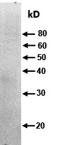
Detection of Human, Mouse, and Rat RAB27A by Western Blot.
Western blot shows lysates of K562 human chronic myelogenous leukemia cell line, 786-O human renal cell adenocarcinoma cell line, MCF-7 human breast cancer cell line, Jurkat human acute T cell leukemia cell line, Rat-2 rat embryonic fibroblast cell line, and Neuro-2A mouse neuroblastoma cell line. PVDF membrane was probed with 0.5 µg/mL of Sheep Anti-Human/Mouse/Rat RAB27A Antigen Affinity-purified Polyclonal Antibody (Catalog # AF7245) followed by HRP-conjugated Anti-Sheep IgG Secondary Antibody (Catalog # HAF016). A specific band was detected for RAB27A at approximately 28 kDa (as indicated). This experiment was conducted under reducing conditions and using Immunoblot Buffer Group 1.
RAB27A in U937 Human Cell Line.
RAB27A was detected in immersion fixed U937 human histiocytic lymphoma cell line using Sheep Anti-Human/Mouse/Rat RAB27A Antigen Affinity-purified Polyclonal Antibody (Catalog # AF7245) at 10 µg/mL for 3 hours at room temperature. Cells were stained using the NorthernLights™ 557-conjugated Anti-Sheep IgG Secondary Antibody (red; Catalog # NL010) and counterstained with DAPI (blue). Specific staining was localized to cytoplasm. View our protocol for Fluorescent ICC Staining of Non-adherent Cells.
Detection of Human Rab27a by Simple WesternTM.
Simple Western lane view shows lysates of K562 human chronic myelogenous leukemia cell line, loaded at 0.2 mg/mL. A specific band was detected for Rab27a at approximately 32 kDa (as indicated) using 5 µg/mL of Sheep Anti-Human/Mouse/Rat Rab27a Antigen Affinity-purified Polyclonal Antibody (Catalog # AF7245) followed by 1:50 dilution of HRP-conjugated Anti-Sheep IgG Secondary Antibody (Catalog # HAF016). This experiment was conducted under reducing conditions and using the 12-230 kDa separation system.
Detection of Rab27a by Immunoprecipitation.
HAP1 near-haploid human cell line lysates were prepared and immunoprecipitation was performed using 2 ug of Sheep Anti-Human/Mouse/Rat Rab27a Antigen Affinity-purified Polyclonal Antibody (Catalog # AF7245) pre-coupled to Dynabeads Protein G. Immunoprecipitated Rab27a was detected in Western Blot with a Rabbit Rab27a antibody. The Ponceau stained transfer of the blot is shown. SM=4% starting material; UB=4% unbound fraction; IP=immunoprecipitate; HC=antibody heavy chain. Image, protocol and testing courtesy of YCharOS Inc. (ycharos.com).
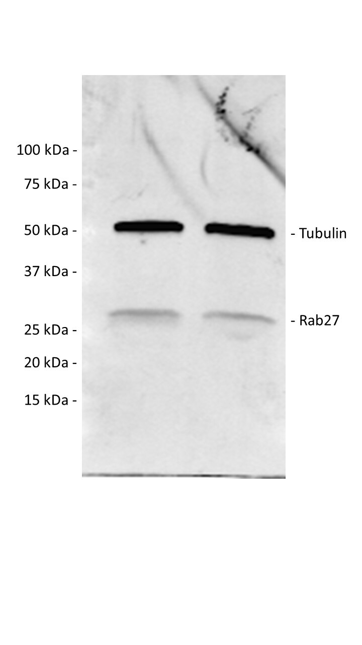
Detection of Rab27a by Western Blot
Analysis of EVs release by drug-sensitive (NCI-H460 and K562) and MDR (NCI-H460/R and K562Dox) counterpart cells. (A) Comparison of the number of EVs released, estimated by NTA measurements of EVs preparations. Left panel: NTA profile of EVs; right panel: quantification of number of particles normalised to the number of donor cells. * p ≤ 0.05. (B) Comparison of the levels of proteins associated to the release of EVs between drug-sensitive (DS: NCI-H460 or K562) and MDR (NCI-H460/R or K562Dox) counterpart cells, analysed by Western blot. Left panel: Representative blots from at least 3 independent experiments; right panel: densitometry analysis expressed after normalisation of the values obtained for each protein with the values obtained for actin (and further expressed in relation to drug-sensitive (DS) cells). * p ≤ 0.05. Image collected and cropped by CiteAb from the following open publication (https://pubmed.ncbi.nlm.nih.gov/34831110), licensed under a CC-BY license. Not internally tested by R&D Systems.Applications for Rab27a Antibody
Application
Recommended Usage
Immunocytochemistry
5-15 µg/mL
Sample: Immersion fixed U937 human histiocytic lymphoma cell line
Sample: Immersion fixed U937 human histiocytic lymphoma cell line
Immunoprecipitation
2 µg/1 mg cell lysate
Sample: Cell lysate of HAP1 near-haploid human cell line
Sample: Cell lysate of HAP1 near-haploid human cell line
Simple Western
5 µg/mL
Sample: K562 human chronic myelogenous leukemia cell line
Sample: K562 human chronic myelogenous leukemia cell line
Western Blot
0.5 µg/mL
Sample: K562 human chronic myelogenous leukemia cell line, 786‑O human renal cell adenocarcinoma cell line, MCF‑7 human breast cancer cell line, Jurkat human acute T cell leukemia cell line, Rat‑2 rat embryonic fibroblast cell line, and Neuro‑2A mouse neuroblastoma cell line
Sample: K562 human chronic myelogenous leukemia cell line, 786‑O human renal cell adenocarcinoma cell line, MCF‑7 human breast cancer cell line, Jurkat human acute T cell leukemia cell line, Rat‑2 rat embryonic fibroblast cell line, and Neuro‑2A mouse neuroblastoma cell line
Reviewed Applications
Read 2 reviews rated 3 using AF7245 in the following applications:
Formulation, Preparation, and Storage
Purification
Antigen Affinity-purified
Reconstitution
Sterile PBS to a final concentration of 0.2 mg/mL. For liquid material, refer to CoA for concentration.
Loading...
Formulation
Lyophilized from a 0.2 μm filtered solution in PBS with Trehalose. See Certificate of Analysis for details.
*Small pack size (-SP) is supplied either lyophilized or as a 0.2 µm filtered solution in PBS.
*Small pack size (-SP) is supplied either lyophilized or as a 0.2 µm filtered solution in PBS.
Shipping
Lyophilized product is shipped at ambient temperature. Liquid small pack size (-SP) is shipped with polar packs. Upon receipt, store immediately at the temperature recommended below.
Stability & Storage
Use a manual defrost freezer and avoid repeated freeze-thaw cycles.
- 12 months from date of receipt, -20 to -70 °C as supplied.
- 1 month, 2 to 8 °C under sterile conditions after reconstitution.
- 6 months, -20 to -70 °C under sterile conditions after reconstitution.
Calculators
Background: Rab27a
Long Name
RAs Genes from Brain Protein 27A
Alternate Names
GS2, HsT18676, RAM
Entrez Gene IDs
5873 (Human)
Gene Symbol
RAB27A
UniProt
Additional Rab27a Products
Product Documents for Rab27a Antibody
Certificate of Analysis
To download a Certificate of Analysis, please enter a lot or batch number in the search box below.
Note: Certificate of Analysis not available for kit components.
Product Specific Notices for Rab27a Antibody
For research use only
Related Research Areas
Citations for Rab27a Antibody
 Powered by Bioz
Customer Reviews for Rab27a Antibody (2)
3 out of 5
2 Customer Ratings
Have you used Rab27a Antibody?
Submit a review and receive an Amazon gift card!
$25/€18/£15/$25CAN/¥2500 Yen for a review with an image
$10/€7/£6/$10CAN/¥1110 Yen for a review without an image
Submit a review
Customer Images
Showing
1
-
2 of
2 reviews
Showing All
Filter By:
-
Application: Western BlotSample Tested: ISE6 CellsSpecies: ixodes scapularisVerified Customer | Posted 07/17/2020This antibody doesn't cross-react well with Rab27a expressed by ISE6 cells.
 Bio-Techne ResponseThis review was submitted through the legacy Novus Innovators Program, reflecting a new species or application tested on a primary antibody.
 -
Application: Western BlotSample Tested: Huh-7 human hepatoma cell lineSpecies: HumanVerified Customer | Posted 08/20/2018
There are no reviews that match your criteria.
Protocols
Find general support by application which include: protocols, troubleshooting, illustrated assays, videos and webinars.
- Appropriate Fixation of IHC/ICC Samples
- Cellular Response to Hypoxia Protocols
- ClariTSA™ Fluorophore Kits
- Detection & Visualization of Antibody Binding
- ICC Cell Smear Protocol for Suspension Cells
- ICC Immunocytochemistry Protocol Videos
- ICC for Adherent Cells
- Immunocytochemistry (ICC) Protocol
- Immunocytochemistry Troubleshooting
- Immunofluorescence of Organoids Embedded in Cultrex Basement Membrane Extract
- Immunohistochemistry (IHC) and Immunocytochemistry (ICC) Protocols
- Immunoprecipitation Protocol
- Preparing Samples for IHC/ICC Experiments
- Preventing Non-Specific Staining (Non-Specific Binding)
- Primary Antibody Selection & Optimization
- Protocol for VisUCyte™ HRP Polymer Detection Reagent
- Protocol for the Fluorescent ICC Staining of Cell Smears - Graphic
- Protocol for the Fluorescent ICC Staining of Cultured Cells on Coverslips - Graphic
- Protocol for the Preparation and Fluorescent ICC Staining of Cells on Coverslips
- Protocol for the Preparation and Fluorescent ICC Staining of Non-adherent Cells
- Protocol for the Preparation and Fluorescent ICC Staining of Stem Cells on Coverslips
- Protocol for the Preparation of a Cell Smear for Non-adherent Cell ICC - Graphic
- R&D Systems Quality Control Western Blot Protocol
- TUNEL and Active Caspase-3 Detection by IHC/ICC Protocol
- The Importance of IHC/ICC Controls
- Troubleshooting Guide: Western Blot Figures
- Western Blot Conditions
- Western Blot Protocol
- Western Blot Protocol for Cell Lysates
- Western Blot Troubleshooting
- Western Blot Troubleshooting Guide
- View all Protocols, Troubleshooting, Illustrated assays and Webinars
Loading...