SH2-containing inositol phosphatase (SHIP), also known as INPP5D, is a negative regulator of signal transduction in hematopoietic cells. Targeted disruption of SHIP in mice leads to a myeloproliferative disorder. Several laboratories have demonstrated the presence of multiple forms of SHIP, including 145 kDa, 135 kDa, and
C‑terminal truncated forms at 125 kDa and 110 kDa in some cell types.

Key Product Details
Validated by
Knockout/Knockdown
Species Reactivity
Human, Mouse, Rat
Applications
Knockout Validated, Western Blot, Immunocytochemistry
Label
Unconjugated
Antibody Source
Monoclonal Rat IgG2A Clone # 257812
Loading...
Product Specifications
Immunogen
E. coli-derived recombinant mouse SHIP
Glu874-Thr941
Accession # Q9ES52
Glu874-Thr941
Accession # Q9ES52
Specificity
Detects human, mouse, and rat SHIP in Western blots.
Clonality
Monoclonal
Host
Rat
Isotype
IgG2A
Scientific Data Images for SHIP Antibody (257812)
Detection of Human SHIP by Western Blot.
Western blot shows lysates of TF-1 human erythroleukemic cell line. Gels were loaded with 10 µg (lane 1), 20 µg (lane 2), and 40 µg (lane 3). PVDF membrane was probed with 1 µg/mL Rat Anti-Human/Mouse/Rat SHIP Monoclonal Antibody (Catalog # MAB2317) followed by HRP-conjugated Anti-Rat IgG Secondary Antibody (HAF005). A specific band for SHIP was detected at approximately 148 kDa (as indicated). This experiment was conducted under reducing conditions and using Immunoblot Buffer Group 3.
SHIP in Mouse Splenocytes.
SHIP was detected in immersion fixed mouse splenocytes using Rat Anti-Human/Mouse/Rat SHIP Monoclonal Antibody (Catalog # MAB2317) at 10 µg/mL for 3 hours at room temperature. Cells were stained using the NorthernLights™ 557-conjugated Anti-Rat IgG Secondary Antibody (red; NL013) and counterstained with DAPI (blue). View our protocol for Fluorescent ICC Staining of Non-adherent Cells.
SHIP Specificity is Shown by Immunocytochemistry in Knockout Cell Line.
U-937 WT and SHIP KO cells were labelled with a green or a far-red fluorescent dye, respectively. Cells were stained with Rat Anti-Human/Mouse/Rat SHIP Monoclonal Antibody (Catalog # MAB2317) followed by incubation with a goat anti-rat Alexa-fluor 555 coupled secondary antibody (upper panel). DAPI-only counterstained cells shown on a lower panel. Acquisition of the blue (nucleus-DAPI), green (identification of WT cells), red (antibody staining) and far-red (identification of KO cells) channels was performed. Representative images of the blue and red (grayscale) channels are shown. WT and KO cells are outlined with green and magenta dashed line, respectively. Primary antibody concentration used: 1 µg/mL. Image, protocol and testing courtesy of YCharOS Inc. (ycharos.com).Applications for SHIP Antibody (257812)
Application
Recommended Usage
Immunocytochemistry
8-25 µg/mL
Sample: Immersion fixed mouse splenocytes
Sample: Immersion fixed mouse splenocytes
Knockout Validated
SHIP is specifically detected in U-937 parental cell line but is not detectable in SHIP knockout U-937 cell line.
Western Blot
1 µg/mL
Sample: TF‑1 human erythroleukemic cell line
Sample: TF‑1 human erythroleukemic cell line
Reviewed Applications
Read 3 reviews rated 3.7 using MAB2317 in the following applications:
Formulation, Preparation, and Storage
Purification
Protein A or G purified from hybridoma culture supernatant
Reconstitution
Reconstitute at 0.5 mg/mL in sterile PBS. For liquid material, refer to CoA for concentration.
Loading...
Formulation
Lyophilized from a 0.2 μm filtered solution in PBS with Trehalose. See Certificate of Analysis for details.
*Small pack size (-SP) is supplied either lyophilized or as a 0.2 µm filtered solution in PBS.
*Small pack size (-SP) is supplied either lyophilized or as a 0.2 µm filtered solution in PBS.
Shipping
Lyophilized product is shipped at ambient temperature. Liquid small pack size (-SP) is shipped with polar packs. Upon receipt, store immediately at the temperature recommended below.
Stability & Storage
Use a manual defrost freezer and avoid repeated freeze-thaw cycles.
- 12 months from date of receipt, -20 to -70 °C as supplied.
- 1 month, 2 to 8 °C under sterile conditions after reconstitution.
- 6 months, -20 to -70 °C under sterile conditions after reconstitution.
Calculators
Background: SHIP
Long Name
SH2-containing Inositol Phosphatase
Alternate Names
INPP5D
Gene Symbol
INPP5D
UniProt
Additional SHIP Products
Product Documents for SHIP Antibody (257812)
Certificate of Analysis
To download a Certificate of Analysis, please enter a lot or batch number in the search box below.
Note: Certificate of Analysis not available for kit components.
Product Specific Notices for SHIP Antibody (257812)
For research use only
Related Research Areas
Customer Reviews for SHIP Antibody (257812) (3)
3.7 out of 5
3 Customer Ratings
Have you used SHIP Antibody (257812)?
Submit a review and receive an Amazon gift card!
$25/€18/£15/$25CAN/¥2500 Yen for a review with an image
$10/€7/£6/$10CAN/¥1110 Yen for a review without an image
Submit a review
Customer Images
Showing
1
-
3 of
3 reviews
Showing All
Filter By:
-
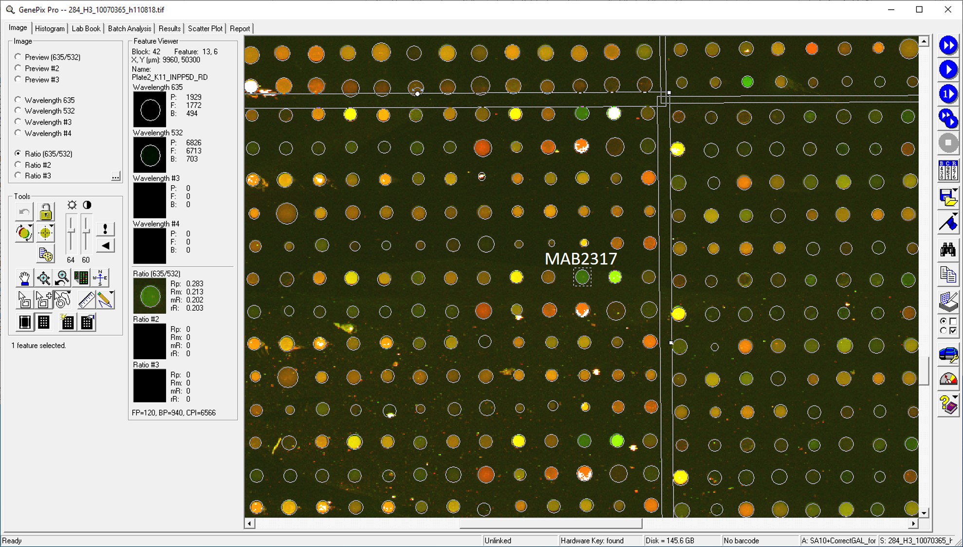
Application: MicroarraysSample Tested: EDTA PlasmaSpecies: HumanVerified Customer | Posted 01/14/2021

-
Application: MicroarraySample Tested: EDTA PlasmaSpecies: HumanVerified Customer | Posted 01/09/2019
-
Application: MicroarraysSample Tested: Platelet-poor EDTA PlasmaSpecies: HumanVerified Customer | Posted 11/14/2018
There are no reviews that match your criteria.
Protocols
Find general support by application which include: protocols, troubleshooting, illustrated assays, videos and webinars.
- Appropriate Fixation of IHC/ICC Samples
- Cellular Response to Hypoxia Protocols
- ClariTSA™ Fluorophore Kits
- Detection & Visualization of Antibody Binding
- ICC Cell Smear Protocol for Suspension Cells
- ICC Immunocytochemistry Protocol Videos
- ICC for Adherent Cells
- Immunocytochemistry (ICC) Protocol
- Immunocytochemistry Troubleshooting
- Immunofluorescence of Organoids Embedded in Cultrex Basement Membrane Extract
- Immunohistochemistry (IHC) and Immunocytochemistry (ICC) Protocols
- Preparing Samples for IHC/ICC Experiments
- Preventing Non-Specific Staining (Non-Specific Binding)
- Primary Antibody Selection & Optimization
- Protocol for VisUCyte™ HRP Polymer Detection Reagent
- Protocol for the Fluorescent ICC Staining of Cell Smears - Graphic
- Protocol for the Fluorescent ICC Staining of Cultured Cells on Coverslips - Graphic
- Protocol for the Preparation and Fluorescent ICC Staining of Cells on Coverslips
- Protocol for the Preparation and Fluorescent ICC Staining of Non-adherent Cells
- Protocol for the Preparation and Fluorescent ICC Staining of Stem Cells on Coverslips
- Protocol for the Preparation of a Cell Smear for Non-adherent Cell ICC - Graphic
- R&D Systems Quality Control Western Blot Protocol
- TUNEL and Active Caspase-3 Detection by IHC/ICC Protocol
- The Importance of IHC/ICC Controls
- Troubleshooting Guide: Western Blot Figures
- Western Blot Conditions
- Western Blot Protocol
- Western Blot Protocol for Cell Lysates
- Western Blot Troubleshooting
- Western Blot Troubleshooting Guide
- View all Protocols, Troubleshooting, Illustrated assays and Webinars
Loading...