Syntaxin 8 (STX8) is a 28 kDa member of the syntaxin family of proteins. It is ubiquitously expressed, embedded in Golgi, endosomal and lysosomal membranes, and serves as a component of the SNARE complex. STX8 is involved in endosomal homotypic fusion events as well as protein trafficking. Human STX8 is a type IV single-pass transmembrane protein (very short exoplasmic C-terminus) that is 236 amino acids (aa) in length. It contains a coiled-coil region (aa 42‑65), a t-SNARE domain (aa 152‑207) that is likely involved in protein-protein interactions, and a short, four amino acid C-terminal luminal sequence. Over aa 1‑215, human STX8 shares 92% aa identity with mouse STX8.

Key Product Details
Species Reactivity
Validated:
Human, Mouse, Rat
Cited:
Human, Mouse, Rat
Applications
Validated:
Immunohistochemistry, Western Blot
Cited:
Western Blot
Label
Unconjugated
Antibody Source
Polyclonal Sheep IgG
Loading...
Product Specifications
Immunogen
E. coli-derived recombinant human Syntaxin 8
Met1-Gly215
Accession # Q9UNK0
Met1-Gly215
Accession # Q9UNK0
Specificity
Detects human, mouse, and rat Syntaxin 8 in Western blots.
Clonality
Polyclonal
Host
Sheep
Isotype
IgG
Scientific Data Images for Syntaxin 8 Antibody
Detection of Human, Mouse, and Rat Syntaxin 8 by Western Blot.
Western blot shows lysates of HeLa human cervical epithelial carcinoma cell line, Jurkat human acute T cell leukemia cell line, L1.2 mouse pro-B cell line, and Rat-2 rat embryonic fibroblast cell line. PVDF membrane was probed with 1 µg/mL Sheep Anti-Human/Mouse/Rat Syntaxin 8 Antigen Affinity-purified Polyclonal Antibody (Catalog # AF5448) followed by HRP-conjugated Anti-Sheep IgG Secondary Antibody (Catalog # HAF016). A specific band for Syntaxin 8 was detected at approximately 28 kDa (as indicated). This experiment was conducted under reducing conditions and using Immunoblot Buffer Group 1.
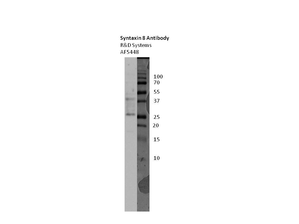
Detection of Human Syntaxin 8 by Western Blot.
Western blot shows recombinant human Syntaxin 12, 16, 1A, 1B2, 5, 6, 7, 8, and 1B1 (5 ng/lane). PVDF membrane was probed with 1 µg/mL Sheep Anti-Human/Mouse/Rat Syntaxin 8 Antigen Affinity-purified Polyclonal Antibody (Catalog # AF5448) followed by HRP-conjugated Anti-Sheep IgG Secondary Antibody (Catalog # HAF016). A specific band for Syntaxin 8 was detected as indicated. This experiment was conducted under reducing conditions and using Immunoblot Buffer Group 1.
Syntaxin 8 in Human Brain.
Syntaxin 8 was detected in immersion fixed paraffin-embedded sections of human brain using Sheep Anti-Human/Mouse/Rat Syntaxin 8 Antigen Affinity-purified Polyclonal Antibody (Catalog # AF5448) at 10 µg/mL overnight at 4 °C. Before incubation with the primary antibody, tissue was subjected to heat-induced epitope retrieval using Antigen Retrieval Reagent-Basic (Catalog # CTS013). Tissue was stained using the Anti-Sheep HRP-DAB Cell & Tissue Staining Kit (brown; Catalog # CTS019) and counterstained with hematoxylin (blue). Specific staining was localized to neuronal cell bodies and processes. View our protocol for Chromogenic IHC Staining of Paraffin-embedded Tissue Sections.Applications for Syntaxin 8 Antibody
Application
Recommended Usage
Immunohistochemistry
5-15 µg/mL
Sample: Immersion fixed paraffin-embedded sections of human brain
Sample: Immersion fixed paraffin-embedded sections of human brain
Western Blot
1 µg/mL
Sample: HeLa human cervical epithelial carcinoma cell line, Jurkat human acute T cell leukemia cell line, L1.2 mouse pro-B cell line, and Rat-2 rat embryonic fibroblast cell line
Sample: HeLa human cervical epithelial carcinoma cell line, Jurkat human acute T cell leukemia cell line, L1.2 mouse pro-B cell line, and Rat-2 rat embryonic fibroblast cell line
Reviewed Applications
Read 1 review rated 3 using AF5448 in the following applications:
Formulation, Preparation, and Storage
Purification
Antigen Affinity-purified
Reconstitution
Reconstitute at 0.2 mg/mL in sterile PBS. For liquid material, refer to CoA for concentration.
Loading...
Formulation
Lyophilized from a 0.2 μm filtered solution in PBS with Trehalose. *Small pack size (SP) is supplied either lyophilized or as a 0.2 µm filtered solution in PBS.
Shipping
Lyophilized product is shipped at ambient temperature. Liquid small pack size (-SP) is shipped with polar packs. Upon receipt, store immediately at the temperature recommended below.
Stability & Storage
Use a manual defrost freezer and avoid repeated freeze-thaw cycles.
- 12 months from date of receipt, -20 to -70 °C as supplied.
- 1 month, 2 to 8 °C under sterile conditions after reconstitution.
- 6 months, -20 to -70 °C under sterile conditions after reconstitution.
Calculators
Background: Syntaxin 8
Alternate Names
CARB, STX8
Gene Symbol
STX8
UniProt
Additional Syntaxin 8 Products
Product Documents for Syntaxin 8 Antibody
Certificate of Analysis
To download a Certificate of Analysis, please enter a lot or batch number in the search box below.
Note: Certificate of Analysis not available for kit components.
Product Specific Notices for Syntaxin 8 Antibody
For research use only
Citations for Syntaxin 8 Antibody
 Powered by Bioz
Customer Reviews for Syntaxin 8 Antibody (1)
3 out of 5
1 Customer Rating
Have you used Syntaxin 8 Antibody?
Submit a review and receive an Amazon gift card!
$25/€18/£15/$25CAN/¥2500 Yen for a review with an image
$10/€7/£6/$10CAN/¥1110 Yen for a review without an image
Submit a review
Customer Images
Showing
1
-
1 of
1 review
Showing All
Filter By:
-
Application: Western BlotSample Tested: T84 human colon carcinoma cell lineSpecies: HumanVerified Customer | Posted 04/25/2017100 ug whole cell lysate, 15% gel additional band at ~40kDa

There are no reviews that match your criteria.
Protocols
Find general support by application which include: protocols, troubleshooting, illustrated assays, videos and webinars.
- Antigen Retrieval Protocol (PIER)
- Antigen Retrieval for Frozen Sections Protocol
- Appropriate Fixation of IHC/ICC Samples
- Cellular Response to Hypoxia Protocols
- Chromogenic IHC Staining of Formalin-Fixed Paraffin-Embedded (FFPE) Tissue Protocol
- Chromogenic Immunohistochemistry Staining of Frozen Tissue
- ClariTSA™ Fluorophore Kits
- Detection & Visualization of Antibody Binding
- Fluorescent IHC Staining of Frozen Tissue Protocol
- Graphic Protocol for Heat-induced Epitope Retrieval
- Graphic Protocol for the Preparation and Fluorescent IHC Staining of Frozen Tissue Sections
- Graphic Protocol for the Preparation and Fluorescent IHC Staining of Paraffin-embedded Tissue Sections
- Graphic Protocol for the Preparation of Gelatin-coated Slides for Histological Tissue Sections
- IHC Sample Preparation (Frozen sections vs Paraffin)
- Immunofluorescent IHC Staining of Formalin-Fixed Paraffin-Embedded (FFPE) Tissue Protocol
- Immunohistochemistry (IHC) and Immunocytochemistry (ICC) Protocols
- Immunohistochemistry Frozen Troubleshooting
- Immunohistochemistry Paraffin Troubleshooting
- Preparing Samples for IHC/ICC Experiments
- Preventing Non-Specific Staining (Non-Specific Binding)
- Primary Antibody Selection & Optimization
- Protocol for Heat-Induced Epitope Retrieval (HIER)
- Protocol for Making a 4% Formaldehyde Solution in PBS
- Protocol for VisUCyte™ HRP Polymer Detection Reagent
- Protocol for the Preparation & Fixation of Cells on Coverslips
- Protocol for the Preparation and Chromogenic IHC Staining of Frozen Tissue Sections
- Protocol for the Preparation and Chromogenic IHC Staining of Frozen Tissue Sections - Graphic
- Protocol for the Preparation and Chromogenic IHC Staining of Paraffin-embedded Tissue Sections
- Protocol for the Preparation and Chromogenic IHC Staining of Paraffin-embedded Tissue Sections - Graphic
- Protocol for the Preparation and Fluorescent IHC Staining of Frozen Tissue Sections
- Protocol for the Preparation and Fluorescent IHC Staining of Paraffin-embedded Tissue Sections
- Protocol for the Preparation of Gelatin-coated Slides for Histological Tissue Sections
- R&D Systems Quality Control Western Blot Protocol
- TUNEL and Active Caspase-3 Detection by IHC/ICC Protocol
- The Importance of IHC/ICC Controls
- Troubleshooting Guide: Immunohistochemistry
- Troubleshooting Guide: Western Blot Figures
- Western Blot Conditions
- Western Blot Protocol
- Western Blot Protocol for Cell Lysates
- Western Blot Troubleshooting
- Western Blot Troubleshooting Guide
- View all Protocols, Troubleshooting, Illustrated assays and Webinars
Loading...