SMAD9 (SMA protein and Mothers Against Decapentaplegic 9; also SMAD8 and MAHD6) is a 55-60 kDa R-Group member of the dwarfin/SMAD family of proteins. It is expressed by BMP-responsive cells, and serves to transmit information from the BMP receptor (BMPR) to the nucleus. In particular, following BMPR activation, SMAD9 is phosphorylated by the BMPR on its C-terminus, promoting heterodimerization with SMAD4 and its translocation into the nucleus. Here, SMAD9 binds to DNA and participates in gene activation. Human SMAD9 is 467 amino acids (aa) in length. It contains a DNA-binding domain termed MH1 (aa 16-140) plus a receptor/transcription factor binding domain termed MH2 (aa 273-467). The C-terminus contains two phosphorylation sites at Ser465 and Ser467. There is one splice form that shows a deletion of aa 224-260. Over aa 158-223, human and mouse SMAD9 share 82% identy in amino acid sequence.

Key Product Details
Species Reactivity
Human, Mouse
Applications
Immunohistochemistry, Immunocytochemistry
Label
Unconjugated
Antibody Source
Polyclonal Sheep IgG
Loading...
Product Specifications
Immunogen
E. coli-derived recombinant human Smad9
Ala158-Ser223
Accession # O15198
Ala158-Ser223
Accession # O15198
Specificity
Detects human Smad9 in direct ELISAs. In direct ELISAs, approximately 5% cross-reactivity with recombinant human Smad1 is observed.
Clonality
Polyclonal
Host
Sheep
Isotype
IgG
Scientific Data Images for Human/Mouse Smad9 Antibody
Smad9 in A172 Human Cell Line.
Smad9 was detected in immersion fixed A172 human glioblastoma cell line using Sheep Anti-Human/Mouse Smad9 Antigen Affinity-purified Polyclonal Antibody (Catalog # AF7215) at 10 µg/mL for 3 hours at room temperature. Cells were stained using the NorthernLights™ 557-conjugated Anti-Sheep IgG Secondary Antibody (red, upper panel; Catalog # NL010) and counterstained with DAPI (blue, lower panel). Specific staining was localized to cytoplasm. View our protocol for Fluorescent ICC Staining of Cells on Coverslips.
Smad9 in Human Prostate Cancer Tissue.
Smad9 was detected in immersion fixed paraffin-embedded sections of human prostate cancer tissue using Sheep Anti-Human Smad9 Antigen Affinity-purified Polyclonal Antibody (Catalog # AF7215) at 3 µg/mL overnight at 4 °C. Before incubation with the primary antibody, tissue was subjected to heat-induced epitope retrieval using Antigen Retrieval Reagent-Basic (Catalog # CTS013). Tissue was stained using the Anti-Sheep HRP-DAB Cell & Tissue Staining Kit (brown; Catalog # CTS019) and counterstained with hematoxylin (blue). Specific staining was localized to nuclei in glandular epithelial cells. View our protocol for Chromogenic IHC Staining of Paraffin-embedded Tissue Sections.
Smad9 in Mouse Embryo.
Smad9 was detected in immersion fixed frozen sections of mouse embryo (13 d.p.c.) using Sheep Anti-Human Smad9 Antigen Affinity-purified Polyclonal Antibody (Catalog # AF7215) at 1.7 µg/mL overnight at 4 °C. Tissue was stained using the Anti-Sheep HRP-DAB Cell & Tissue Staining Kit (brown; Catalog # CTS019) and counterstained with hematoxylin (blue). Specific staining was localized to developing muscle cells. View our protocol for Chromogenic IHC Staining of Frozen Tissue Sections.Applications for Human/Mouse Smad9 Antibody
Application
Recommended Usage
Immunocytochemistry
5-15 µg/mL
Sample: Immersion fixed A172 human glioblastoma cell line
Sample: Immersion fixed A172 human glioblastoma cell line
Immunohistochemistry
5-15 µg/mL
Sample: Immersion fixed paraffin-embedded sections of human prostate cancer tissue and immersion fixed frozen sections of mouse embryo (13 d.p.c.)
Sample: Immersion fixed paraffin-embedded sections of human prostate cancer tissue and immersion fixed frozen sections of mouse embryo (13 d.p.c.)
Reviewed Applications
Read 1 review rated 3 using AF7215 in the following applications:
Formulation, Preparation, and Storage
Purification
Antigen Affinity-purified
Reconstitution
Sterile PBS to a final concentration of 0.2 mg/mL. For liquid material, refer to CoA for concentration.
Loading...
Formulation
Lyophilized from a 0.2 μm filtered solution in PBS with Trehalose. *Small pack size (SP) is supplied either lyophilized or as a 0.2 µm filtered solution in PBS.
Shipping
Lyophilized product is shipped at ambient temperature. Liquid small pack size (-SP) is shipped with polar packs. Upon receipt, store immediately at the temperature recommended below.
Stability & Storage
Use a manual defrost freezer and avoid repeated freeze-thaw cycles.
- 12 months from date of receipt, -20 to -70 °C as supplied.
- 1 month, 2 to 8 °C under sterile conditions after reconstitution.
- 6 months, -20 to -70 °C under sterile conditions after reconstitution.
Calculators
Background: Smad9
Long Name
Mothers Against DPP Homolog 9
Alternate Names
Madh6, MADH9
Gene Symbol
SMAD9
UniProt
Additional Smad9 Products
Product Documents for Human/Mouse Smad9 Antibody
Certificate of Analysis
To download a Certificate of Analysis, please enter a lot or batch number in the search box below.
Note: Certificate of Analysis not available for kit components.
Product Specific Notices for Human/Mouse Smad9 Antibody
For research use only
Customer Reviews for Human/Mouse Smad9 Antibody (1)
3 out of 5
1 Customer Rating
Have you used Human/Mouse Smad9 Antibody?
Submit a review and receive an Amazon gift card!
$25/€18/£15/$25CAN/¥2500 Yen for a review with an image
$10/€7/£6/$10CAN/¥1110 Yen for a review without an image
Submit a review
Customer Images
Showing
1
-
1 of
1 review
Showing All
Filter By:
-
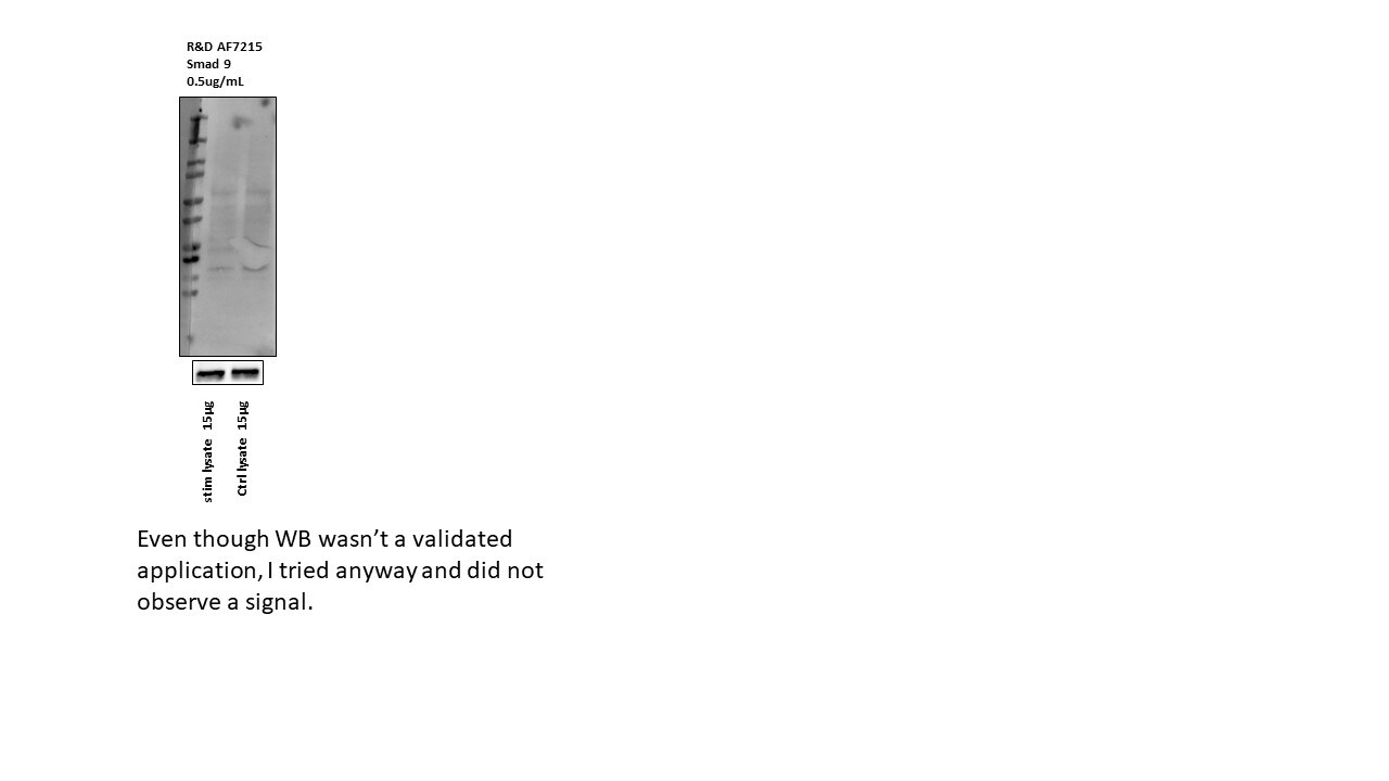
Application: Western BlotSample Tested: Mv1Lu mink lung epithelial cell lineSpecies: MinkVerified Customer | Posted 11/20/2019

There are no reviews that match your criteria.
Protocols
Find general support by application which include: protocols, troubleshooting, illustrated assays, videos and webinars.
- Antigen Retrieval Protocol (PIER)
- Antigen Retrieval for Frozen Sections Protocol
- Appropriate Fixation of IHC/ICC Samples
- Cellular Response to Hypoxia Protocols
- Chromogenic IHC Staining of Formalin-Fixed Paraffin-Embedded (FFPE) Tissue Protocol
- Chromogenic Immunohistochemistry Staining of Frozen Tissue
- ClariTSA™ Fluorophore Kits
- Detection & Visualization of Antibody Binding
- Fluorescent IHC Staining of Frozen Tissue Protocol
- Graphic Protocol for Heat-induced Epitope Retrieval
- Graphic Protocol for the Preparation and Fluorescent IHC Staining of Frozen Tissue Sections
- Graphic Protocol for the Preparation and Fluorescent IHC Staining of Paraffin-embedded Tissue Sections
- Graphic Protocol for the Preparation of Gelatin-coated Slides for Histological Tissue Sections
- ICC Cell Smear Protocol for Suspension Cells
- ICC Immunocytochemistry Protocol Videos
- ICC for Adherent Cells
- IHC Sample Preparation (Frozen sections vs Paraffin)
- Immunocytochemistry (ICC) Protocol
- Immunocytochemistry Troubleshooting
- Immunofluorescence of Organoids Embedded in Cultrex Basement Membrane Extract
- Immunofluorescent IHC Staining of Formalin-Fixed Paraffin-Embedded (FFPE) Tissue Protocol
- Immunohistochemistry (IHC) and Immunocytochemistry (ICC) Protocols
- Immunohistochemistry Frozen Troubleshooting
- Immunohistochemistry Paraffin Troubleshooting
- Preparing Samples for IHC/ICC Experiments
- Preventing Non-Specific Staining (Non-Specific Binding)
- Primary Antibody Selection & Optimization
- Protocol for Heat-Induced Epitope Retrieval (HIER)
- Protocol for Making a 4% Formaldehyde Solution in PBS
- Protocol for VisUCyte™ HRP Polymer Detection Reagent
- Protocol for the Fluorescent ICC Staining of Cell Smears - Graphic
- Protocol for the Fluorescent ICC Staining of Cultured Cells on Coverslips - Graphic
- Protocol for the Preparation & Fixation of Cells on Coverslips
- Protocol for the Preparation and Chromogenic IHC Staining of Frozen Tissue Sections
- Protocol for the Preparation and Chromogenic IHC Staining of Frozen Tissue Sections - Graphic
- Protocol for the Preparation and Chromogenic IHC Staining of Paraffin-embedded Tissue Sections
- Protocol for the Preparation and Chromogenic IHC Staining of Paraffin-embedded Tissue Sections - Graphic
- Protocol for the Preparation and Fluorescent ICC Staining of Cells on Coverslips
- Protocol for the Preparation and Fluorescent ICC Staining of Non-adherent Cells
- Protocol for the Preparation and Fluorescent ICC Staining of Stem Cells on Coverslips
- Protocol for the Preparation and Fluorescent IHC Staining of Frozen Tissue Sections
- Protocol for the Preparation and Fluorescent IHC Staining of Paraffin-embedded Tissue Sections
- Protocol for the Preparation of Gelatin-coated Slides for Histological Tissue Sections
- Protocol for the Preparation of a Cell Smear for Non-adherent Cell ICC - Graphic
- TUNEL and Active Caspase-3 Detection by IHC/ICC Protocol
- The Importance of IHC/ICC Controls
- Troubleshooting Guide: Immunohistochemistry
- View all Protocols, Troubleshooting, Illustrated assays and Webinars
Loading...