
Key Product Details
Species Reactivity
Validated:
Human
Cited:
Frog - Xenopus (African Clawed Frog), Primate
Applications
Validated:
Immunohistochemistry, Western Blot, Immunocytochemistry
Cited:
Immunohistochemistry, Immunohistochemistry-Frozen
Label
Unconjugated
Antibody Source
Polyclonal Sheep IgG
Loading...
Product Specifications
Immunogen
Chinese hamster ovary cell line CHO-derived recombinant human Protocadherin-15
Gln27-Ala1376
Accession # Q96QU1
Gln27-Ala1376
Accession # Q96QU1
Specificity
Detects human Protocadherin-15 in direct ELISAs.
Clonality
Polyclonal
Host
Sheep
Isotype
IgG
Scientific Data Images for Human Protocadherin-15 Antibody
Detection of Human Protocadherin-15 by Western Blot.
Western blot shows lysates of HEK293T human embryonic kidney cell line and HEK293T cell line spiked with Recombinant Human Protocadherin-15 (5 ng/lane). PVDF membrane was probed with 2 µg/mL of Sheep Anti-Human Protocadherin-15 Antigen Affinity-purified Polyclonal Antibody (Catalog # AF6729) followed by HRP-conjugated Anti-Sheep IgG Secondary Antibody (Catalog # HAF016). A specific band was detected for Protocadherin-15 at approximately 190 kDa (as indicated). This experiment was conducted under reducing conditions and using Immunoblot Buffer Group 8.
Protocadherin-15 in YT Human Cell Line.
Protocadherin-15 was detected in immersion fixed YT human leukemia natural killer-like cell line using Sheep Anti-Human Protocadherin-15 Antigen Affinity-purified Polyclonal Antibody (Catalog # AF6729) at 10 µg/mL for 3 hours at room temperature. Cells were stained using the NorthernLights™ 557-conjugated Anti-Sheep IgG Secondary Antibody (red; Catalog # NL010) and counterstained with DAPI (blue). Specific staining was localized to cell surfaces and cytoplasm. View our protocol for Fluorescent ICC Staining of Non-adherent Cells.
Protocadherin‑15 in Human Kidney.
Protocadherin-15 was detected in immersion fixed paraffin-embedded sections of human kidney using Sheep Anti-Human Protocadherin-15 Antigen Affinity-purified Polyclonal Antibody (Catalog # AF6729) at 10 µg/mL overnight at 4 °C. Tissue was stained using the Anti-Sheep HRP-DAB Cell & Tissue Staining Kit (brown; Catalog # CTS019) and counterstained with hematoxylin (blue). Specific staining was localized to cytoplasm in convoluted tubules. View our protocol for Chromogenic IHC Staining of Paraffin-embedded Tissue Sections.Applications for Human Protocadherin-15 Antibody
Application
Recommended Usage
Immunocytochemistry
5-15 µg/mL
Sample: Immersion fixed YT human leukemia natural killer-like cell line
Sample: Immersion fixed YT human leukemia natural killer-like cell line
Immunohistochemistry
5-15 µg/mL
Sample: Immersion fixed paraffin-embedded sections of human kidney
Sample: Immersion fixed paraffin-embedded sections of human kidney
Western Blot
2 µg/mL
Sample: HEK293T human embryonic kidney cell line spiked with Recombinant Human Protocadherin-15
Sample: HEK293T human embryonic kidney cell line spiked with Recombinant Human Protocadherin-15
Reviewed Applications
Read 2 reviews rated 5 using AF6729 in the following applications:
Formulation, Preparation, and Storage
Purification
Antigen Affinity-purified
Reconstitution
Sterile PBS to a final concentration of 0.2 mg/mL. For liquid material, refer to CoA for concentration.
Loading...
Formulation
Lyophilized from a 0.2 μm filtered solution in PBS with Trehalose. *Small pack size (SP) is supplied either lyophilized or as a 0.2 µm filtered solution in PBS.
Shipping
Lyophilized product is shipped at ambient temperature. Liquid small pack size (-SP) is shipped with polar packs. Upon receipt, store immediately at the temperature recommended below.
Stability & Storage
Use a manual defrost freezer and avoid repeated freeze-thaw cycles.
- 12 months from date of receipt, -20 to -70 °C as supplied.
- 1 month, 2 to 8 °C under sterile conditions after reconstitution.
- 6 months, -20 to -70 °C under sterile conditions after reconstitution.
Calculators
Background: Protocadherin-15
Alternate Names
CDHR15, DFNB23, PCDH15, Protocadherin15, USH1F
Gene Symbol
PCDH15
UniProt
Additional Protocadherin-15 Products
Product Documents for Human Protocadherin-15 Antibody
Certificate of Analysis
To download a Certificate of Analysis, please enter a lot or batch number in the search box below.
Note: Certificate of Analysis not available for kit components.
Product Specific Notices for Human Protocadherin-15 Antibody
For research use only
Related Research Areas
Citations for Human Protocadherin-15 Antibody
 Powered by Bioz
Customer Reviews for Human Protocadherin-15 Antibody (2)
5 out of 5
2 Customer Ratings
Have you used Human Protocadherin-15 Antibody?
Submit a review and receive an Amazon gift card!
$25/€18/£15/$25CAN/¥2500 Yen for a review with an image
$10/€7/£6/$10CAN/¥1110 Yen for a review without an image
Submit a review
Customer Images
Showing
1
-
2 of
2 reviews
Showing All
Filter By:
-
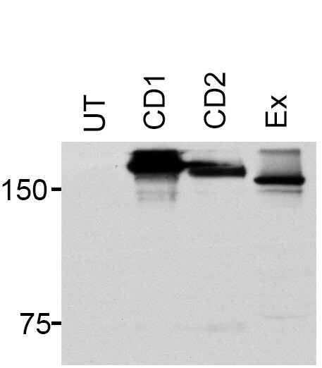
Application: Western BlotSample Tested: HEK293 transfected cells and HEK 293 transfectedSpecies: HumanVerified Customer | Posted 10/02/2018HEK 293 cells untransfected ;UT; or expressing Pcdh15-CD1 or Pcdh15-CD2 isoforms. A Pcdh15 construct the cytoplasmic tail was also expressed in these cells Ex;.PVDF membrane was blocked with 3% milk for 1 hour at room temperature. Primary antibody sheep anti-Pcdh15 was used at a concentration 2ug/mL in 3% milk and the membrane incubated overnight at 4C. Secondary antibody anti-sheep-HRP was used at a dilution 1:2500 in 3% milk and incubated for 1 hour at room temperature.

-
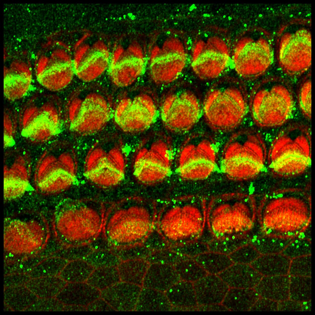
Application: Immunohistochemistry-FrozenSample Tested: Organ of Corti (inner ear)Species: MouseVerified Customer | Posted 09/25/2018P1 organ of Corti stained with sheep anti-Pcdh15 (green) and counterstained with phalloidin (red). Outer hair cells show a strong immunereactivity for Pcdh15 at the hair cell bundle level.P1 mouse organ of Corti was paraformaldehyde fixed (4%) for 30 min, blocked 1 hour in PBS Triton X-100 0.1% containing 10% fetal calf serum and then incubated overnight with sheep anti-Pcdh15 (1:200, green) in blocking solution. Secondary antibody was Alexa488 anti-sheep (1:200). Tissue was counterstained with phalloidin (red).
There are no reviews that match your criteria.
Protocols
Find general support by application which include: protocols, troubleshooting, illustrated assays, videos and webinars.
- Antigen Retrieval Protocol (PIER)
- Antigen Retrieval for Frozen Sections Protocol
- Appropriate Fixation of IHC/ICC Samples
- Cellular Response to Hypoxia Protocols
- Chromogenic IHC Staining of Formalin-Fixed Paraffin-Embedded (FFPE) Tissue Protocol
- Chromogenic Immunohistochemistry Staining of Frozen Tissue
- ClariTSA™ Fluorophore Kits
- Detection & Visualization of Antibody Binding
- Fluorescent IHC Staining of Frozen Tissue Protocol
- Graphic Protocol for Heat-induced Epitope Retrieval
- Graphic Protocol for the Preparation and Fluorescent IHC Staining of Frozen Tissue Sections
- Graphic Protocol for the Preparation and Fluorescent IHC Staining of Paraffin-embedded Tissue Sections
- Graphic Protocol for the Preparation of Gelatin-coated Slides for Histological Tissue Sections
- ICC Cell Smear Protocol for Suspension Cells
- ICC Immunocytochemistry Protocol Videos
- ICC for Adherent Cells
- IHC Sample Preparation (Frozen sections vs Paraffin)
- Immunocytochemistry (ICC) Protocol
- Immunocytochemistry Troubleshooting
- Immunofluorescence of Organoids Embedded in Cultrex Basement Membrane Extract
- Immunofluorescent IHC Staining of Formalin-Fixed Paraffin-Embedded (FFPE) Tissue Protocol
- Immunohistochemistry (IHC) and Immunocytochemistry (ICC) Protocols
- Immunohistochemistry Frozen Troubleshooting
- Immunohistochemistry Paraffin Troubleshooting
- Preparing Samples for IHC/ICC Experiments
- Preventing Non-Specific Staining (Non-Specific Binding)
- Primary Antibody Selection & Optimization
- Protocol for Heat-Induced Epitope Retrieval (HIER)
- Protocol for Making a 4% Formaldehyde Solution in PBS
- Protocol for VisUCyte™ HRP Polymer Detection Reagent
- Protocol for the Fluorescent ICC Staining of Cell Smears - Graphic
- Protocol for the Fluorescent ICC Staining of Cultured Cells on Coverslips - Graphic
- Protocol for the Preparation & Fixation of Cells on Coverslips
- Protocol for the Preparation and Chromogenic IHC Staining of Frozen Tissue Sections
- Protocol for the Preparation and Chromogenic IHC Staining of Frozen Tissue Sections - Graphic
- Protocol for the Preparation and Chromogenic IHC Staining of Paraffin-embedded Tissue Sections
- Protocol for the Preparation and Chromogenic IHC Staining of Paraffin-embedded Tissue Sections - Graphic
- Protocol for the Preparation and Fluorescent ICC Staining of Cells on Coverslips
- Protocol for the Preparation and Fluorescent ICC Staining of Non-adherent Cells
- Protocol for the Preparation and Fluorescent ICC Staining of Stem Cells on Coverslips
- Protocol for the Preparation and Fluorescent IHC Staining of Frozen Tissue Sections
- Protocol for the Preparation and Fluorescent IHC Staining of Paraffin-embedded Tissue Sections
- Protocol for the Preparation of Gelatin-coated Slides for Histological Tissue Sections
- Protocol for the Preparation of a Cell Smear for Non-adherent Cell ICC - Graphic
- R&D Systems Quality Control Western Blot Protocol
- TUNEL and Active Caspase-3 Detection by IHC/ICC Protocol
- The Importance of IHC/ICC Controls
- Troubleshooting Guide: Immunohistochemistry
- Troubleshooting Guide: Western Blot Figures
- Western Blot Conditions
- Western Blot Protocol
- Western Blot Protocol for Cell Lysates
- Western Blot Troubleshooting
- Western Blot Troubleshooting Guide
- View all Protocols, Troubleshooting, Illustrated assays and Webinars
Loading...