
Key Product Details
Species Reactivity
Validated:
Human, Rat
Cited:
Human, Mouse, Rat, Transgenic Mouse
Applications
Validated:
Immunohistochemistry, Western Blot, Immunocytochemistry, Immunoprecipitation
Cited:
Immunohistochemistry, Immunohistochemistry-Frozen, Western Blot, ELISA Development
Label
Unconjugated
Antibody Source
Polyclonal Sheep IgG
Loading...
Product Specifications
Immunogen
Mouse myeloma cell line NS0-derived recombinant human Brevican
Asp23-Pro911
Accession # AAH27971
Asp23-Pro911
Accession # AAH27971
Specificity
Detects human Brevican in direct ELISAs and Western blots. In direct ELISAs, less than 20% cross-reactivity with recombinant mouse Brevican is observed.
Clonality
Polyclonal
Host
Sheep
Isotype
IgG
Scientific Data Images for Brevican Antibody
Detection of Human Brevican by Western Blot.
Western blot shows lysates of human brain (cerebellum) tissue and human brain (motor cortex) tissue. PVDF membrane was probed with 0.1 µg/mL of Sheep Anti-Human/Rat Brevican Antigen Affinity-purified Polyclonal Antibody (Catalog # AF4009) followed by Donkey Anti-Sheep IgG HRP-conjugated Antigen Affinity-purified Polyclonal Antibody(Catalog # HAF016). Specific bands were detected for Brevican at approximately 60 and 90 kDa (as indicated). This experiment was conducted under reducing conditions and using Immunoblot Buffer Group 1.
Brevican in Rat Cortical Stem Cells.
Brevican was detected in immersion fixed rat cortical stem cells differentiated for 7 days by growth factor withdrawal using Sheep Anti-Human/Rat Brevican Antigen Affinity-purified Polyclonal Antibody (Catalog # AF4009) at 10 µg/mL for 3 hours at room temperature. Cells were stained using the NorthernLights™ 557-conjugated Anti-Sheep IgG Secondary Antibody (red; Catalog # NL010) and counterstained with DAPI (blue). Specific staining was localized to cytoplasm. View our protocol for Fluorescent ICC Staining of Stem Cells on Coverslips.
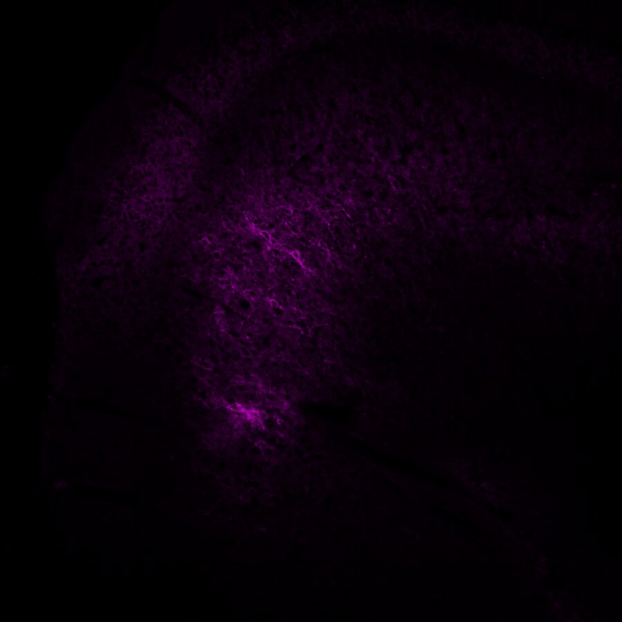
Brevican in Human Brain.
Brevican was detected in immersion fixed paraffin-embedded sections of human brain (cortex) using Sheep Anti-Human/Rat Brevican Antigen Affinity-purified Polyclonal Antibody (Catalog # AF4009) at 5 µg/mL overnight at 4 °C. Tissue was stained using the Anti-Sheep HRP-DAB Cell & Tissue Staining Kit (brown; Catalog # CTS019) and counterstained with hematoxylin (blue). Specific staining was localized to neuropil. View our protocol for Chromogenic IHC Staining of Paraffin-embedded Tissue Sections.
Detection of Brevican by Western Blot
Brevican labeling in histological sections and western blots of human and rat prefrontal samples. (A) Micrographs and charts illustrating the average area fraction of brevican immunoreactivity in control and CUS exposed rats (four micrographs to the left and underlying charts) and in control and MDD human subjects (four micrographs to the right and underlying charts) showing no differences in overall brevican immunoreactivity and within 8 µm2 rectangles (see legends referring to rectangles in Figs. 2 And 3) around NRs both in animals with stress and in human subjects with MDD. Arrows point to nodes of Ranvier identified in the left two pictures using triple immunofluorescent staining for CASPR (labels paranodes in green) Neurofascin (red) and Brevican (blue in all the micrographs). Calibration bar is 5 µm for rat and 8 µm for human micrographs. (B) Representative western blot lanes and quantification of optical density of Brevican bands (relative to housekeeping protein actin) in the frontal pole of control and CUS rats, and in the PFC white matter of human subjects. The pictures present bands from 2 representative rats subjected to stress as compared to 2 controls (picture to the left) and two human MDD subjects against two non-psychiatric controls (picture to the right). Quantification for bands for all subjects in each group in the study at approximately 140 and 80 kD is presented in the charts. All lanes and bands in the pictures appear as they were positioned and developed in the Western blot PVDF membrane. Image collected and cropped by CiteAb from the following open publication (https://pubmed.ncbi.nlm.nih.gov/37775676), licensed under a CC-BY license. Not internally tested by R&D Systems.Applications for Brevican Antibody
Application
Recommended Usage
Immunocytochemistry
5-15 µg/mL
Sample: Immersion fixed rat cortical stem cells differentiated for 7 days by growth factor withdrawal
Sample: Immersion fixed rat cortical stem cells differentiated for 7 days by growth factor withdrawal
Immunohistochemistry
5-15 µg/mL
Sample: Immersion fixed paraffin-embedded sections of human brain (cortex)
Sample: Immersion fixed paraffin-embedded sections of human brain (cortex)
Immunoprecipitation
25 µg/mL
Sample: Conditioned cell culture medium spiked with Recombinant Human Brevican (Catalog # 4009‑BC), see our available Western blot detection antibodies
Sample: Conditioned cell culture medium spiked with Recombinant Human Brevican (Catalog # 4009‑BC), see our available Western blot detection antibodies
Western Blot
0.1 µg/mL
Sample: Human brain (cerebellum) tissue and human brain (motor cortex) tissue
Sample: Human brain (cerebellum) tissue and human brain (motor cortex) tissue
Reviewed Applications
Read 1 review rated 5 using AF4009 in the following applications:
Formulation, Preparation, and Storage
Purification
Antigen Affinity-purified
Reconstitution
Reconstitute at 0.2 mg/mL in sterile PBS. For liquid material, refer to CoA for concentration.
Loading...
Formulation
Lyophilized from a 0.2 μm filtered solution in PBS with Trehalose. *Small pack size (SP) is supplied either lyophilized or as a 0.2 µm filtered solution in PBS.
Shipping
Lyophilized product is shipped at ambient temperature. Liquid small pack size (-SP) is shipped with polar packs. Upon receipt, store immediately at the temperature recommended below.
Stability & Storage
Use a manual defrost freezer and avoid repeated freeze-thaw cycles.
- 12 months from date of receipt, -20 to -70 °C as supplied.
- 1 month, 2 to 8 °C under sterile conditions after reconstitution.
- 6 months, -20 to -70 °C under sterile conditions after reconstitution.
Calculators
Background: Brevican
References
- Viapiano, M.S. and R.T. Matthews (2006) Trends Mol. Med. 12:488.
- Gary, S.C. et al. (2000) Gene 256:139.
- Jaworski, D.M. et al. (1994) J. Cell Biol. 125:495.
- Seidenbecher, C.I. et al. (1995) J. Biol. Chem. 270:27206.
- Hamel, M.G. et al. (2005) J. Neurochem. 93:1533.
- Ogawa, T. et al. (2001) J. Comp. Neurol. 432:285
- Yamada, H. et al. (1997) J. Neurosci. 17:7784.
- Seidenbecher, C.I. et al. (2002) J. Neurochem. 83:738.
- Matthews, R.T. et al. (2000) J. Biol. Chem. 275:22695.
- Nakamura, H. et al. (2000) J. Biol. Chem. 275:38885.
- Mayer, J. et al. (2005) BMC Neurosci. 6:52.
- Yuan, W. et al. (2002) Neuroscience 114:1091.
- Deepa, S.S. et al. (2006) J. Biol. Chem. 281:17789.
- Jaworski, D.M. et al. (1999) Exp. Neurol. 157:327.
- Viapiano, M.S. et al. (2005) Cancer Res. 65:6726.
- Viapiano, M.S. et al. (2003) J. Biol. Chem. 278:33239.
Long Name
Brain Specific Proteoglycan in the Aggrecan Family
Alternate Names
ALPBRE, BCAN, BEHAB, CSPG7
Gene Symbol
BCAN
UniProt
Additional Brevican Products
Product Documents for Brevican Antibody
Certificate of Analysis
To download a Certificate of Analysis, please enter a lot or batch number in the search box below.
Note: Certificate of Analysis not available for kit components.
Product Specific Notices for Brevican Antibody
For research use only
Related Research Areas
Citations for Brevican Antibody
 Powered by Bioz
Customer Reviews for Brevican Antibody (1)
5 out of 5
1 Customer Rating
Have you used Brevican Antibody?
Submit a review and receive an Amazon gift card!
$25/€18/£15/$25CAN/¥2500 Yen for a review with an image
$10/€7/£6/$10CAN/¥1110 Yen for a review without an image
Submit a review
Customer Images
Showing
1
-
1 of
1 review
Showing All
Filter By:
-
Application: Immunocytochemistry/ImmunofluorescenceSample Tested: Adult brainSpecies: RatVerified Customer | Posted 04/10/2018Used at a 1:500 but will need to increase to 1:100 for better staining

There are no reviews that match your criteria.
Protocols
Find general support by application which include: protocols, troubleshooting, illustrated assays, videos and webinars.
- Antigen Retrieval Protocol (PIER)
- Antigen Retrieval for Frozen Sections Protocol
- Appropriate Fixation of IHC/ICC Samples
- Cellular Response to Hypoxia Protocols
- Chromogenic IHC Staining of Formalin-Fixed Paraffin-Embedded (FFPE) Tissue Protocol
- Chromogenic Immunohistochemistry Staining of Frozen Tissue
- ClariTSA™ Fluorophore Kits
- Detection & Visualization of Antibody Binding
- Fluorescent IHC Staining of Frozen Tissue Protocol
- Graphic Protocol for Heat-induced Epitope Retrieval
- Graphic Protocol for the Preparation and Fluorescent IHC Staining of Frozen Tissue Sections
- Graphic Protocol for the Preparation and Fluorescent IHC Staining of Paraffin-embedded Tissue Sections
- Graphic Protocol for the Preparation of Gelatin-coated Slides for Histological Tissue Sections
- ICC Cell Smear Protocol for Suspension Cells
- ICC Immunocytochemistry Protocol Videos
- ICC for Adherent Cells
- IHC Sample Preparation (Frozen sections vs Paraffin)
- Immunocytochemistry (ICC) Protocol
- Immunocytochemistry Troubleshooting
- Immunofluorescence of Organoids Embedded in Cultrex Basement Membrane Extract
- Immunofluorescent IHC Staining of Formalin-Fixed Paraffin-Embedded (FFPE) Tissue Protocol
- Immunohistochemistry (IHC) and Immunocytochemistry (ICC) Protocols
- Immunohistochemistry Frozen Troubleshooting
- Immunohistochemistry Paraffin Troubleshooting
- Immunoprecipitation Protocol
- Preparing Samples for IHC/ICC Experiments
- Preventing Non-Specific Staining (Non-Specific Binding)
- Primary Antibody Selection & Optimization
- Protocol for Heat-Induced Epitope Retrieval (HIER)
- Protocol for Making a 4% Formaldehyde Solution in PBS
- Protocol for VisUCyte™ HRP Polymer Detection Reagent
- Protocol for the Fluorescent ICC Staining of Cell Smears - Graphic
- Protocol for the Fluorescent ICC Staining of Cultured Cells on Coverslips - Graphic
- Protocol for the Preparation & Fixation of Cells on Coverslips
- Protocol for the Preparation and Chromogenic IHC Staining of Frozen Tissue Sections
- Protocol for the Preparation and Chromogenic IHC Staining of Frozen Tissue Sections - Graphic
- Protocol for the Preparation and Chromogenic IHC Staining of Paraffin-embedded Tissue Sections
- Protocol for the Preparation and Chromogenic IHC Staining of Paraffin-embedded Tissue Sections - Graphic
- Protocol for the Preparation and Fluorescent ICC Staining of Cells on Coverslips
- Protocol for the Preparation and Fluorescent ICC Staining of Non-adherent Cells
- Protocol for the Preparation and Fluorescent ICC Staining of Stem Cells on Coverslips
- Protocol for the Preparation and Fluorescent IHC Staining of Frozen Tissue Sections
- Protocol for the Preparation and Fluorescent IHC Staining of Paraffin-embedded Tissue Sections
- Protocol for the Preparation of Gelatin-coated Slides for Histological Tissue Sections
- Protocol for the Preparation of a Cell Smear for Non-adherent Cell ICC - Graphic
- R&D Systems Quality Control Western Blot Protocol
- TUNEL and Active Caspase-3 Detection by IHC/ICC Protocol
- The Importance of IHC/ICC Controls
- Troubleshooting Guide: Immunohistochemistry
- Troubleshooting Guide: Western Blot Figures
- Western Blot Conditions
- Western Blot Protocol
- Western Blot Protocol for Cell Lysates
- Western Blot Troubleshooting
- Western Blot Troubleshooting Guide
- View all Protocols, Troubleshooting, Illustrated assays and Webinars
Loading...