
Key Product Details
Validated by
Biological Validation
Species Reactivity
Validated:
Human, Rat
Cited:
Human, Rat
Applications
Validated:
Immunohistochemistry, Western Blot, ELISA Capture (Matched Antibody Pair), Neutralization, Immunocytochemistry
Cited:
Immunohistochemistry, Immunohistochemistry-Paraffin, Neutralization, Immunocytochemistry, ELISA Development
Label
Unconjugated
Antibody Source
Polyclonal Goat IgG
Loading...
Product Specifications
Immunogen
E. coli-derived recombinant rat IL-2
Ala21-Gln155
Accession # P17108
Ala21-Gln155
Accession # P17108
Specificity
Detects rat IL-2 in ELISAs. Detects human and rat IL-2 in Western blots. In sandwich immunoassays, less than 0.2% cross-reactivity with recombinant mouse IL‑2 is observed.
Clonality
Polyclonal
Host
Goat
Isotype
IgG
Endotoxin Level
<0.10 EU per 1 μg of the antibody by the LAL method.
Scientific Data Images for IL-2 Antibody
Detection of Human IL‑2 by Western Blot.
Western blot shows lysates of monensin treated human peripheral blood mononuclear cells (PBMCs) with no additional treatment (-) or additionally treated (+) with 0.5 μg/mL calcium ionomycin (Iono) and 50 ng/mL PMA overnight. PVDF membrane was probed with 2 µg/mL of Goat Anti-Human/Rat IL-2 Antigen Affinity-purified Polyclonal Antibody (Catalog # AF-502-NA) followed by HRP-conjugated Anti-Goat IgG Secondary Antibody (HAF017). A specific band was detected for IL-2 at approximately 14 kDa (as indicated). This experiment was conducted under reducing conditions and using Immunoblot Buffer Group 1.
IL‑2 in Rat Splenocytes.
IL-2 was detected in immersion fixed rat splenocytes stimulated with calcium ionomycin and PMA using Goat Anti-Human/Rat IL-2 Antigen Affinity-purified Polyclonal Antibody (Catalog # AF-502-NA) at 5 µg/mL for 3 hours at room temperature. Cells were stained using the NorthernLights™ 557-conjugated Anti-Goat IgG Secondary Antibody (red; NL001) and counterstained with DAPI (blue). Specific staining was localized to cytoplasm. View our protocol for Fluorescent ICC Staining of Non-adherent Cells.
IL‑2 in Rat Spleen.
IL-2 was detected in immersion fixed frozen sections of rat spleen using Goat Anti-Human/Rat IL-2 Antigen Affinity-purified Polyclonal Antibody (Catalog # AF-502-NA) at 5 µg/mL overnight at 4 °C. Tissue was stained using the Anti-Goat HRP-DAB Cell & Tissue Staining Kit (brown; CTS008) and counterstained with hematoxylin (blue). Specific staining was localized to cytoplasm in lymphocytes. View our protocol for Chromogenic IHC Staining of Frozen Tissue Sections.
Cell Proliferation Induced by IL‑2 and Neutralization by Rat IL‑2 Antibody.
Recombinant Rat IL-2 (502-RL) stimulates proliferation in the CTLL-2 mouse cytotoxic T cell line in a dose-dependent manner (orange line). Proliferation elicited by Recombinant Rat IL-2 (2 ng/mL) is neutralized (green line) by increasing concentrations of Goat Anti-Human/Rat IL-2 Antigen Affinity-purified Polyclonal Antibody (Catalog # AF-502-NA). The ND50 is typically 0.15-0.75 µg/mL.
Detection of IL‑2 in Human Tonsil.
IL‑2 was detected in immersion fixed paraffin-embedded sections of Human Tonsil using Goat Anti-Human/Rat IL‑2 Antigen Affinity-purified Polyclonal Antibody (Catalog # AF-502-NA) at 3 µg/mL for 1 hour at room temperature followed by incubation with the Anti-Goat IgG VisUCyte™ HRP Polymer Antibody (Catalog # VC004). Before incubation with the primary antibody, tissue was subjected to heat-induced epitope retrieval using VisUCyte Antigen Retrieval Reagent-Basic (Catalog # VCTS021). Tissue was stained using DAB (brown) and counterstained with hematoxylin (blue). Specific staining was localized to cytoplasm in lymphocytes. View our protocol for IHC Staining with VisUCyte HRP Polymer Detection Reagents.
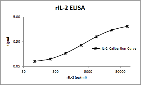
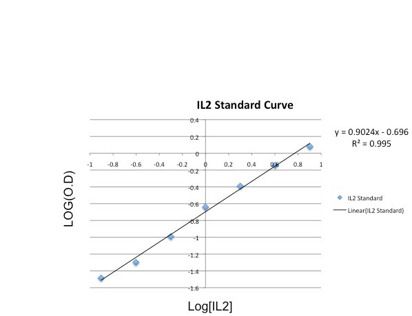
Rat IL-2 ELISA Standard Curve
Recombinant Rat IL‑2 (Catalog # 502-RL) was serially diluted and captured by Goat Anti-Human/Rat IL‑2 Antigen Affinity-purified Polyclonal Antibody (Catalog # AF-502-NA) coated on a Clear Polystyrene Microplate (Catalog # DY990). Goat Anti-Human/Rat IL‑2 Antigen Affinity-purified Polyclonal Antibody (Catalog # AF-502-NA) was biotinylated and incubated with the protein captured on the plate. Detection of the standard curve was achieved by incubating Streptavidin-HRP (Catalog # DY998)
Rat IL-2 ELISA Standard Curve
Recombinant Rat IL‑2 (Catalog # 502-RL) was serially diluted and captured by Goat Anti-Human/Rat IL‑2 Antigen Affinity-purified Polyclonal Antibody (Catalog # AF-502-NA) coated on a Clear Polystyrene Microplate (Catalog # DY990). Goat Anti-Human/Rat IL‑2 Antigen Affinity-purified Polyclonal Antibody (Catalog # AF-502-NA) was biotinylated and incubated with the protein captured on the plate. Detection of the standard curve was achieved by incubating Streptavidin-HRP (Catalog # DY998)Applications for IL-2 Antibody
Application
Recommended Usage
Immunocytochemistry
5-15 µg/mL
Sample: Immersion fixed rat splenocytes stimulated with calcium ionomycin and PMA
Sample: Immersion fixed rat splenocytes stimulated with calcium ionomycin and PMA
Immunohistochemistry
3-15 µg/mL
Sample: Immersion fixed frozen sections of Rat Spleen and Human Tonsil
Sample: Immersion fixed frozen sections of Rat Spleen and Human Tonsil
Western Blot
2 µg/mL
Sample: Human peripheral blood mononuclear cells (PBMCs) treated with monensin, 0.5ug/mL calcium ionomycin and 50ng/mL PMA overnight
Sample: Human peripheral blood mononuclear cells (PBMCs) treated with monensin, 0.5ug/mL calcium ionomycin and 50ng/mL PMA overnight
Neutralization
Measured by its ability to neutralize IL‑2-induced proliferation in the CTLL‑2 mouse cytotoxic T cell line. Gearing, A.J.H. and C.B. Bird (1987) in Lymphokines and Interferons, A Practical Approach. Clemens, M.J. et al. (eds): IRL Press. 276. The Neutralization Dose (ND50) is typically 0.15-0.75 µg/mL in the presence of 2 ng/mL Recombinant Rat IL‑2.
Rat IL-2 Sandwich Immunoassay
Please Note: Optimal dilutions of this antibody should be experimentally determined.
Reviewed Applications
Read 2 reviews rated 4.5 using AF-502-NA in the following applications:
Formulation, Preparation, and Storage
Purification
Antigen Affinity-purified
Reconstitution
Reconstitute at 0.2 mg/mL in sterile PBS. For liquid material, refer to CoA for concentration.
Loading...
Formulation
Lyophilized from a 0.2 μm filtered solution in PBS with Trehalose. *Small pack size (SP) is supplied either lyophilized or as a 0.2 µm filtered solution in PBS.
Shipping
Lyophilized product is shipped at ambient temperature. Liquid small pack size (-SP) is shipped with polar packs. Upon receipt, store immediately at the temperature recommended below.
Stability & Storage
Use a manual defrost freezer and avoid repeated freeze-thaw cycles.
- 12 months from date of receipt, -20 to -70 °C as supplied.
- 1 month, 2 to 8 °C under sterile conditions after reconstitution.
- 6 months, -20 to -70 °C under sterile conditions after reconstitution.
Calculators
Background: IL-2
References
- Ma, A. et al. (2006) Annu. Rev. Immunol. 24:657.
- Gaffen, S.L. and K.D. Liu (2004) Cytokine 28:109.
- McKnight, A. et al. (1989) Immunogenetics 30:145.
- Liparoto, S.F. et al. (2002) Biochemistry 41:2543.
- Wang, X. et al. (2005) Science 310:1159.
- Bodnar, A. et al. (2008) Immunol. Lett. 116:117.
- Jaleco, S. et al. (2003) J. Immunol. 171:61.
- Malek, T.R. (2003) J. Leukoc. Biol. 74:961.
- Laurence, A. et al. (2007) Immunity 26:371.
- Kryczek, I. et al. (2007) J. Immunol. 178:6730.
- Afzali, B. et al. (2007) Clin. Exp. Immunol. 148:32.
- Fehervari, Z. et al. (2006) Trends Immunol. 27:109.
Long Name
Interleukin 2
Alternate Names
Aldesleukin, IL2, Proleukin, TCGF
Entrez Gene IDs
Gene Symbol
IL2
UniProt
Additional IL-2 Products
Product Documents for IL-2 Antibody
Certificate of Analysis
To download a Certificate of Analysis, please enter a lot or batch number in the search box below.
Note: Certificate of Analysis not available for kit components.
Product Specific Notices for IL-2 Antibody
For research use only
Related Research Areas
Citations for IL-2 Antibody
 Powered by Bioz
Customer Reviews for IL-2 Antibody (2)
4.5 out of 5
2 Customer Ratings
Have you used IL-2 Antibody?
Submit a review and receive an Amazon gift card!
$25/€18/£15/$25CAN/¥2500 Yen for a review with an image
$10/€7/£6/$10CAN/¥1110 Yen for a review without an image
Submit a review
Customer Images
Showing
1
-
2 of
2 reviews
Showing All
Filter By:
-
Application: ELISASample Tested: SerumSpecies: RatVerified Customer | Posted 11/08/2017The rat polyclonal AF502 was paired with BAF502 to build an ELISA for the measurement of Rat IL-2 in serum samples.

-
Application: ELISASample Tested: Cell culture supernatantVerified Customer | Posted 10/26/2015ELISA plates were coated by incubating the diluted capture antibody (AF-502-NA in PBS at 1.6ug/mL) overnight at 4C. To detect the captured IL2 from the supernatants being tested, the detection antibody BAF502 was used at 400ng/mL followed by avidin-HRP. <br />Specificity: Specific<br />Sensitivity: Sensitive<br />Buffer: Wash and blocking buffers<br />Dilution: 1.6ug/mL
There are no reviews that match your criteria.
Protocols
Find general support by application which include: protocols, troubleshooting, illustrated assays, videos and webinars.
- Antigen Retrieval Protocol (PIER)
- Antigen Retrieval for Frozen Sections Protocol
- Appropriate Fixation of IHC/ICC Samples
- Cellular Response to Hypoxia Protocols
- Chromogenic IHC Staining of Formalin-Fixed Paraffin-Embedded (FFPE) Tissue Protocol
- Chromogenic Immunohistochemistry Staining of Frozen Tissue
- ClariTSA™ Fluorophore Kits
- Detection & Visualization of Antibody Binding
- Fluorescent IHC Staining of Frozen Tissue Protocol
- Graphic Protocol for Heat-induced Epitope Retrieval
- Graphic Protocol for the Preparation and Fluorescent IHC Staining of Frozen Tissue Sections
- Graphic Protocol for the Preparation and Fluorescent IHC Staining of Paraffin-embedded Tissue Sections
- Graphic Protocol for the Preparation of Gelatin-coated Slides for Histological Tissue Sections
- ICC Cell Smear Protocol for Suspension Cells
- ICC Immunocytochemistry Protocol Videos
- ICC for Adherent Cells
- IHC Sample Preparation (Frozen sections vs Paraffin)
- Immunocytochemistry (ICC) Protocol
- Immunocytochemistry Troubleshooting
- Immunofluorescence of Organoids Embedded in Cultrex Basement Membrane Extract
- Immunofluorescent IHC Staining of Formalin-Fixed Paraffin-Embedded (FFPE) Tissue Protocol
- Immunohistochemistry (IHC) and Immunocytochemistry (ICC) Protocols
- Immunohistochemistry Frozen Troubleshooting
- Immunohistochemistry Paraffin Troubleshooting
- Preparing Samples for IHC/ICC Experiments
- Preventing Non-Specific Staining (Non-Specific Binding)
- Primary Antibody Selection & Optimization
- Protocol for Heat-Induced Epitope Retrieval (HIER)
- Protocol for Making a 4% Formaldehyde Solution in PBS
- Protocol for VisUCyte™ HRP Polymer Detection Reagent
- Protocol for the Fluorescent ICC Staining of Cell Smears - Graphic
- Protocol for the Fluorescent ICC Staining of Cultured Cells on Coverslips - Graphic
- Protocol for the Preparation & Fixation of Cells on Coverslips
- Protocol for the Preparation and Chromogenic IHC Staining of Frozen Tissue Sections
- Protocol for the Preparation and Chromogenic IHC Staining of Frozen Tissue Sections - Graphic
- Protocol for the Preparation and Chromogenic IHC Staining of Paraffin-embedded Tissue Sections
- Protocol for the Preparation and Chromogenic IHC Staining of Paraffin-embedded Tissue Sections - Graphic
- Protocol for the Preparation and Fluorescent ICC Staining of Cells on Coverslips
- Protocol for the Preparation and Fluorescent ICC Staining of Non-adherent Cells
- Protocol for the Preparation and Fluorescent ICC Staining of Stem Cells on Coverslips
- Protocol for the Preparation and Fluorescent IHC Staining of Frozen Tissue Sections
- Protocol for the Preparation and Fluorescent IHC Staining of Paraffin-embedded Tissue Sections
- Protocol for the Preparation of Gelatin-coated Slides for Histological Tissue Sections
- Protocol for the Preparation of a Cell Smear for Non-adherent Cell ICC - Graphic
- R&D Systems Quality Control Western Blot Protocol
- TUNEL and Active Caspase-3 Detection by IHC/ICC Protocol
- The Importance of IHC/ICC Controls
- Troubleshooting Guide: Immunohistochemistry
- Troubleshooting Guide: Western Blot Figures
- Western Blot Conditions
- Western Blot Protocol
- Western Blot Protocol for Cell Lysates
- Western Blot Troubleshooting
- Western Blot Troubleshooting Guide
- View all Protocols, Troubleshooting, Illustrated assays and Webinars
Loading...
Associated Pathways


Innate Lymphoid Cell Differentiation Pathways
 Jak/STAT Signaling Pathway
 Th1 Differentiation Pathway
 Th2 Differentiation Pathway
