Reg1A, also known as PTP, PSP, and lithostathine, is a member of the Reg family of secreted proteins with a C-type lectin domain. Due to variable glycosylation, pancreatic Reg1A exists as multiple species of 16‑18 kDa. Reg1A promotes the maintenance and growth of pancreatic islet beta -cells and intestinal villi. It is up‑regulated in pancreatitis and some carcinomas. Reg1A is an antigenic target in autoimmune diabetes. Human Reg1A shares 65%‑68% aa sequence identity with mouse and rat Reg1A.
Reg1B, also known as secretory pancreatic stone protein 2 and lithostathine 1 beta, is a type I subclass member of the Reg family, which comprises secreted proteins with a C-type lectin domain. Reg1B is highly related to Reg1A, a protein that is implicated in islet cell regeneration and diabetogenesis.Human Reg1A/B Antibody (431211)
R&D Systems | Catalog # MAB49371

Key Product Details
Species Reactivity
Human
Applications
Immunohistochemistry, Western Blot
Label
Unconjugated
Antibody Source
Monoclonal Rat IgG2A Clone # 431211
Loading...
Product Specifications
Immunogen
E. coli-derived recombinant human Reg1A
Gln23-Asn166
Accession # P05451
Gln23-Asn166
Accession # P05451
Specificity
Detects human Reg1A in ELISAs. 100% cross-reactivity with recombinant human (rh) Reg1B in Western blot is observed. In direct ELISAs no
cross-reactivity with rhReg3A, recombinant mouse Reg1, or recombinant rat Reg1A
is observed.
Clonality
Monoclonal
Host
Rat
Isotype
IgG2A
Scientific Data Images for Human Reg1A/B Antibody (431211)
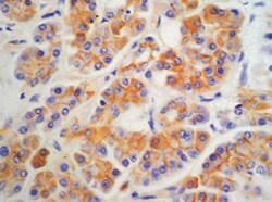
Reg1A in Human Pancreas.
Reg1A was detected in immersion fixed paraffin-embedded sections of human pancreas using Rat Anti-Human Reg1A Monoclonal Antibody (Catalog # MAB49371) at 15 µg/mL overnight at 4 °C. Before incubation with the primary antibody, tissue was subjected to heat-induced epitope retrieval using Antigen Retrieval Reagent-Basic (Catalog # CTS013). Tissue was stained using the Anti-Rat HRP-DAB Cell & Tissue Staining Kit (brown; Catalog # CTS017) and counterstained with hematoxylin (blue). Specific staining was localized to exocrine cells. View our protocol for Chromogenic IHC Staining of Paraffin-embedded Tissue Sections.Applications for Human Reg1A/B Antibody (431211)
Application
Recommended Usage
Immunohistochemistry
8-25 µg/mL
Sample: Immersion fixed paraffin-embedded sections of human pancreas
Sample: Immersion fixed paraffin-embedded sections of human pancreas
Western Blot
1 µg/mL
Sample: Recombinant Human Reg1A
Sample: Recombinant Human Reg1A
Reviewed Applications
Read 2 reviews rated 4.5 using MAB49371 in the following applications:
Formulation, Preparation, and Storage
Purification
Protein A or G purified from hybridoma culture supernatant
Reconstitution
Sterile PBS to a final concentration of 0.5 mg/mL. For liquid material, refer to CoA for concentration.
Loading...
Formulation
Lyophilized from a 0.2 μm filtered solution in PBS with Trehalose. *Small pack size (SP) is supplied either lyophilized or as a 0.2 µm filtered solution in PBS.
Shipping
Lyophilized product is shipped at ambient temperature. Liquid small pack size (-SP) is shipped with polar packs. Upon receipt, store immediately at the temperature recommended below.
Stability & Storage
Use a manual defrost freezer and avoid repeated freeze-thaw cycles.
- 12 months from date of receipt, -20 to -70 °C as supplied.
- 1 month, 2 to 8 °C under sterile conditions after reconstitution.
- 6 months, -20 to -70 °C under sterile conditions after reconstitution.
Calculators
Background: Reg1A/B
Long Name
Regenerating Islet-derived 1A/B
UniProt
Additional Reg1A/B Products
Product Documents for Human Reg1A/B Antibody (431211)
Certificate of Analysis
To download a Certificate of Analysis, please enter a lot or batch number in the search box below.
Note: Certificate of Analysis not available for kit components.
Product Specific Notices for Human Reg1A/B Antibody (431211)
For research use only
Related Research Areas
Citations for Human Reg1A/B Antibody (431211)
 Powered by Bioz
Customer Reviews for Human Reg1A/B Antibody (431211) (2)
4.5 out of 5
2 Customer Ratings
Have you used Human Reg1A/B Antibody (431211)?
Submit a review and receive an Amazon gift card!
$25/€18/£15/$25CAN/¥2500 Yen for a review with an image
$10/€7/£6/$10CAN/¥1110 Yen for a review without an image
Submit a review
Customer Images
Showing
1
-
2 of
2 reviews
Showing All
Filter By:
-
Application: ImmunohistochemistrySample Tested: Pancreas tissueSpecies: HumanVerified Customer | Posted 05/17/2022

-
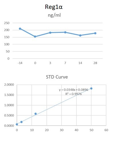
Application: ELISASample Tested: Serum and PlasmaSpecies: HumanVerified Customer | Posted 06/09/2020We used this antibody in an in-house ELISA along with pAb (AF4937) and protein standard to quantify Reg1 alpha in human serum and plasma. This combination detected Reg1alpha in human serum and plasma.
There are no reviews that match your criteria.
Protocols
Find general support by application which include: protocols, troubleshooting, illustrated assays, videos and webinars.
- Antigen Retrieval Protocol (PIER)
- Antigen Retrieval for Frozen Sections Protocol
- Appropriate Fixation of IHC/ICC Samples
- Cellular Response to Hypoxia Protocols
- Chromogenic IHC Staining of Formalin-Fixed Paraffin-Embedded (FFPE) Tissue Protocol
- Chromogenic Immunohistochemistry Staining of Frozen Tissue
- ClariTSA™ Fluorophore Kits
- Detection & Visualization of Antibody Binding
- Fluorescent IHC Staining of Frozen Tissue Protocol
- Graphic Protocol for Heat-induced Epitope Retrieval
- Graphic Protocol for the Preparation and Fluorescent IHC Staining of Frozen Tissue Sections
- Graphic Protocol for the Preparation and Fluorescent IHC Staining of Paraffin-embedded Tissue Sections
- Graphic Protocol for the Preparation of Gelatin-coated Slides for Histological Tissue Sections
- IHC Sample Preparation (Frozen sections vs Paraffin)
- Immunofluorescent IHC Staining of Formalin-Fixed Paraffin-Embedded (FFPE) Tissue Protocol
- Immunohistochemistry (IHC) and Immunocytochemistry (ICC) Protocols
- Immunohistochemistry Frozen Troubleshooting
- Immunohistochemistry Paraffin Troubleshooting
- Preparing Samples for IHC/ICC Experiments
- Preventing Non-Specific Staining (Non-Specific Binding)
- Primary Antibody Selection & Optimization
- Protocol for Heat-Induced Epitope Retrieval (HIER)
- Protocol for Making a 4% Formaldehyde Solution in PBS
- Protocol for VisUCyte™ HRP Polymer Detection Reagent
- Protocol for the Preparation & Fixation of Cells on Coverslips
- Protocol for the Preparation and Chromogenic IHC Staining of Frozen Tissue Sections
- Protocol for the Preparation and Chromogenic IHC Staining of Frozen Tissue Sections - Graphic
- Protocol for the Preparation and Chromogenic IHC Staining of Paraffin-embedded Tissue Sections
- Protocol for the Preparation and Chromogenic IHC Staining of Paraffin-embedded Tissue Sections - Graphic
- Protocol for the Preparation and Fluorescent IHC Staining of Frozen Tissue Sections
- Protocol for the Preparation and Fluorescent IHC Staining of Paraffin-embedded Tissue Sections
- Protocol for the Preparation of Gelatin-coated Slides for Histological Tissue Sections
- R&D Systems Quality Control Western Blot Protocol
- TUNEL and Active Caspase-3 Detection by IHC/ICC Protocol
- The Importance of IHC/ICC Controls
- Troubleshooting Guide: Immunohistochemistry
- Troubleshooting Guide: Western Blot Figures
- Western Blot Conditions
- Western Blot Protocol
- Western Blot Protocol for Cell Lysates
- Western Blot Troubleshooting
- Western Blot Troubleshooting Guide
- View all Protocols, Troubleshooting, Illustrated assays and Webinars
Loading...