Reg1A (Regenerating gene IA/ alpha ; also REG, ICRF, PSPS, PTP and Lithostathine-1 alpha ) is a secreted, variably glycosylated 15-22 kDa type I member of the REG gene family of proteins. It is induced in proliferating cell types such as colonic epithelium, islet beta -cells, and multiple tumor types, and is constitutively expressed by pancreatic acinar cells. Thus, Reg1A is found both in blood (ng/mL quantities) and pancreatic secretions. It would appear that Reg1A plays a role in blocking apoptosis, and is upregulated by both IL-22 and IL-6. Human Reg1A is synthesized as a 166 amino acid (aa) precursor. It contains a 22 aa signal sequence plus a 144 aa mature region that is characterized by the presence of three intrachain disulfide bonds, one C-type lectin domain (aa 34-164), and a utilized N-terminal O-linked glycosylation site (Thr27). Variablility in the glycosylation pattern, plus proteolytic cleavage after Arg11 generates Reg1A isoforms in the 15-22 kDa range. There is one potential isoform variant that shows an alternative start site at Met9. Mature (aa 23-166) human Reg1A shares 76% and 69% aa sequence identity with mouse and rat Reg1A, respectively.
Reg1B, also known as secretory pancreatic stone protein 2 and lithostathine 1 beta, is a type I subclass member of the Reg family, which comprises secreted proteins with a C-type lectin domain. Reg1B is highly related to Reg1A, a protein that is implicated in islet cell regeneration and diabetogenesis.
Key Product Details
Species Reactivity
Human
Applications
Immunohistochemistry, Western Blot
Label
Unconjugated
Antibody Source
Polyclonal Sheep IgG
Loading...
Product Specifications
Immunogen
Chinese hamster ovary cell line CHO-derived recombinant human Reg1A
Gln23-Asn166
Accession # P05451
Gln23-Asn166
Accession # P05451
Specificity
Detects human Reg1A in direct ELISAs. In direct ELISAs, approximately 80% cross-reactivity with recombinant human (rh) Reg1B is observed, approximately 20% cross-reactivity with recombinant mouse Reg1, and less than 5% cross-reactivity with rhReg3A and recombinant rat Reg1A is observed. 100% cross-reactivity with rhReg1B in Western blot is observed.
Clonality
Polyclonal
Host
Sheep
Isotype
IgG
Scientific Data Images for Human Reg1A/B Antibody
Detection of Human Reg1A by Western Blot.
Western blot shows lysates of human pancreas tissue. PVDF membrane was probed with 1 µg/mL of Sheep Anti-Human Reg1A Antigen Affinity-purified Polyclonal Antibody (Catalog # AF4937) followed by HRP-conjugated Anti-Sheep IgG Secondary Antibody (Catalog # HAF016). Specific bands were detected for Reg1A at approximately 20-22 kDa (as indicated). This experiment was conducted under reducing conditions and using Immunoblot Buffer Group 1.
Reg1A in Human Pancreas.
Reg1A was detected in immersion fixed paraffin-embedded sections of human pancreas using Sheep Anti-Human Reg1A Antigen Affinity-purified Polyclonal Antibody (Catalog # AF4937) at 3 µg/mL overnight at 4 °C. Before incubation with the primary antibody, tissue was subjected to heat-induced epitope retrieval using Antigen Retrieval Reagent-Basic (Catalog # CTS013). Tissue was stained using the Anti-Sheep HRP-DAB Cell & Tissue Staining Kit (brown; Catalog # CTS019) and counterstained with hematoxylin (blue). Specific staining was localized to cytoplasm in exocrine cells. View our protocol for Chromogenic IHC Staining of Paraffin-embedded Tissue Sections.Applications for Human Reg1A/B Antibody
Application
Recommended Usage
Immunohistochemistry
5-15 µg/mL
Sample: Immersion fixed paraffin-embedded sections of human pancreas
Sample: Immersion fixed paraffin-embedded sections of human pancreas
Western Blot
1 µg/mL
Sample: Human pancreas tissue
Sample: Human pancreas tissue
Reviewed Applications
Read 1 review rated 4 using AF4937 in the following applications:
Formulation, Preparation, and Storage
Purification
Antigen Affinity-purified
Reconstitution
Sterile PBS to a final concentration of 0.2 mg/mL. For liquid material, refer to CoA for concentration.
Loading...
Formulation
Lyophilized from a 0.2 μm filtered solution in PBS with Trehalose. *Small pack size (SP) is supplied either lyophilized or as a 0.2 µm filtered solution in PBS.
Shipping
Lyophilized product is shipped at ambient temperature. Liquid small pack size (-SP) is shipped with polar packs. Upon receipt, store immediately at the temperature recommended below.
Stability & Storage
Use a manual defrost freezer and avoid repeated freeze-thaw cycles.
- 12 months from date of receipt, -20 to -70 °C as supplied.
- 1 month, 2 to 8 °C under sterile conditions after reconstitution.
- 6 months, -20 to -70 °C under sterile conditions after reconstitution.
Calculators
Background: Reg1A/B
Long Name
Regenerating Islet-derived 1A/B
UniProt
Additional Reg1A/B Products
Product Documents for Human Reg1A/B Antibody
Certificate of Analysis
To download a Certificate of Analysis, please enter a lot or batch number in the search box below.
Note: Certificate of Analysis not available for kit components.
Product Specific Notices for Human Reg1A/B Antibody
For research use only
Related Research Areas
Customer Reviews for Human Reg1A/B Antibody (1)
4 out of 5
1 Customer Rating
Have you used Human Reg1A/B Antibody?
Submit a review and receive an Amazon gift card!
$25/€18/£15/$25CAN/¥2500 Yen for a review with an image
$10/€7/£6/$10CAN/¥1110 Yen for a review without an image
Submit a review
Customer Images
Showing
1
-
1 of
1 review
Showing All
Filter By:
-
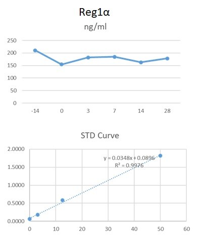
Application: ELISASample Tested: Serum and PlasmaSpecies: HumanVerified Customer | Posted 06/09/2020We used this antibody in an in-house ELISA along with mAb (49371) and protein standard to quantify Reg1 alpha in human serum and plasma. This combination detected Reg1alpha in human serum and plasma.

There are no reviews that match your criteria.
Protocols
Find general support by application which include: protocols, troubleshooting, illustrated assays, videos and webinars.
- Antigen Retrieval Protocol (PIER)
- Antigen Retrieval for Frozen Sections Protocol
- Appropriate Fixation of IHC/ICC Samples
- Cellular Response to Hypoxia Protocols
- Chromogenic IHC Staining of Formalin-Fixed Paraffin-Embedded (FFPE) Tissue Protocol
- Chromogenic Immunohistochemistry Staining of Frozen Tissue
- ClariTSA™ Fluorophore Kits
- Detection & Visualization of Antibody Binding
- Fluorescent IHC Staining of Frozen Tissue Protocol
- Graphic Protocol for Heat-induced Epitope Retrieval
- Graphic Protocol for the Preparation and Fluorescent IHC Staining of Frozen Tissue Sections
- Graphic Protocol for the Preparation and Fluorescent IHC Staining of Paraffin-embedded Tissue Sections
- Graphic Protocol for the Preparation of Gelatin-coated Slides for Histological Tissue Sections
- IHC Sample Preparation (Frozen sections vs Paraffin)
- Immunofluorescent IHC Staining of Formalin-Fixed Paraffin-Embedded (FFPE) Tissue Protocol
- Immunohistochemistry (IHC) and Immunocytochemistry (ICC) Protocols
- Immunohistochemistry Frozen Troubleshooting
- Immunohistochemistry Paraffin Troubleshooting
- Preparing Samples for IHC/ICC Experiments
- Preventing Non-Specific Staining (Non-Specific Binding)
- Primary Antibody Selection & Optimization
- Protocol for Heat-Induced Epitope Retrieval (HIER)
- Protocol for Making a 4% Formaldehyde Solution in PBS
- Protocol for VisUCyte™ HRP Polymer Detection Reagent
- Protocol for the Preparation & Fixation of Cells on Coverslips
- Protocol for the Preparation and Chromogenic IHC Staining of Frozen Tissue Sections
- Protocol for the Preparation and Chromogenic IHC Staining of Frozen Tissue Sections - Graphic
- Protocol for the Preparation and Chromogenic IHC Staining of Paraffin-embedded Tissue Sections
- Protocol for the Preparation and Chromogenic IHC Staining of Paraffin-embedded Tissue Sections - Graphic
- Protocol for the Preparation and Fluorescent IHC Staining of Frozen Tissue Sections
- Protocol for the Preparation and Fluorescent IHC Staining of Paraffin-embedded Tissue Sections
- Protocol for the Preparation of Gelatin-coated Slides for Histological Tissue Sections
- R&D Systems Quality Control Western Blot Protocol
- TUNEL and Active Caspase-3 Detection by IHC/ICC Protocol
- The Importance of IHC/ICC Controls
- Troubleshooting Guide: Immunohistochemistry
- Troubleshooting Guide: Western Blot Figures
- Western Blot Conditions
- Western Blot Protocol
- Western Blot Protocol for Cell Lysates
- Western Blot Troubleshooting
- Western Blot Troubleshooting Guide
- View all Protocols, Troubleshooting, Illustrated assays and Webinars
Loading...