Survivin is a member of the inhibitor of apoptosis (IAP) family and can inhibit apoptosis induced by a variety of factors. It is expressed in most human cancers but not in normal adult tissues. Survivin is expressed in a cell cycle-dependent manner and associates with the mitotic apparatus.

Key Product Details
Species Reactivity
Validated:
Human
Cited:
Human, Mouse
Applications
Validated:
Flow Cytometry, CyTOF-ready
Cited:
Immunohistochemistry, Flow Cytometry, CyTof
Label
Unconjugated
Antibody Source
Monoclonal Mouse IgG1 Clone # 91630
Loading...
Product Specifications
Immunogen
E. coli-derived recombinant human Survivin
Met1-Asp142
Accession # O15392
Met1-Asp142
Accession # O15392
Specificity
Detects human Survivin in direct ELISAs.
Clonality
Monoclonal
Host
Mouse
Isotype
IgG1
Scientific Data Images for Human Survivin Antibody (91630)
Detection of Survivin in Jurkat cells by Flow Cytometry
Jurkat cells were stained with Mouse Anti-Human Survivin Monoclonal Antibody (Catalog # MAB886, filled histogram) or isotype control antibody (Catalog # MAB002, open histogram) followed by Phycoerythrin-conjugated Anti-Mouse IgG Secondary Antibody (Catalog # F0102B). To facilitate intracellular staining, cells were fixed with Flow Cytometry Fixation Buffer (Catalog # FC004) and permeabilized with Flow Cytometry Permeabilization/Wash Buffer I (Catalog # FC005). View our protocol for Staining Intracellular Molecules.
Detection of Survivin by Flow Cytometry
Exposure to survivin enriched PD-1+ Bcl-6+ subset of Tfh via STAT3 dependent mechanismsDBA/1 mice were immunized with survivin-derived peptide (100 μg/mouse × 4, subcutaneously). Control mice were immunized with ovalbumin-derived peptide (OVA). Both groups were then subjected to collagen-induced arthritis. Anti-survivin IgG antibodies, anti-Fcg antibodies, & survivin levels in serum were measured by ELISA A. Flow cytometry analysis of the expression of survivin B. in CD44hiCD4+ lymphocytes, & expression of PD-1 C. & Bcl-6 E. in survivin+CXCR5+ CD4+ lymphocytes in spleen (SPL) & lymph nodes (LN). PD-1 expression correlated to the size of CXCR5+survivin+ population D. Dots represent individual mice & the horizontal line shows median of the group. Protein levels of active STAT3 phosphorylated at Y705 (pStat3), total Stat3, Bcl-6 & actin in spleen were analyzed by the Western blot F. The levels of each protein were quantified in ratio to actin of each sample. Quantification of the detected bands is presented as box plots. Transcription of Bcl-6, cMaf & IL-21 in the spleen was analyzed by RT-PCR & presented in relative quantity (RQ) to the median of the control group G. Anti-survivin antibodies in serum correlated with the size of survivin+ CD4 population in spleen of survivin-immunized mice H. Comparison of the correlations in the survivin- & OVA-immunized groups is done by the Fisher r-to-z transformation analysis. Anti-Fcg antibodies correlated with the intensity of PD-1 on CXCR5+survivin+ CD4 lymphocytes I., as measured by flow cytometry. Box plots with line represent IQR of the group & median, respectively, & error lines indicate min & max values. The Mann-Whitney U-test was used to compare differences between groups. Correlation analyses were performed using Spearman's test. Image collected & cropped by CiteAb from the following open publication (https://pubmed.ncbi.nlm.nih.gov/26343374), licensed under a CC-BY license. Not internally tested by R&D Systems.
Detection of Survivin by Flow Cytometry
Survivin positive subset of CD44hi CD4 lymphocytes in mouse possess a complete phenotype of Tfh cells Spleen and lymph nodes from collagen II immunized arthritic (CIA) mice were analyzed for expression of survivin and Bcl-6 using flow cytometry A. Cells were gated on memory CD44hiCD4+ lymphocytes. Expression of CXCR5 B. and PD-1 C. was investigated within Bcl-6+ survivin+ and Bcl-6− survivin+ cells. Dots represent individual mice and the horizontal line shows median of the group. Survivin translation in CIA mice was inhibited by shRNA-producing constructs provided as a single intra-peritoneal injection (shSurv16, n = 10, or shSurv13+16, n = 10). Control mice were treated with a non-targeting RNA construct (shNT, n = 9). Survivin expression in spleen was analyzed by flow cytometry 12 days after the injection. Cells were gated on CD4+ lymphocytes. Intensity of survivin expression (MFI) within the groups is shown by a representative histogram and summarized in a box plot D. Survivin expression (MFI) on CD4 lymphocytes correlated to the size of CD44hiCD62L+ population E. Expression of CXCR5 on CD44hiCD4+ lymphocytes in the groups is shown as box plot F. CXCR5+ population correlates with the intensity of survivin G. and with Bcl-6 mRNA H. Boxes and lines represent IQR and median, respectively, and error lines indicate min and max values. The Mann-Whitney U-test was used to compare differences between groups. Correlation analyses were performed using Spearman's test. Image collected and cropped by CiteAb from the following open publication (https://pubmed.ncbi.nlm.nih.gov/26343374), licensed under a CC-BY license. Not internally tested by R&D Systems.
Detection of Survivin by Flow Cytometry
Survivin expression is an essential feature of human CXCR5+ Tfh cell phenotypeIntracellular expression of survivin was investigated in memory (CD45RA−) or naïve (CD45RA+) CD4+ T cells of RA patients (n = 21) and healthy controls (n = 10) using flow cytometry. Cells are gated on CD4+ lymphocytes. Box plots show the frequency of survivin+ cells A. and the mean fluorescence intensity (MFI) of survivin B. Expression of CXCR5 C. within survivin+ and survivin− CD4+ cells, and Bcl-6 D. within survivin+ and survivin− memory (CD45RA−) CD4+ cells of RA patients. The intensity of survivin expression E. within Bcl-6+ and Bcl-6− survivin+ CXCR5+ CD4 cells. The Mann-Whitney U-test was used to compare differences between groups. PBMCs of healthy subjects (1 × 10⁶/ml, n = 6) were cultured with anti-CD3 (0.25 μg/ml) alone or in combination with IL-12 (20 ng/ml) or IL-21 (50 ng/ml). On day 5, the formation of Tfh cells was recognized by expression of CXCR5 and intracellular production of IL-21. Cells were gated on viable CD4+ lymphocytes. Intensity of CXCR5 expression on survivin+ CD4 cells is shown F. The frequency of CXCR5+ cells within survivin+ and survivin− CD4 subsets stimulated with alpha CD3 + IL-12 G. Intracellular production of IL-21 within the CXCR5+survivin+ and CXCR5+survivin− CD4 cells stimulated with alpha CD3 + IL-12 is shown by histogram H. Frequency of PD-1+ IL-21+ cells is shown by box plots I. The Wilcoxon matched-pairs signed rank test to compare differences. Boxes and lines represent IQR and median, respectively, and error lines indicate min and max values. Image collected and cropped by CiteAb from the following open publication (https://pubmed.ncbi.nlm.nih.gov/26343374), licensed under a CC-BY license. Not internally tested by R&D Systems.
Detection of Survivin by Flow Cytometry
Survivin positive subset of CD44hi CD4 lymphocytes in mouse possess a complete phenotype of Tfh cells Spleen and lymph nodes from collagen II immunized arthritic (CIA) mice were analyzed for expression of survivin and Bcl-6 using flow cytometry A. Cells were gated on memory CD44hiCD4+ lymphocytes. Expression of CXCR5 B. and PD-1 C. was investigated within Bcl-6+ survivin+ and Bcl-6− survivin+ cells. Dots represent individual mice and the horizontal line shows median of the group. Survivin translation in CIA mice was inhibited by shRNA-producing constructs provided as a single intra-peritoneal injection (shSurv16, n = 10, or shSurv13+16, n = 10). Control mice were treated with a non-targeting RNA construct (shNT, n = 9). Survivin expression in spleen was analyzed by flow cytometry 12 days after the injection. Cells were gated on CD4+ lymphocytes. Intensity of survivin expression (MFI) within the groups is shown by a representative histogram and summarized in a box plot D. Survivin expression (MFI) on CD4 lymphocytes correlated to the size of CD44hiCD62L+ population E. Expression of CXCR5 on CD44hiCD4+ lymphocytes in the groups is shown as box plot F. CXCR5+ population correlates with the intensity of survivin G. and with Bcl-6 mRNA H. Boxes and lines represent IQR and median, respectively, and error lines indicate min and max values. The Mann-Whitney U-test was used to compare differences between groups. Correlation analyses were performed using Spearman's test. Image collected and cropped by CiteAb from the following open publication (https://pubmed.ncbi.nlm.nih.gov/26343374), licensed under a CC-BY license. Not internally tested by R&D Systems.Applications for Human Survivin Antibody (91630)
Application
Recommended Usage
CyTOF-ready
Ready to be labeled using established conjugation methods. No BSA or other carrier proteins that could interfere with conjugation.
Flow Cytometry
0.25 µg/106 cells
Sample: Jurkat human acute T cell leukemia cell line
Sample: Jurkat human acute T cell leukemia cell line
Reviewed Applications
Read 1 review rated 5 using MAB886 in the following applications:
Flow Cytometry Panel Builder
Bio-Techne Knows Flow Cytometry
Save time and reduce costly mistakes by quickly finding compatible reagents using the Panel Builder Tool.
Advanced Features
- Spectra Viewer - Custom analysis of spectra from multiple fluorochromes
- Spillover Popups - Visualize the spectra of individual fluorochromes
- Antigen Density Selector - Match fluorochrome brightness with antigen density

Formulation, Preparation, and Storage
Purification
Protein A or G purified from hybridoma culture supernatant
Reconstitution
Reconstitute at 0.5 mg/mL in sterile PBS. For liquid material, refer to CoA for concentration.
Loading...
Formulation
Lyophilized from a 0.2 μm filtered solution in PBS with Trehalose. See Certificate of Analysis for details.
*Small pack size (-SP) is supplied either lyophilized or as a 0.2 µm filtered solution in PBS.
*Small pack size (-SP) is supplied either lyophilized or as a 0.2 µm filtered solution in PBS.
Shipping
Lyophilized product is shipped at ambient temperature. Liquid small pack size (-SP) is shipped with polar packs. Upon receipt, store immediately at the temperature recommended below.
Stability & Storage
Use a manual defrost freezer and avoid repeated freeze-thaw cycles.
- 12 months from date of receipt, -20 to -70 °C as supplied.
- 1 month, 2 to 8 °C under sterile conditions after reconstitution.
- 6 months, -20 to -70 °C under sterile conditions after reconstitution.
Calculators
Background: Survivin
Additional Survivin Products
Product Documents for Human Survivin Antibody (91630)
Certificate of Analysis
To download a Certificate of Analysis, please enter a lot or batch number in the search box below.
Note: Certificate of Analysis not available for kit components.
Product Specific Notices for Human Survivin Antibody (91630)
For research use only
Related Research Areas
Citations for Human Survivin Antibody (91630)
 Powered by Bioz
Customer Reviews for Human Survivin Antibody (91630) (1)
5 out of 5
1 Customer Rating
Have you used Human Survivin Antibody (91630)?
Submit a review and receive an Amazon gift card!
$25/€18/£15/$25CAN/¥2500 Yen for a review with an image
$10/€7/£6/$10CAN/¥1110 Yen for a review without an image
Submit a review
Customer Images
Showing
1
-
1 of
1 review
Showing All
Filter By:
-
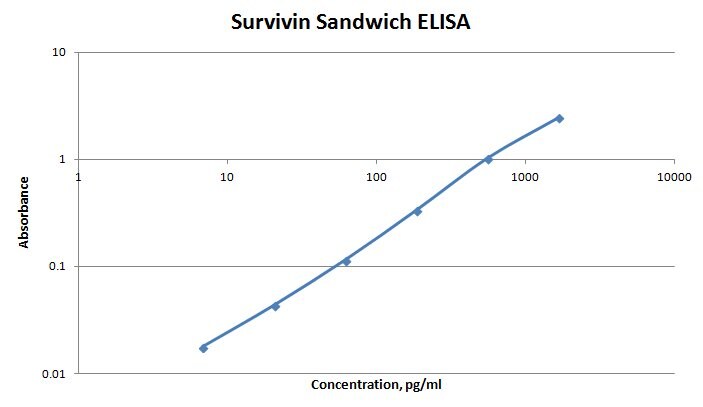
Application: ELISASample Tested: Serum and PlasmaSpecies: HumanVerified Customer | Posted 11/07/2017The antibody MAB886 was used as the capture in a sandwich ELISA along with AF6471 as detection. The ELISA worked very well. Healthy human serum and plasma samples were not detectable although some disease state samples are detectable.

There are no reviews that match your criteria.
Protocols
Find general support by application which include: protocols, troubleshooting, illustrated assays, videos and webinars.
- 7-Amino Actinomycin D (7-AAD) Cell Viability Flow Cytometry Protocol
- Extracellular Membrane Flow Cytometry Protocol
- Flow Cytometry Protocol for Cell Surface Markers
- Flow Cytometry Protocol for Staining Membrane Associated Proteins
- Flow Cytometry Staining Protocols
- Flow Cytometry Troubleshooting Guide
- Intracellular Flow Cytometry Protocol Using Alcohol (Methanol)
- Intracellular Flow Cytometry Protocol Using Detergents
- Intracellular Nuclear Staining Flow Cytometry Protocol Using Detergents
- Intracellular Staining Flow Cytometry Protocol Using Alcohol Permeabilization
- Intracellular Staining Flow Cytometry Protocol Using Detergents to Permeabilize Cells
- Propidium Iodide Cell Viability Flow Cytometry Protocol
- Protocol for Liperfluo
- Protocol for the Characterization of Human Th22 Cells
- Protocol for the Characterization of Human Th9 Cells
- Protocol: Annexin V and PI Staining by Flow Cytometry
- Protocol: Annexin V and PI Staining for Apoptosis by Flow Cytometry
- Troubleshooting Guide: Fluorokine Flow Cytometry Kits
- View all Protocols, Troubleshooting, Illustrated assays and Webinars
Loading...
Associated Pathways
