
IL-1 beta/IL-1F2 Antibody (A7F7-R)
Novus Biologicals | Catalog # NBP3-32464
Recombinant Monoclonal Antibody

Scientific images may be resized, cropped, or adjusted for brightness or contrast for presentation purposes. These updates are limited to presentation and do not affect the underlying scientific interpretation of the data. Where available, additional source data may be provided upon request.
Loading...
Key Product Details
Species Reactivity
Human, Mouse, Rat
Applications
Immunohistochemistry, Immunohistochemistry-Paraffin, Western Blot, ELISA
Label
Unconjugated
Antibody Source
Recombinant Monoclonal Mouse IgG1 Clone # A7F7-R expressed in HEK293
Loading...
Product Specifications
Immunogen
Recombinant protein within Human IL-1 beta/IL-1F2 aa 117-269 / 269. (Uniprot: P01584)
Localization
Extracellular exosome, Secreted, Lysosome, Cytosol.
Clonality
Monoclonal
Host
Mouse
Isotype
IgG1
Theoretical MW
31 kDa.
Disclaimer note: The observed molecular weight of the protein may vary from the listed predicted molecular weight due to post translational modifications, post translation cleavages, relative charges, and other experimental factors.
Disclaimer note: The observed molecular weight of the protein may vary from the listed predicted molecular weight due to post translational modifications, post translation cleavages, relative charges, and other experimental factors.
Scientific Data Images for IL-1 beta/IL-1F2 Antibody (A7F7-R)
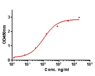
ELISA: IL-1 beta/IL-1F2 Antibody (A7F7-R) [NBP3-32464]
ELISA: IL-1 beta/IL-1F2 Antibody (A7F7-R) [NBP3-32464] - IL-1 beta/IL-1F2 Antibody (NBP3-32464) in indirect ELISA.Indirect ELISA analysis of IL-1 beta was performed by coating wells of a 96-well plate with 50 µl per well of IL-1 beta antigen diluted in carbonate/bicarbonate buffer, at a concentration of 1 µg/mL overnight at 4℃. Wells of the plate were washed, blocked with blocking buffer, and incubated with 50 µl per well of a mouse IL-1 beta monoclonal antibody starting at a concentration of 20 µg/mL and serially diluting it to a concentration of 1.28 ng/mL for 2 hours at room temperature. The plate was washed and incubated with 50 µl per well of an HRP-conjugated goat anti-mouse IgG secondary antibody at a dilution of 1:10,000 for one hour at room temperature. Detection was performed using an Ultra TMB Substrate for 5 minutes at room temperature in the dark. The reaction was stopped with sulfuric acid and absorbances were read on a spectrophotometer at 450 nm.
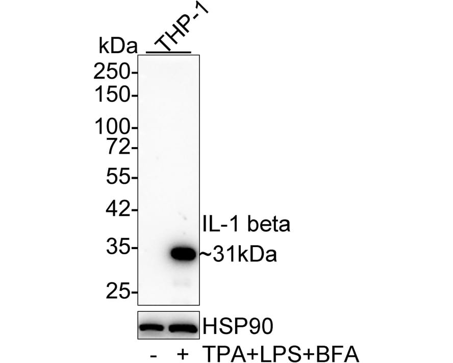
Western Blot: IL-1 beta/IL-1F2 Antibody (A7F7-R) [NBP3-32464]
Western Blot: IL-1 beta/IL-1F2 Antibody (A7F7-R) [NBP3-32464] - Western blot analysis of IL-1 beta/IL-1F2 on different lysates with Mouse anti-IL-1 beta/IL-1F2 antibody (NBP3-32464) at 1/1,000 dilution.Lane 1: THP-1 cell lysate
Lane 2: THP-1 treated with 80nM TPA overnight then treated with 100ng/mL LPS for 6 hours add 300ng/mL BFA for 3 hours cell lysate
Lysates/proteins at 10 ug/Lane.
Predicted band size: 31 kDa
Observed band size: 31 kDa
Exposure time: 1 minute;
4-20% SDS-PAGE gel.
Proteins were transferred to a PVDF membrane and blocked with 5% NFDM/TBST for 1 hour at room temperature. The primary antibody (NBP3-32464) at 1/1,000 dilution was used in 5% NFDM/TBST at 4 overnight. Goat Anti-Mouse IgG - HRP Secondary Antibody at 1/50,000 dilution was used for 1 hour at room temperature.
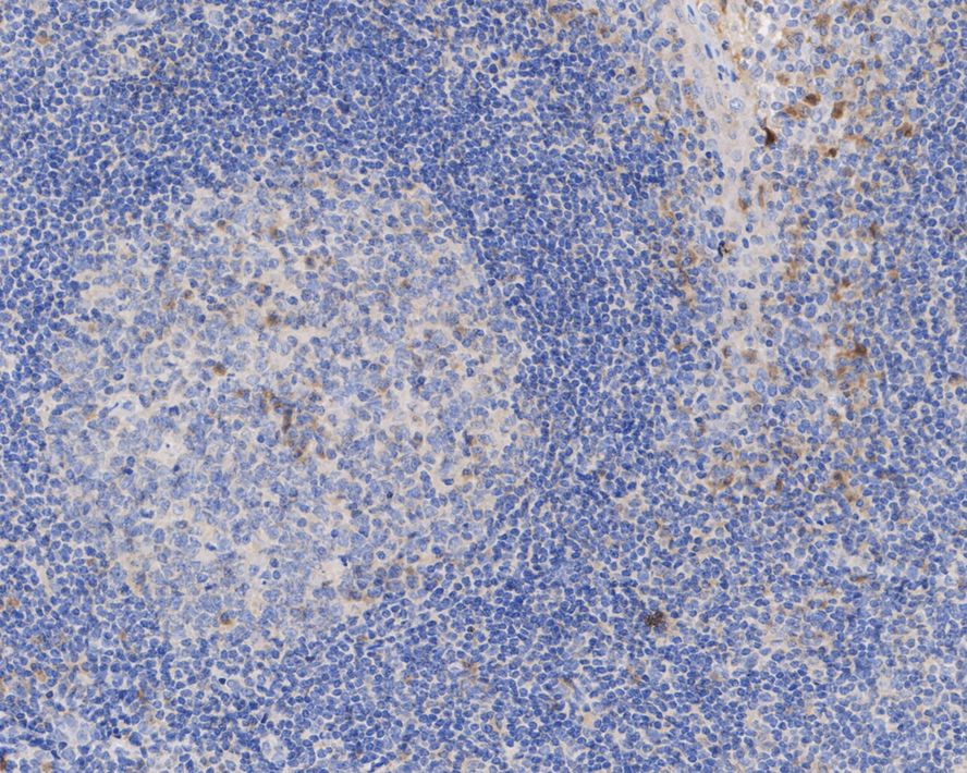
Immunohistochemistry: IL-1 beta/IL-1F2 Antibody (A7F7-R) [NBP3-32464]
Immunohistochemistry: IL-1 beta/IL-1F2 Antibody (A7F7-R) [NBP3-32464] - Immunohistochemical analysis of paraffin-embedded human tonsil tissue with Mouse anti-IL-1 beta/IL-1F2 antibody (NBP3-32464) at 1/100 dilution.The section was pre-treated using heat mediated antigen retrieval with Tris-EDTA buffer (pH 9.0) for 20 minutes. The tissues were blocked in 1% BSA for 20 minutes at room temperature, washed with ddH2O and PBS, and then probed with the primary antibody (NBP3-32464) at 1/100 dilution for 1 hour at room temperature. The detection was performed using an HRP conjugated compact polymer system. DAB was used as the chromogen. Tissues were counterstained with hematoxylin and mounted with DPX.
![IL-1 beta/IL-1F2 Antibody (A7F7-R) Western Blot: IL-1 beta/IL-1F2 Antibody (A7F7-R) [NBP3-32464]](https://resources.rndsystems.com/images/products/nbp3-32464_-western-blot-639204590073356618.jpg "Western Blot: IL-1 beta/IL-1F2 Antibody (A7F7-R) [NBP3-32464]")
Western Blot: IL-1 beta/IL-1F2 Antibody (A7F7-R) [NBP3-32464]
Western blot analysis on different lysates with NBP3-32464 at 1/2,000 dilution. Lane 1: THP-1 cell lysate Lane 2: THP-1 treated with 80nM TPA overnight then replaced with 100ng/mL LPS for 6 hours add 300ng/mL BFA for last 3 hours cell lysate Lysates/proteins at 20 µg/Lane. Predicted band size: 31 kDa Observed band size: 35/17 kDa Exposure time: 4 seconds; 4-20% SDS-PAGE gel. Proteins were transferred to a PVDF membrane and blocked with 5% NFDM/TBST for 1 hour at room temperature. NBP3-32464 at 1/2,000 dilution was used in 5% NFDM/TBST at 4℃ overnight. Goat anti-Mouse IgG [HRP] at 1/50,000 dilution was used for 1 hour at room temperature.![IL-1 beta/IL-1F2 Antibody (A7F7-R) Western Blot: IL-1 beta/IL-1F2 Antibody (A7F7-R) [NBP3-32464]](https://resources.rndsystems.com/images/products/nbp3-32464_-western-blot-639204597792672447.jpg "Western Blot: IL-1 beta/IL-1F2 Antibody (A7F7-R) [NBP3-32464]")
Western Blot: IL-1 beta/IL-1F2 Antibody (A7F7-R) [NBP3-32464]
Western blot analysis of different proteins with NBP3-32464 at 1/2,000 dilution. Lane 1: Recombinant mouse IL-1 beta Lane 2: Recombinant rat IL-1 beta Lane 3: Recombinant human IL-1 beta Lysates/proteins at 50 ng/Lane. Exposure time: 30 seconds; 4-20% SDS-PAGE gel. Proteins were transferred to a PVDF membrane and blocked with 5% NFDM/TBST for 1 hour at room temperature. NBP3-32464 at 1/2,000 dilution was used in 5% NFDM/TBST at room temperature for 2 hours. Goat anti-Mouse IgG [HRP] at 1/50,000 dilution was used for 1 hour at room temperature.Applications for IL-1 beta/IL-1F2 Antibody (A7F7-R)
Application
Recommended Usage
ELISA
1:20000
Immunohistochemistry-Paraffin
1:100
Western Blot
1:1000-1:2000
Formulation, Preparation, and Storage
Purification
Protein A purified
Formulation
PBS (pH7.4), 0.1% BSA and 40% Glycerol
Preservative
0.05% Sodium Azide
Concentration
1 mg/ml
Shipping
The product is shipped with polar packs. Upon receipt, store it immediately at the temperature recommended below.
Stability & Storage
Store at 4C short term. Aliquot and store at -20C long term. Avoid freeze-thaw cycles.
Background: IL-1 beta/IL-1F2
IL-1 beta binding to its receptor IL-1RI and the downstream signaling contributes to a dual pathophysiological role (3). On one hand, IL-1 beta signaling activates immune cells and drives CD4+ T cell polarization to T helper type 1 (Th1) and Th17 cells, resulting in anti-tumor responses and mediation of acute inflammation (2,3). However, IL-1 beta also supports tumor growth and metastasis driven by multiple mechanisms including chronic inflammation, an immunosuppressive tumor microenvironment (TME), and angiogenesis (3). Additionally, IL-1 beta signaling been implicated in the pathogenesis of neuroinflammatory diseases of the central nervous system (CNS) such as multiple sclerosis (MS), Alzheimer's disease, and diabetic retinopathy (DR) (2). Mouse studies have shown regression of tumors treated with IL-1 as well as protective effects of IL-1 beta in instances of induced colitis and colon carcinoma (3). Conversely, blocking IL-1 beta has also shown promising effect in cancer treatment, especially when combined with chemotherapeutics (2,3). Approved IL-1 beta monoclonal antibody canakinumab has shown significant therapeutic promise in the treatment of DR (2). Given its multifaceted role in disease, IL-1 beta is a promising therapeutic target.
References
1. Lopez-Castejon G, Brough D. Understanding the mechanism of IL-1beta secretion. Cytokine Growth Factor Rev. 2011;22(4):189-195. https://doi.org/10.1016/j.cytogfr.2011.10.001
2. Mendiola AS, Cardona AE. The IL-1beta phenomena in neuroinflammatory diseases. J Neural Transm (Vienna). 2018;125(5):781-795. https://doi.org/10.1007/s00702-017-1732-9
3. Bent R, Moll L, Grabbe S, Bros M. Interleukin-1 Beta-A Friend or Foe in Malignancies?. Int J Mol Sci. 2018;19(8):2155. https://doi.org/doi:10.3390/ijms19082155
4. Krumm B, Xiang Y, Deng J. Structural biology of the IL-1 superfamily: key cytokines in the regulation of immune and inflammatory responses. Protein Sci. 2014;23(5):526-538. https://doi.org/10.1002/pro.2441
5. He Y, Hara H, Nunez G. Mechanism and Regulation of NLRP3 Inflammasome Activation. Trends Biochem Sci. 2016;41(12):1012-1021. https://doi.org/10.1016/j.tibs.2016.09.002
6. Uniprot (P01584)
Long Name
Interleukin 1 beta
Alternate Names
IL-1b, IL-1F2, IL1 beta, IL1B
Gene Symbol
IL1B
Additional IL-1 beta/IL-1F2 Products
Product Documents for IL-1 beta/IL-1F2 Antibody (A7F7-R)
Certificate of Analysis
To download a Certificate of Analysis, please enter a lot or batch number in the search box below.
Product Specific Notices for IL-1 beta/IL-1F2 Antibody (A7F7-R)
This product is for research use only and is not approved for use in humans or in clinical diagnosis. Primary Antibodies are guaranteed for 1 year from date of receipt.
Related Research Areas
Customer Reviews for IL-1 beta/IL-1F2 Antibody (A7F7-R)
There are currently no reviews for this product. Be the first to review IL-1 beta/IL-1F2 Antibody (A7F7-R) and earn rewards!
Have you used IL-1 beta/IL-1F2 Antibody (A7F7-R)?
Submit a review and receive an Amazon gift card!
$25/€18/£15/$25CAN/¥2500 for a review with an image
$10/€7/£6/$10CAN/¥1110 for a review without an image
Submit a review
Protocols
Find general support by application which include: protocols, troubleshooting, illustrated assays, videos and webinars.
- Antigen Retrieval Protocol (PIER)
- Antigen Retrieval for Frozen Sections Protocol
- Appropriate Fixation of IHC/ICC Samples
- Cellular Response to Hypoxia Protocols
- Chromogenic IHC Staining of Formalin-Fixed Paraffin-Embedded (FFPE) Tissue Protocol
- Chromogenic Immunohistochemistry Staining of Frozen Tissue
- ClariTSA™ Fluorophore Kits
- Detection & Visualization of Antibody Binding
- ELISA Sample Preparation & Collection Guide
- ELISA Troubleshooting Guide
- Fluorescent IHC Staining of Frozen Tissue Protocol
- Graphic Protocol for Heat-induced Epitope Retrieval
- Graphic Protocol for the Preparation and Fluorescent IHC Staining of Frozen Tissue Sections
- Graphic Protocol for the Preparation and Fluorescent IHC Staining of Paraffin-embedded Tissue Sections
- Graphic Protocol for the Preparation of Gelatin-coated Slides for Histological Tissue Sections
- How to Run an R&D Systems DuoSet ELISA
- How to Run an R&D Systems Quantikine ELISA
- How to Run an R&D Systems Quantikine™ QuicKit™ ELISA
- IHC Sample Preparation (Frozen sections vs Paraffin)
- Immunofluorescent IHC Staining of Formalin-Fixed Paraffin-Embedded (FFPE) Tissue Protocol
- Immunohistochemistry (IHC) and Immunocytochemistry (ICC) Protocols
- Immunohistochemistry Frozen Troubleshooting
- Immunohistochemistry Paraffin Troubleshooting
- Preparing Samples for IHC/ICC Experiments
- Preventing Non-Specific Staining (Non-Specific Binding)
- Primary Antibody Selection & Optimization
- Protocol for Heat-Induced Epitope Retrieval (HIER)
- Protocol for Making a 4% Formaldehyde Solution in PBS
- Protocol for VisUCyte™ HRP Polymer Detection Reagent
- Protocol for the Preparation & Fixation of Cells on Coverslips
- Protocol for the Preparation and Chromogenic IHC Staining of Frozen Tissue Sections
- Protocol for the Preparation and Chromogenic IHC Staining of Frozen Tissue Sections - Graphic
- Protocol for the Preparation and Chromogenic IHC Staining of Paraffin-embedded Tissue Sections
- Protocol for the Preparation and Chromogenic IHC Staining of Paraffin-embedded Tissue Sections - Graphic
- Protocol for the Preparation and Fluorescent IHC Staining of Frozen Tissue Sections
- Protocol for the Preparation and Fluorescent IHC Staining of Paraffin-embedded Tissue Sections
- Protocol for the Preparation of Gelatin-coated Slides for Histological Tissue Sections
- Quantikine HS ELISA Kit Assay Principle, Alkaline Phosphatase
- Quantikine HS ELISA Kit Principle, Streptavidin-HRP Polymer
- R&D Systems Quality Control Western Blot Protocol
- Sandwich ELISA (Colorimetric) – Biotin/Streptavidin Detection Protocol
- Sandwich ELISA (Colorimetric) – Direct Detection Protocol
- TUNEL and Active Caspase-3 Detection by IHC/ICC Protocol
- The Importance of IHC/ICC Controls
- Troubleshooting Guide: ELISA
- Troubleshooting Guide: Immunohistochemistry
- Troubleshooting Guide: Western Blot Figures
- Western Blot Conditions
- Western Blot Protocol
- Western Blot Protocol for Cell Lysates
- Western Blot Troubleshooting
- Western Blot Troubleshooting Guide
- View all Protocols, Troubleshooting, Illustrated assays and Webinars
Loading...
Associated Pathways



Innate Lymphoid Cell Differentiation Pathways
 NOD-like Receptor Signaling Pathways
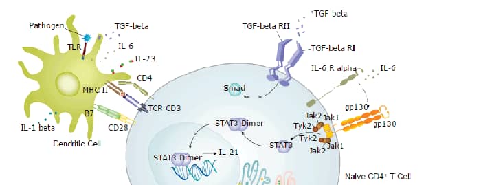
 Th17 Differentiation Pathway
 Toll-Like Receptor Signaling Pathways
