JNK1 [p Thr183, p Tyr185] Antibody (HL1008) - Azide and BSA Free
Novus Biologicals | Catalog # NBP3-13678
Recombinant Monoclonal Antibody
Loading...
Key Product Details
Validated by
Independent Antibodies
Species Reactivity
Human, Mouse
Applications
Immunohistochemistry, Immunohistochemistry-Paraffin, Western Blot
Label
Unconjugated
Antibody Source
Recombinant Monoclonal Rabbit IgG Clone # HL1008 expressed in HEK293
Format
Azide and BSA Free
Loading...
Product Specifications
Immunogen
Carrier-protein conjugated synthetic peptide surrounding phospho Thr183/Tyr185 of human JNK1. The exact sequence is proprietary.
Modification
p Thr183, p Tyr185
Clonality
Monoclonal
Host
Rabbit
Isotype
IgG
Description
Centrifuge briefly prior to opening.
Scientific Data Images for JNK1 [p Thr183, p Tyr185] Antibody (HL1008) - Azide and BSA Free
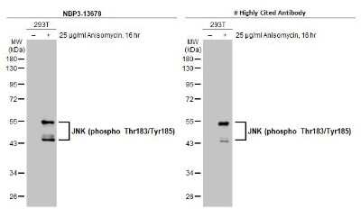
Western Blot: JNK1 [p Thr183, p Tyr185] Antibody (HL1008) [NBP3-13678] - Untreated (-) and treated (+) 293T whole cell extracts (30 ug) were separated by 10% SDS-PAGE, and the membranes were blotted with JNK1 (phospho Thr183/Tyr185) antibody [HL1008] (NBP3-13678) diluted at 1:1000 and competitor's antibody diluted at 1:500. The HRP-conjugated anti-rabbit IgG antibody (NBP2-19301) was used to detect the primary antibody.
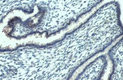
Immunohistochemistry-Paraffin: JNK1 [p Thr183, p Tyr185] Antibody (HL1008) [NBP3-13678] - JNK1 (phospho Thr183/Tyr185) antibody [HL1008] detects JNK1 (phospho Thr183/Tyr185) protein at nucleus by immunohistochemical analysis. Sample: Paraffin-embedded human breast carcinoma. JNK1 (phospho Thr183/Tyr185) stained by JNK1 (phospho Thr183/Tyr185) antibody [HL1008] (NBP3-13678) diluted at 1:100. Antigen Retrieval: Citrate buffer, pH 6.0, 15 min
![JNK1 [p Thr183, p Tyr185] Antibody (HL1008)](https://resources.rndsystems.com/images/products/nbp3-13678_rabbit-jnk1-p-thr-183-p-tyr-185-mab-hl1008-9102024204127.jpg "Western Blot: JNK1 [p Thr183, p Tyr185] Antibody (HL1008) [NBP3-13678] -")
Western Blot: JNK1 [p Thr183, p Tyr185] Antibody (HL1008) [NBP3-13678] -
Western Blot: JNK1 [p Thr183, p Tyr185] Antibody (HL1008) [NBP3-13678] - Untreated (-) and treated (+) 293T whole cell extract (30 ug) were separated by 10% SDS-PAGE, and the membrane was blotted with JNK1 antibody [HL1008] (NBP3-13678) diluted at 1:1000. The HRP-conjugated anti-rabbit IgG antibody was used to detect the primary antibody.![JNK1 [p Thr183, p Tyr185] Antibody (HL1008)](https://resources.rndsystems.com/images/products/nbp3-13678_rabbit-jnk1-p-thr-183-p-tyr-185-mab-hl1008-91020242013406.jpg "Western Blot: JNK1 [p Thr183, p Tyr185] Antibody (HL1008) [NBP3-13678] -")
Western Blot: JNK1 [p Thr183, p Tyr185] Antibody (HL1008) [NBP3-13678] -
Western Blot: JNK1 [p Thr183, p Tyr185] Antibody (HL1008) [NBP3-13678] - Untreated (-) and treated (+) Raw264.7 whole cell extract (30 ug) were separated by 10% SDS-PAGE, and the membrane was blotted with JNK1 antibody [HL1008] (NBP3-13678) diluted at 1:1000. The HRP-conjugated anti-rabbit IgG antibody was used to detect the primary antibody.![JNK1 [p Thr183, p Tyr185] Antibody (HL1008)](https://resources.rndsystems.com/images/products/nbp3-13678_rabbit-jnk1-p-thr-183-p-tyr-185-mab-hl1008-91020241959351.jpg "Western Blot: JNK1 [p Thr183, p Tyr185] Antibody (HL1008) [NBP3-13678] -")
Western Blot: JNK1 [p Thr183, p Tyr185] Antibody (HL1008) [NBP3-13678] -
Western Blot: JNK1 [p Thr183, p Tyr185] Antibody (HL1008) [NBP3-13678] - Untreated (-) and treated (+) PC-12 whole cell extract (50 ug) were separated by 10% SDS-PAGE, and the membrane was blotted with JNK1 antibody [HL1008] (NBP3-13678) diluted at 1:1000. The HRP-conjugated anti-rabbit IgG antibody was used to detect the primary antibody.![JNK1 [p Thr183, p Tyr185] Antibody (HL1008) - Azide and BSA Free](https://resources.rndsystems.com/images/products/nbp3-13678_rabbit-jnk1-p-thr-183-p-tyr-185-mab-hl1008-western-blot-261120251837507.jpg "Western Blot: JNK1 [p Thr183, p Tyr185] Antibody (HL1008) [NBP3-13678] -")
Western Blot: JNK1 [p Thr183, p Tyr185] Antibody (HL1008) [NBP3-13678] -
Untreated (-) and treated (+) 293T whole cell extracts (30 ug) were separated by 10% SDS-PAGE, and the membrane was blotted with JNK (phospho Thr183/Tyr185) antibody [HL1008] (NBP3-13678) diluted at 1:500. The HRP-conjugated anti-rabbit IgG antibody was used to detect the primary antibody.Applications for JNK1 [p Thr183, p Tyr185] Antibody (HL1008) - Azide and BSA Free
Application
Recommended Usage
Immunohistochemistry
Optimal dilutions of this antibody should be experimentally determined.
Immunohistochemistry-Paraffin
Optimal dilutions of this antibody should be experimentally determined.
Western Blot
Optimal dilutions of this antibody should be experimentally determined.
Formulation, Preparation, and Storage
Purification
Protein A purified
Formulation
PBS
Format
Azide and BSA Free
Preservative
No Preservative
Concentration
Concentrations vary lot to lot. See vial label for concentration. If unlisted please contact technical services.
Shipping
The product is shipped with polar packs. Upon receipt, store it immediately at the temperature recommended below.
Stability & Storage
Store at 4C short term. Aliquot and store at -20C long term. Avoid freeze-thaw cycles.
Background: JNK1
Long Name
C-Jun N-terminal Kinase 1
Alternate Names
MAPK8, PRKM8, SAPK1
Gene Symbol
MAPK8
Additional JNK1 Products
Product Documents for JNK1 [p Thr183, p Tyr185] Antibody (HL1008) - Azide and BSA Free
Certificate of Analysis
To download a Certificate of Analysis, please enter a lot or batch number in the search box below.
Product Specific Notices for JNK1 [p Thr183, p Tyr185] Antibody (HL1008) - Azide and BSA Free
This product is for research use only and is not approved for use in humans or in clinical diagnosis. Primary Antibodies are guaranteed for 1 year from date of receipt.
Customer Reviews for JNK1 [p Thr183, p Tyr185] Antibody (HL1008) - Azide and BSA Free
There are currently no reviews for this product. Be the first to review JNK1 [p Thr183, p Tyr185] Antibody (HL1008) - Azide and BSA Free and earn rewards!
Have you used JNK1 [p Thr183, p Tyr185] Antibody (HL1008) - Azide and BSA Free?
Submit a review and receive an Amazon gift card!
$25/€18/£15/$25CAN/¥2500 Yen for a review with an image
$10/€7/£6/$10CAN/¥1110 Yen for a review without an image
Submit a review
Protocols
Find general support by application which include: protocols, troubleshooting, illustrated assays, videos and webinars.
- Antigen Retrieval Protocol (PIER)
- Antigen Retrieval for Frozen Sections Protocol
- Appropriate Fixation of IHC/ICC Samples
- Cellular Response to Hypoxia Protocols
- Chromogenic IHC Staining of Formalin-Fixed Paraffin-Embedded (FFPE) Tissue Protocol
- Chromogenic Immunohistochemistry Staining of Frozen Tissue
- ClariTSA™ Fluorophore Kits
- Detection & Visualization of Antibody Binding
- Fluorescent IHC Staining of Frozen Tissue Protocol
- Graphic Protocol for Heat-induced Epitope Retrieval
- Graphic Protocol for the Preparation and Fluorescent IHC Staining of Frozen Tissue Sections
- Graphic Protocol for the Preparation and Fluorescent IHC Staining of Paraffin-embedded Tissue Sections
- Graphic Protocol for the Preparation of Gelatin-coated Slides for Histological Tissue Sections
- IHC Sample Preparation (Frozen sections vs Paraffin)
- Immunofluorescent IHC Staining of Formalin-Fixed Paraffin-Embedded (FFPE) Tissue Protocol
- Immunohistochemistry (IHC) and Immunocytochemistry (ICC) Protocols
- Immunohistochemistry Frozen Troubleshooting
- Immunohistochemistry Paraffin Troubleshooting
- Preparing Samples for IHC/ICC Experiments
- Preventing Non-Specific Staining (Non-Specific Binding)
- Primary Antibody Selection & Optimization
- Protocol for Heat-Induced Epitope Retrieval (HIER)
- Protocol for Making a 4% Formaldehyde Solution in PBS
- Protocol for VisUCyte™ HRP Polymer Detection Reagent
- Protocol for the Preparation & Fixation of Cells on Coverslips
- Protocol for the Preparation and Chromogenic IHC Staining of Frozen Tissue Sections
- Protocol for the Preparation and Chromogenic IHC Staining of Frozen Tissue Sections - Graphic
- Protocol for the Preparation and Chromogenic IHC Staining of Paraffin-embedded Tissue Sections
- Protocol for the Preparation and Chromogenic IHC Staining of Paraffin-embedded Tissue Sections - Graphic
- Protocol for the Preparation and Fluorescent IHC Staining of Frozen Tissue Sections
- Protocol for the Preparation and Fluorescent IHC Staining of Paraffin-embedded Tissue Sections
- Protocol for the Preparation of Gelatin-coated Slides for Histological Tissue Sections
- R&D Systems Quality Control Western Blot Protocol
- TUNEL and Active Caspase-3 Detection by IHC/ICC Protocol
- The Importance of IHC/ICC Controls
- Troubleshooting Guide: Immunohistochemistry
- Troubleshooting Guide: Western Blot Figures
- Western Blot Conditions
- Western Blot Protocol
- Western Blot Protocol for Cell Lysates
- Western Blot Troubleshooting
- Western Blot Troubleshooting Guide
- View all Protocols, Troubleshooting, Illustrated assays and Webinars
Loading...
Associated Pathways


TGF-beta Signaling Pathways
 Toll-Like Receptor Signaling Pathways
 Wnt Signaling Pathways: beta-Catenin-dependent Wnt Signaling
