Key Product Details
Species Reactivity
Human, Mouse, Rat, Canine
Applications
Immunohistochemistry, Immunohistochemistry-Paraffin, Western Blot, Flow Cytometry, Immunocytochemistry/ Immunofluorescence, Immunoprecipitation
Label
Unconjugated
Antibody Source
Monoclonal Mouse IgG2B Clone # OTI8A8
Loading...
Product Specifications
Immunogen
Full-length protein expressed in 293T cell transfected with human MAP2K4 expression vector
Reactivity Notes
Please note that this antibody is reactive to Mouse and derived from the same host, Mouse. Mouse-On-Mouse blocking reagent may be needed for IHC and ICC experiments to reduce high background signal. You can find these reagents under catalog numbers PK-2200-NB and MP-2400-NB. Please contact Technical Support if you have any questions.
Specificity
This antibody is specific for Homo sapiens mitogen-activated protein kinase kinase 4 (MAP2K4).
Clonality
Monoclonal
Host
Mouse
Isotype
IgG2B
Theoretical MW
44.3 kDa.
Disclaimer note: The observed molecular weight of the protein may vary from the listed predicted molecular weight due to post translational modifications, post translation cleavages, relative charges, and other experimental factors.
Disclaimer note: The observed molecular weight of the protein may vary from the listed predicted molecular weight due to post translational modifications, post translation cleavages, relative charges, and other experimental factors.
Scientific Data Images for MKK4/MEK4 Antibody (OTI8A8)
![Western Blot: MKK4/MEK4 Antibody (OTI8A8) [NBP1-47839]](https://resources.rndsystems.com/images/products/MKK4-MEK4-Antibody-OTI8A8-Western-Blot-NBP1-47839-img0020.jpg "Western Blot: MKK4/MEK4 Antibody (OTI8A8) [NBP1-47839]")
Western Blot: MKK4/MEK4 Antibody (OTI8A8) [NBP1-47839]
Western Blot: MKK4/MEK4 Antibody (OTI8A8) [NBP1-47839] - Analysis of extracts (35ug) from 9 different cell lines by using anti-MEK4 monoclonal antibody.![Immunocytochemistry/ Immunofluorescence: MKK4/MEK4 Antibody (OTI8A8) [NBP1-47839]](https://resources.rndsystems.com/images/products/MKK4-MEK4-Antibody-OTI8A8-Immunocytochemistry-Immunofluorescence-NBP1-47839-img0013.jpg "Immunocytochemistry/ Immunofluorescence: MKK4/MEK4 Antibody (OTI8A8) [NBP1-47839]")
Immunocytochemistry/ Immunofluorescence: MKK4/MEK4 Antibody (OTI8A8) [NBP1-47839]
Immunocytochemistry/Immunofluorescence: MKK4/MEK4 Antibody (OTI8A8) [NBP1-47839] - Staining of COS7 cells transiently transfected by pCMV6-ENTRY MEK4.![Immunohistochemistry-Paraffin: MKK4/MEK4 Antibody (OTI8A8) [NBP1-47839]](https://resources.rndsystems.com/images/products/MKK4-MEK4-Antibody-OTI8A8-Immunohistochemistry-Paraffin-NBP1-47839-img0022.jpg "Immunohistochemistry-Paraffin: MKK4/MEK4 Antibody (OTI8A8) [NBP1-47839]")
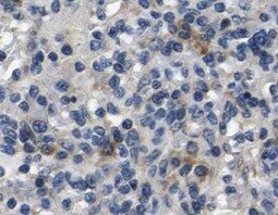
Immunohistochemistry-Paraffin: MKK4/MEK4 Antibody (OTI8A8) [NBP1-47839]
Immunohistochemistry-Paraffin: MKK4/MEK4 Antibody (OTI8A8) [NBP1-47839] - Staining of paraffin-embedded mouse spleen tissue using MKK4/MEK4 antibody (OTI8A8). Image from verified customer review.![Flow Cytometry: MKK4/MEK4 Antibody (OTI8A8) [NBP1-47839]](https://resources.rndsystems.com/images/products/MKK4-MEK4-Antibody-OTI8A8-Flow-Cytometry-NBP1-47839-img0011.jpg "Flow Cytometry: MKK4/MEK4 Antibody (OTI8A8) [NBP1-47839]")
Flow Cytometry: MKK4/MEK4 Antibody (OTI8A8) [NBP1-47839]
Flow Cytometry: MKK4/MEK4 Antibody (OTI8A8) [NBP1-47839] - HEK293T cells transfected with either overexpression plasmid (Red) or empty vector control plasmid (Blue) were immunostained by anti-MEK4 antibody, and then analyzed by flow cytometry.![Immunocytochemistry/ Immunofluorescence: MKK4/MEK4 Antibody (OTI8A8) [NBP1-47839]](https://resources.rndsystems.com/images/products/MKK4-MEK4-Antibody-OTI8A8-Immunocytochemistry-Immunofluorescence-NBP1-47839-img0012.jpg "Immunocytochemistry/ Immunofluorescence: MKK4/MEK4 Antibody (OTI8A8) [NBP1-47839]")
Immunocytochemistry/ Immunofluorescence: MKK4/MEK4 Antibody (OTI8A8) [NBP1-47839]
Immunocytochemistry/Immunofluorescence: MKK4/MEK4 Antibody (OTI8A8) [NBP1-47839] - Staining of HepG2 cells using anti-MEK4 mouse monoclonal antibody.![Immunohistochemistry-Paraffin: MKK4/MEK4 Antibody (OTI8A8) [NBP1-47839]](https://resources.rndsystems.com/images/products/MKK4-MEK4-Antibody-OTI8A8-Immunohistochemistry-Paraffin-NBP1-47839-img0014.jpg "Immunohistochemistry-Paraffin: MKK4/MEK4 Antibody (OTI8A8) [NBP1-47839]")
Immunohistochemistry-Paraffin: MKK4/MEK4 Antibody (OTI8A8) [NBP1-47839]
Immunohistochemistry-Paraffin: MKK4/MEK4 Antibody (OTI8A8) [NBP1-47839] - Staining of paraffin-embedded Carcinoma of Human kidney tissue using anti-MEK4 mouse monoclonal antibody.![Immunohistochemistry-Paraffin: MKK4/MEK4 Antibody (OTI8A8) [NBP1-47839]](https://resources.rndsystems.com/images/products/MKK4-MEK4-Antibody-OTI8A8-Immunohistochemistry-Paraffin-NBP1-47839-img0015.jpg "Immunohistochemistry-Paraffin: MKK4/MEK4 Antibody (OTI8A8) [NBP1-47839]")
Immunohistochemistry-Paraffin: MKK4/MEK4 Antibody (OTI8A8) [NBP1-47839]
Immunohistochemistry-Paraffin: MKK4/MEK4 Antibody (OTI8A8) [NBP1-47839] - Staining of paraffin-embedded Carcinoma of Human thyroid tissue using anti-MEK4 mouse monoclonal antibody.![Immunohistochemistry-Paraffin: MKK4/MEK4 Antibody (OTI8A8) [NBP1-47839]](https://resources.rndsystems.com/images/products/MKK4-MEK4-Antibody-OTI8A8-Immunohistochemistry-Paraffin-NBP1-47839-img0017.jpg "Immunohistochemistry-Paraffin: MKK4/MEK4 Antibody (OTI8A8) [NBP1-47839]")
Immunohistochemistry-Paraffin: MKK4/MEK4 Antibody (OTI8A8) [NBP1-47839]
Immunohistochemistry-Paraffin: MKK4/MEK4 Antibody (OTI8A8) [NBP1-47839] - Staining of paraffin-embedded Human pancreas tissue using anti-MEK4 mouse monoclonal antibody.![Immunohistochemistry-Paraffin: MKK4/MEK4 Antibody (OTI8A8) [NBP1-47839]](https://resources.rndsystems.com/images/products/MKK4-MEK4-Antibody-OTI8A8-Immunohistochemistry-Paraffin-NBP1-47839-img0018.jpg "Immunohistochemistry-Paraffin: MKK4/MEK4 Antibody (OTI8A8) [NBP1-47839]")
Immunohistochemistry-Paraffin: MKK4/MEK4 Antibody (OTI8A8) [NBP1-47839]
Immunohistochemistry-Paraffin: MKK4/MEK4 Antibody (OTI8A8) [NBP1-47839] - Staining of paraffin-embedded Human thyroid tissue using anti-MEK4 mouse monoclonal antibody.![Immunoprecipitation: MKK4/MEK4 Antibody (OTI8A8) [NBP1-47839]](https://resources.rndsystems.com/images/products/MKK4-MEK4-Antibody-OTI8A8-Immunoprecipitation-NBP1-47839-img0021.jpg "Immunoprecipitation: MKK4/MEK4 Antibody (OTI8A8) [NBP1-47839]")
Immunoprecipitation: MKK4/MEK4 Antibody (OTI8A8) [NBP1-47839]
Immunoprecipitation: MKK4/MEK4 Antibody (OTI8A8) [NBP1-47839] - (Negative control: IP without adding anti-MAP2K4 antibody.). For each experiment, 500ul of DDK tagged MAP2K4 overexpression lysates (at 1:5 dilution with HEK293T lysate), 2ug of anti-MAP2K4 antibody and 20ul (0.1mg) of goat anti-mouse conjugated magnetic beads were mixed and incubated overnight. After extensive wash to remove any non-specific binding, the immuno-precipitated products were analyzed with rabbit anti-DDK polyclonal antibody.Applications for MKK4/MEK4 Antibody (OTI8A8)
Application
Recommended Usage
Flow Cytometry
1:100
Immunocytochemistry/ Immunofluorescence
1:50-100
Immunohistochemistry
1:150
Immunohistochemistry-Paraffin
1:150
Immunoprecipitation
2ug/500ul
Western Blot
1:500-1000
Reviewed Applications
Read 1 review rated 5 using NBP1-47839 in the following applications:
Flow Cytometry Panel Builder
Bio-Techne Knows Flow Cytometry
Save time and reduce costly mistakes by quickly finding compatible reagents using the Panel Builder Tool.
Advanced Features
- Spectra Viewer - Custom analysis of spectra from multiple fluorochromes
- Spillover Popups - Visualize the spectra of individual fluorochromes
- Antigen Density Selector - Match fluorochrome brightness with antigen density

Formulation, Preparation, and Storage
Purification
Immunogen affinity purified
Formulation
PBS (pH 7.3), 1.0% BSA and 50% Glycerol
Preservative
0.02% Sodium Azide
Concentration
2.36 mg/ml
Shipping
The product is shipped with polar packs. Upon receipt, store it immediately at the temperature recommended below.
Stability & Storage
Store at -20C. Avoid freeze-thaw cycles.
Background: MKK4
Long Name
Mitogen-activated Protein Kinase Kinase 4
Alternate Names
JNKK1, MAP2K4, MAPKK4, MEK4, PRKMK4, SERK1
Entrez Gene IDs
6416 (Human)
Gene Symbol
MAP2K4
Additional MKK4 Products
Product Documents for MKK4/MEK4 Antibody (OTI8A8)
Certificate of Analysis
To download a Certificate of Analysis, please enter a lot or batch number in the search box below.
Product Specific Notices for MKK4/MEK4 Antibody (OTI8A8)
This product is for research use only and is not approved for use in humans or in clinical diagnosis. Primary Antibodies are guaranteed for 1 year from date of receipt.
Customer Reviews for MKK4/MEK4 Antibody (OTI8A8) (1)
5 out of 5
1 Customer Rating
Have you used MKK4/MEK4 Antibody (OTI8A8)?
Submit a review and receive an Amazon gift card!
$25/€18/£15/$25CAN/¥2500 Yen for a review with an image
$10/€7/£6/$10CAN/¥1110 Yen for a review without an image
Submit a review
Customer Images
Showing
1
-
1 of
1 review
Showing All
Filter By:
-
Application: Immunohistochemistry-ParaffinSample Tested: Spleen tissueSpecies: MouseVerified Customer | Posted 12/21/2021Mouse spleen

There are no reviews that match your criteria.
Protocols
Find general support by application which include: protocols, troubleshooting, illustrated assays, videos and webinars.
- 7-Amino Actinomycin D (7-AAD) Cell Viability Flow Cytometry Protocol
- Antigen Retrieval Protocol (PIER)
- Antigen Retrieval for Frozen Sections Protocol
- Appropriate Fixation of IHC/ICC Samples
- Cellular Response to Hypoxia Protocols
- Chromogenic IHC Staining of Formalin-Fixed Paraffin-Embedded (FFPE) Tissue Protocol
- Chromogenic Immunohistochemistry Staining of Frozen Tissue
- ClariTSA™ Fluorophore Kits
- Detection & Visualization of Antibody Binding
- Extracellular Membrane Flow Cytometry Protocol
- Flow Cytometry Protocol for Cell Surface Markers
- Flow Cytometry Protocol for Staining Membrane Associated Proteins
- Flow Cytometry Staining Protocols
- Flow Cytometry Troubleshooting Guide
- Fluorescent IHC Staining of Frozen Tissue Protocol
- Graphic Protocol for Heat-induced Epitope Retrieval
- Graphic Protocol for the Preparation and Fluorescent IHC Staining of Frozen Tissue Sections
- Graphic Protocol for the Preparation and Fluorescent IHC Staining of Paraffin-embedded Tissue Sections
- Graphic Protocol for the Preparation of Gelatin-coated Slides for Histological Tissue Sections
- ICC Cell Smear Protocol for Suspension Cells
- ICC Immunocytochemistry Protocol Videos
- ICC for Adherent Cells
- IHC Sample Preparation (Frozen sections vs Paraffin)
- Immunocytochemistry (ICC) Protocol
- Immunocytochemistry Troubleshooting
- Immunofluorescence of Organoids Embedded in Cultrex Basement Membrane Extract
- Immunofluorescent IHC Staining of Formalin-Fixed Paraffin-Embedded (FFPE) Tissue Protocol
- Immunohistochemistry (IHC) and Immunocytochemistry (ICC) Protocols
- Immunohistochemistry Frozen Troubleshooting
- Immunohistochemistry Paraffin Troubleshooting
- Immunoprecipitation Protocol
- Intracellular Flow Cytometry Protocol Using Alcohol (Methanol)
- Intracellular Flow Cytometry Protocol Using Detergents
- Intracellular Nuclear Staining Flow Cytometry Protocol Using Detergents
- Intracellular Staining Flow Cytometry Protocol Using Alcohol Permeabilization
- Intracellular Staining Flow Cytometry Protocol Using Detergents to Permeabilize Cells
- Preparing Samples for IHC/ICC Experiments
- Preventing Non-Specific Staining (Non-Specific Binding)
- Primary Antibody Selection & Optimization
- Propidium Iodide Cell Viability Flow Cytometry Protocol
- Protocol for Heat-Induced Epitope Retrieval (HIER)
- Protocol for Liperfluo
- Protocol for Making a 4% Formaldehyde Solution in PBS
- Protocol for VisUCyte™ HRP Polymer Detection Reagent
- Protocol for the Characterization of Human Th22 Cells
- Protocol for the Characterization of Human Th9 Cells
- Protocol for the Fluorescent ICC Staining of Cell Smears - Graphic
- Protocol for the Fluorescent ICC Staining of Cultured Cells on Coverslips - Graphic
- Protocol for the Preparation & Fixation of Cells on Coverslips
- Protocol for the Preparation and Chromogenic IHC Staining of Frozen Tissue Sections
- Protocol for the Preparation and Chromogenic IHC Staining of Frozen Tissue Sections - Graphic
- Protocol for the Preparation and Chromogenic IHC Staining of Paraffin-embedded Tissue Sections
- Protocol for the Preparation and Chromogenic IHC Staining of Paraffin-embedded Tissue Sections - Graphic
- Protocol for the Preparation and Fluorescent ICC Staining of Cells on Coverslips
- Protocol for the Preparation and Fluorescent ICC Staining of Non-adherent Cells
- Protocol for the Preparation and Fluorescent ICC Staining of Stem Cells on Coverslips
- Protocol for the Preparation and Fluorescent IHC Staining of Frozen Tissue Sections
- Protocol for the Preparation and Fluorescent IHC Staining of Paraffin-embedded Tissue Sections
- Protocol for the Preparation of Gelatin-coated Slides for Histological Tissue Sections
- Protocol for the Preparation of a Cell Smear for Non-adherent Cell ICC - Graphic
- Protocol: Annexin V and PI Staining by Flow Cytometry
- Protocol: Annexin V and PI Staining for Apoptosis by Flow Cytometry
- R&D Systems Quality Control Western Blot Protocol
- TUNEL and Active Caspase-3 Detection by IHC/ICC Protocol
- The Importance of IHC/ICC Controls
- Troubleshooting Guide: Fluorokine Flow Cytometry Kits
- Troubleshooting Guide: Immunohistochemistry
- Troubleshooting Guide: Western Blot Figures
- Western Blot Conditions
- Western Blot Protocol
- Western Blot Protocol for Cell Lysates
- Western Blot Troubleshooting
- Western Blot Troubleshooting Guide
- View all Protocols, Troubleshooting, Illustrated assays and Webinars


