ESGP (ES cell and germ cell-specific protein) is a presumably secreted polypeptide that belongs to no known protein family. It is expressed specifically in pluripotential cell types, and does not appear to be involved in differentiation. Mouse ESGP precursor is 84 amino acids (aa) in length. It contains a 25 aa signal sequence with a 59 aa mature segment. There are no N-linked glycosylation sites. One potential 108 aa variant shows an alternate start site 24 aa upstream of the standard start site.

Key Product Details
Species Reactivity
Validated:
Mouse
Cited:
Human, Mouse, Golden Syrian Hamster, Transgenic Mouse
Applications
Validated:
Immunohistochemistry, Western Blot, Immunocytochemistry
Cited:
Immunohistochemistry, Western Blot, Immunocytochemistry
Label
Unconjugated
Antibody Source
Polyclonal Sheep IgG
Loading...
Product Specifications
Immunogen
E. coli-derived recombinant mouse ESGP
Leu24-Lys84
Accession # Q2Q5T5
Leu24-Lys84
Accession # Q2Q5T5
Specificity
Detects mouse ESGP in direct ELISAs and Western blots.
Clonality
Polyclonal
Host
Sheep
Isotype
IgG
Scientific Data Images for Mouse ESGP Antibody
Detection of Mouse ESGP by Western Blot.
Western blot shows lysates of D3 mouse embryonic stem cell line. PVDF membrane was probed with 0.1 µg/mL of Sheep Anti-Mouse ESGP Antigen Affinity-purified Polyclonal Antibody (Catalog # AF4580) followed by HRP-conjugated Anti-Sheep IgG Secondary Antibody (Catalog # HAF016). A specific band was detected for ESGP at approximately 9 kDa (as indicated). This experiment was conducted under reducing conditions and using Immunoblot Buffer Group 8.
ESGP in Mouse Testis.
ESGP was detected in perfusion fixed frozen sections of mouse testis using Sheep Anti-Mouse ESGP Antigen Affinity-purified Polyclonal Antibody (Catalog # AF4580) at 1.7 µg/mL overnight at 4 °C. Tissue was stained using the Northern-Lights™ 557-conjugated Anti-Sheep IgG Secondary Antibody (red; Catalog # NL010) and counterstained with DAPI (blue). Specific staining was localized to late spermatids. View our protocol for Fluorescent IHC Staining of Frozen Tissue Sections.
ESGP in D3 Mouse Cell Line.
ESGP was detected in immersion fixed D3 mouse embryonic stem cell line using Sheep Anti-Mouse ESGP Antigen Affinity-purified Polyclonal Antibody (Catalog # AF4580) at 10 µg/mL for 3 hours at room temperature. Cells were stained using the NorthernLights™ 557-conjugated Anti-Sheep IgG Secondary Antibody (red; Catalog # NL010) and counterstained with DAPI (blue). Specific staining was localized to cytoplasm and cell secretion. View our protocol for Fluorescent ICC Staining of Stem Cells on Coverslips.Applications for Mouse ESGP Antibody
Application
Recommended Usage
Immunocytochemistry
5-15 µg/mL
Sample: Immersion fixed D3 mouse embryonic stem cell line
Sample: Immersion fixed D3 mouse embryonic stem cell line
Immunohistochemistry
5-15 µg/mL
Sample: Perfusion fixed frozen sections of mouse testes
Sample: Perfusion fixed frozen sections of mouse testes
Western Blot
0.1 µg/mL
Sample: D3 mouse embryonic stem cell line
Sample: D3 mouse embryonic stem cell line
Reviewed Applications
Read 1 review rated 5 using AF4580 in the following applications:
Formulation, Preparation, and Storage
Purification
Antigen Affinity-purified
Reconstitution
Reconstitute at 0.2 mg/mL in sterile PBS. For liquid material, refer to CoA for concentration.
Loading...
Formulation
Lyophilized from a 0.2 μm filtered solution in PBS with Trehalose. *Small pack size (SP) is supplied either lyophilized or as a 0.2 µm filtered solution in PBS.
Shipping
Lyophilized product is shipped at ambient temperature. Liquid small pack size (-SP) is shipped with polar packs. Upon receipt, store immediately at the temperature recommended below.
Stability & Storage
Use a manual defrost freezer and avoid repeated freeze-thaw cycles.
- 12 months from date of receipt, -20 to -70 °C as supplied.
- 1 month, 2 to 8 °C under sterile conditions after reconstitution.
- 6 months, -20 to -70 °C under sterile conditions after reconstitution.
Calculators
Background: ESGP
Long Name
Embryonic Stem Cell and Germ Cell Specific Protein
Alternate Names
EG653016
Entrez Gene IDs
653016 (Mouse)
Gene Symbol
MYMX
UniProt
Additional ESGP Products
Product Documents for Mouse ESGP Antibody
Certificate of Analysis
To download a Certificate of Analysis, please enter a lot or batch number in the search box below.
Note: Certificate of Analysis not available for kit components.
Product Specific Notices for Mouse ESGP Antibody
For research use only
Related Research Areas
Citations for Mouse ESGP Antibody
 Powered by Bioz
Customer Reviews for Mouse ESGP Antibody (1)
5 out of 5
1 Customer Rating
Have you used Mouse ESGP Antibody?
Submit a review and receive an Amazon gift card!
$25/€18/£15/$25CAN/¥2500 Yen for a review with an image
$10/€7/£6/$10CAN/¥1110 Yen for a review without an image
Submit a review
Customer Images
Showing
1
-
1 of
1 review
Showing All
Filter By:
-
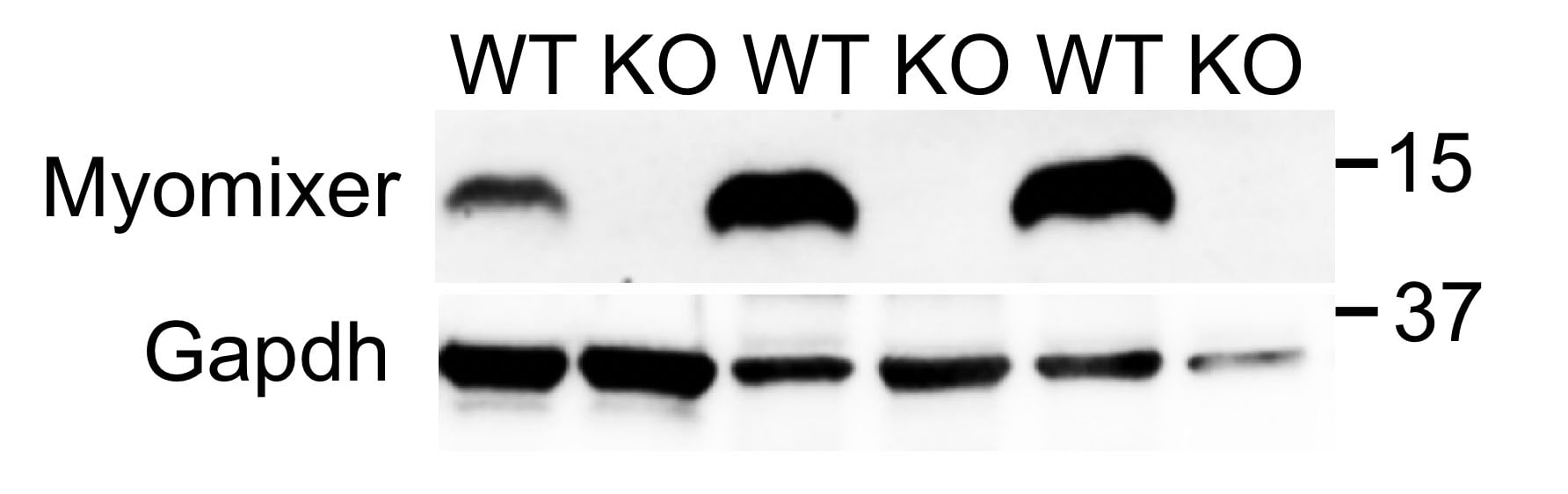
Application: Knockout ValidatedSample Tested: Muscle tissueSpecies: MouseVerified Customer | Posted 10/16/2020Western blotting analysis of Myomixer expression in gastrocnemius muscles at day 3 post-injury. Cat# AF4580, 1:1,000 dilution.Specificity validated by KO samples
 Bio-Techne ResponseThis review was submitted through the legacy Novus Innovators Program, reflecting a new species or application tested on a primary antibody.
There are no reviews that match your criteria.
Protocols
Find general support by application which include: protocols, troubleshooting, illustrated assays, videos and webinars.
- Antigen Retrieval Protocol (PIER)
- Antigen Retrieval for Frozen Sections Protocol
- Appropriate Fixation of IHC/ICC Samples
- Cellular Response to Hypoxia Protocols
- Chromogenic IHC Staining of Formalin-Fixed Paraffin-Embedded (FFPE) Tissue Protocol
- Chromogenic Immunohistochemistry Staining of Frozen Tissue
- ClariTSA™ Fluorophore Kits
- Detection & Visualization of Antibody Binding
- Fluorescent IHC Staining of Frozen Tissue Protocol
- Graphic Protocol for Heat-induced Epitope Retrieval
- Graphic Protocol for the Preparation and Fluorescent IHC Staining of Frozen Tissue Sections
- Graphic Protocol for the Preparation and Fluorescent IHC Staining of Paraffin-embedded Tissue Sections
- Graphic Protocol for the Preparation of Gelatin-coated Slides for Histological Tissue Sections
- ICC Cell Smear Protocol for Suspension Cells
- ICC Immunocytochemistry Protocol Videos
- ICC for Adherent Cells
- IHC Sample Preparation (Frozen sections vs Paraffin)
- Immunocytochemistry (ICC) Protocol
- Immunocytochemistry Troubleshooting
- Immunofluorescence of Organoids Embedded in Cultrex Basement Membrane Extract
- Immunofluorescent IHC Staining of Formalin-Fixed Paraffin-Embedded (FFPE) Tissue Protocol
- Immunohistochemistry (IHC) and Immunocytochemistry (ICC) Protocols
- Immunohistochemistry Frozen Troubleshooting
- Immunohistochemistry Paraffin Troubleshooting
- Preparing Samples for IHC/ICC Experiments
- Preventing Non-Specific Staining (Non-Specific Binding)
- Primary Antibody Selection & Optimization
- Protocol for Heat-Induced Epitope Retrieval (HIER)
- Protocol for Making a 4% Formaldehyde Solution in PBS
- Protocol for VisUCyte™ HRP Polymer Detection Reagent
- Protocol for the Fluorescent ICC Staining of Cell Smears - Graphic
- Protocol for the Fluorescent ICC Staining of Cultured Cells on Coverslips - Graphic
- Protocol for the Preparation & Fixation of Cells on Coverslips
- Protocol for the Preparation and Chromogenic IHC Staining of Frozen Tissue Sections
- Protocol for the Preparation and Chromogenic IHC Staining of Frozen Tissue Sections - Graphic
- Protocol for the Preparation and Chromogenic IHC Staining of Paraffin-embedded Tissue Sections
- Protocol for the Preparation and Chromogenic IHC Staining of Paraffin-embedded Tissue Sections - Graphic
- Protocol for the Preparation and Fluorescent ICC Staining of Cells on Coverslips
- Protocol for the Preparation and Fluorescent ICC Staining of Non-adherent Cells
- Protocol for the Preparation and Fluorescent ICC Staining of Stem Cells on Coverslips
- Protocol for the Preparation and Fluorescent IHC Staining of Frozen Tissue Sections
- Protocol for the Preparation and Fluorescent IHC Staining of Paraffin-embedded Tissue Sections
- Protocol for the Preparation of Gelatin-coated Slides for Histological Tissue Sections
- Protocol for the Preparation of a Cell Smear for Non-adherent Cell ICC - Graphic
- R&D Systems Quality Control Western Blot Protocol
- TUNEL and Active Caspase-3 Detection by IHC/ICC Protocol
- The Importance of IHC/ICC Controls
- Troubleshooting Guide: Immunohistochemistry
- Troubleshooting Guide: Western Blot Figures
- Western Blot Conditions
- Western Blot Protocol
- Western Blot Protocol for Cell Lysates
- Western Blot Troubleshooting
- Western Blot Troubleshooting Guide
- View all Protocols, Troubleshooting, Illustrated assays and Webinars
Loading...