The Flt-3 (fms-like tyrosine kinase) receptor, also named Flk-2 (fetal liver kinase) and Stk-1 (stem cell tyrosine kinase) and designated CD135, is a member of the class III subfamily of receptor tyrosine kinases. This familay includes KIT, the receptor for SCF, and C-FMS, the receptor for M-CSF. The extracellular region of these receptors contains five immunoglobulin-like domains and the intracellular region contains a split kinase domain. Mouse Flt-3 cDNA encodes a 992 amino acid (aa) residue type I membrane protein with a 27 aa residue signal peptide, a 517 aa extracellular domain with 10 potential N-linked glycosylation sites, a 20 aa residue transmembrane domain and a 428 aa residue cytoplasmic domain. Mouse Flt-3 shares 85% amino acid sequence identity with human Flt-3. Flt-3 expression has been detected in various tissues, including placenta, gonads, and tissues of nervous and hematopoietic origin. Among hematopoietic cells, the expression of Flt-3 was found to be restricted to the highly enriched stem/progenitor cell populations. The ligand for Flt-3 (FL) has been identified to be a transmembrane protein with structural homology to M-CSF and SCF. Recombinant soluble Flt-3 Fc chimeric protein has been shown to bind FL with high affinity and is a potent FL antagonist.
Mouse Flt-3/Flk-2 Antibody (113308)
R&D Systems | Catalog # MAB7681

Key Product Details
Species Reactivity
Mouse
Applications
Flow Cytometry, Immunocytochemistry, CyTOF-ready
Label
Unconjugated
Antibody Source
Monoclonal Rat IgG2A Clone # 113308
Loading...
Product Specifications
Immunogen
Mouse myeloma cell line NS0-derived recombinant mouse Flt‑3/Flk‑2
Asn28-Ser544 (predicted)
Accession # Q00342
Asn28-Ser544 (predicted)
Accession # Q00342
Specificity
Detects mouse Flt‑3/Flk‑2 in direct ELISAs. In direct ELISAs, no cross-reactivity with recombinant human Flt-3 is observed.
Clonality
Monoclonal
Host
Rat
Isotype
IgG2A
Scientific Data Images for Mouse Flt-3/Flk-2 Antibody (113308)
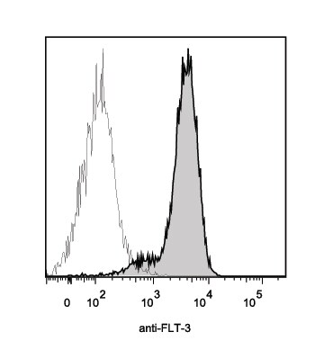
Detection of Flt‑3/Flk‑2 in M1 cells by Flow Cytometry
M1 cells were stained with Rat Anti-Mouse Flt‑3/Flk‑2 Monoclonal Antibody (Catalog # mab7681, filled histogram) or isotype control antibody (Catalog # MAB006, open histogram) followed by Allophycocyanin-conjugated Anti-Rat IgG Secondary Antibody (Catalog # F0113). View our protocol for Staining Membrane-associated Proteins.
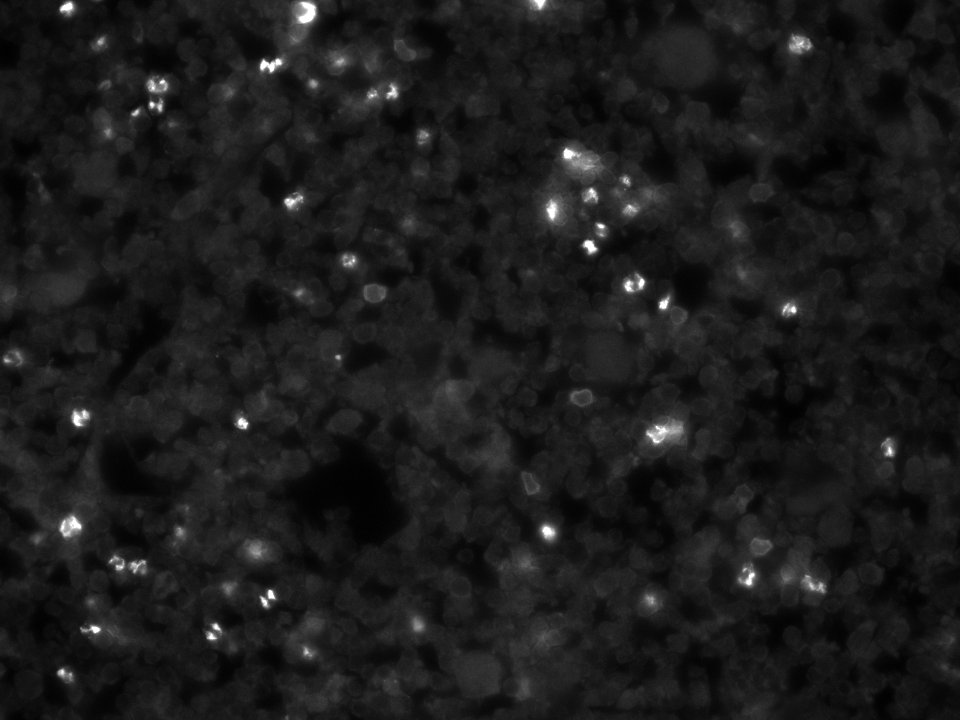
Flt‑3/Flk‑2 in M1 Mouse Cell Line.
Flt-3/Flk-2 was detected in immersion fixed M1 mouse myeloid leukemia cell line using Rat Anti-Mouse Flt-3/Flk-2 Monoclonal Antibody (Catalog # MAB7681) at 10 µg/mL for 3 hours at room temperature. Cells were stained using the NorthernLights™ 557-conjugated Anti-Rat IgG Secondary Antibody (red; Catalog # NL013) and counterstained with DAPI (blue). View our protocol for Fluorescent ICC Staining of Cells on Coverslips.Applications for Mouse Flt-3/Flk-2 Antibody (113308)
Application
Recommended Usage
CyTOF-ready
Ready to be labeled using established conjugation methods. No BSA or other carrier proteins that could interfere with conjugation.
Flow Cytometry
0.25 µg/106 cells
Sample: M1 mouse myeloid leukemia cell line
Sample: M1 mouse myeloid leukemia cell line
Immunocytochemistry
8-25 µg/mL
Sample: Immersion fixed M1 mouse myeloid leukemia cell line
Sample: Immersion fixed M1 mouse myeloid leukemia cell line
Reviewed Applications
Read 2 reviews rated 3.5 using MAB7681 in the following applications:
Flow Cytometry Panel Builder
Bio-Techne Knows Flow Cytometry
Save time and reduce costly mistakes by quickly finding compatible reagents using the Panel Builder Tool.
Advanced Features
- Spectra Viewer - Custom analysis of spectra from multiple fluorochromes
- Spillover Popups - Visualize the spectra of individual fluorochromes
- Antigen Density Selector - Match fluorochrome brightness with antigen density

Formulation, Preparation, and Storage
Purification
Protein A or G purified from hybridoma culture supernatant
Reconstitution
Reconstitute at 0.5 mg/mL in sterile PBS. For liquid material, refer to CoA for concentration.
Loading...
Formulation
Lyophilized from a 0.2 μm filtered solution in PBS with Trehalose. See Certificate of Analysis for details.
*Small pack size (-SP) is supplied either lyophilized or as a 0.2 µm filtered solution in PBS.
*Small pack size (-SP) is supplied either lyophilized or as a 0.2 µm filtered solution in PBS.
Shipping
Lyophilized product is shipped at ambient temperature. Liquid small pack size (-SP) is shipped with polar packs. Upon receipt, store immediately at the temperature recommended below.
Stability & Storage
Use a manual defrost freezer and avoid repeated freeze-thaw cycles.
- 12 months from date of receipt, -20 to -70 °C as supplied.
- 1 month, 2 to 8 °C under sterile conditions after reconstitution.
- 6 months, -20 to -70 °C under sterile conditions after reconstitution.
Calculators
Background: Flt-3/Flk-2
References
- Rosnet, O. et al. (1996) Acta. Haemato. 95:218.
- Drexler, H.G. (1996) Leukemia 10:588.
Long Name
fms-like Tyrosine Kinase 3
Alternate Names
CD135, Flk-2, Flt3, STK-1
Gene Symbol
FLT3
UniProt
Additional Flt-3/Flk-2 Products
Product Documents for Mouse Flt-3/Flk-2 Antibody (113308)
Certificate of Analysis
To download a Certificate of Analysis, please enter a lot or batch number in the search box below.
Note: Certificate of Analysis not available for kit components.
Product Specific Notices for Mouse Flt-3/Flk-2 Antibody (113308)
For research use only
Citations for Mouse Flt-3/Flk-2 Antibody (113308)
 Powered by Bioz
Customer Reviews for Mouse Flt-3/Flk-2 Antibody (113308) (2)
3.5 out of 5
2 Customer Ratings
Have you used Mouse Flt-3/Flk-2 Antibody (113308)?
Submit a review and receive an Amazon gift card!
$25/€18/£15/$25CAN/¥2500 Yen for a review with an image
$10/€7/£6/$10CAN/¥1110 Yen for a review without an image
Submit a review
Customer Images
Showing
1
-
2 of
2 reviews
Showing All
Filter By:
-
Application: Immunofluorescence - Fixed-frozenSample Tested: bone marrowSpecies: MouseVerified Customer | Posted 10/21/2018few CD135+ cells with membranous staining. Rather high background, especially in granulocytes.Whole Femur Bone Marrow Sections, Fixed-Frozen / Decalcified

-
Application: Flow CytometrySample Tested: mouse AML cellsSpecies: MouseVerified Customer | Posted 11/08/2016
There are no reviews that match your criteria.
Protocols
Find general support by application which include: protocols, troubleshooting, illustrated assays, videos and webinars.
- 7-Amino Actinomycin D (7-AAD) Cell Viability Flow Cytometry Protocol
- Appropriate Fixation of IHC/ICC Samples
- Cellular Response to Hypoxia Protocols
- ClariTSA™ Fluorophore Kits
- Detection & Visualization of Antibody Binding
- Extracellular Membrane Flow Cytometry Protocol
- Flow Cytometry Protocol for Cell Surface Markers
- Flow Cytometry Protocol for Staining Membrane Associated Proteins
- Flow Cytometry Staining Protocols
- Flow Cytometry Troubleshooting Guide
- ICC Cell Smear Protocol for Suspension Cells
- ICC Immunocytochemistry Protocol Videos
- ICC for Adherent Cells
- Immunocytochemistry (ICC) Protocol
- Immunocytochemistry Troubleshooting
- Immunofluorescence of Organoids Embedded in Cultrex Basement Membrane Extract
- Immunohistochemistry (IHC) and Immunocytochemistry (ICC) Protocols
- Intracellular Flow Cytometry Protocol Using Alcohol (Methanol)
- Intracellular Flow Cytometry Protocol Using Detergents
- Intracellular Nuclear Staining Flow Cytometry Protocol Using Detergents
- Intracellular Staining Flow Cytometry Protocol Using Alcohol Permeabilization
- Intracellular Staining Flow Cytometry Protocol Using Detergents to Permeabilize Cells
- Preparing Samples for IHC/ICC Experiments
- Preventing Non-Specific Staining (Non-Specific Binding)
- Primary Antibody Selection & Optimization
- Propidium Iodide Cell Viability Flow Cytometry Protocol
- Protocol for Liperfluo
- Protocol for VisUCyte™ HRP Polymer Detection Reagent
- Protocol for the Characterization of Human Th22 Cells
- Protocol for the Characterization of Human Th9 Cells
- Protocol for the Fluorescent ICC Staining of Cell Smears - Graphic
- Protocol for the Fluorescent ICC Staining of Cultured Cells on Coverslips - Graphic
- Protocol for the Preparation and Fluorescent ICC Staining of Cells on Coverslips
- Protocol for the Preparation and Fluorescent ICC Staining of Non-adherent Cells
- Protocol for the Preparation and Fluorescent ICC Staining of Stem Cells on Coverslips
- Protocol for the Preparation of a Cell Smear for Non-adherent Cell ICC - Graphic
- Protocol: Annexin V and PI Staining by Flow Cytometry
- Protocol: Annexin V and PI Staining for Apoptosis by Flow Cytometry
- TUNEL and Active Caspase-3 Detection by IHC/ICC Protocol
- The Importance of IHC/ICC Controls
- Troubleshooting Guide: Fluorokine Flow Cytometry Kits
- View all Protocols, Troubleshooting, Illustrated assays and Webinars
Loading...
Associated Pathways

