Interleukin 6 (IL-6) is a multifunctional cytokine that exerts its activities by binding to a high-affinity receptor complex consisting of two membrane glycoproteins: an 80 kDa ligand binding subunit (IL-6 R alpha /CD126) and a 130 kDa nonligand-binding signal-transducing subunit (gp130/CD130) (1‑4). The mouse IL-6 R alpha cDNA encodes a precursor type I transmembrane protein of 460 amino acids (aa) that contains a 19 aa signal sequence, a 345 aa extracellular ligand binding domain, a 21 aa transmembrane region, and a 75 aa cytoplasmic segment (2). The extracellular segment contains an Ig-like and a fibronectin-type III domain, plus a membrane proximal WSXWS motif. In their extracellular regions, mouse IL‑6 R alpha shares 89%, 51% and 50% aa identity with rat, human and porcine IL‑6 R alpha, respectively. Unlike gp130 that is expressed ubiquitously, the cellular distribution of IL-6 R alpha is predominantly limited to hepatocytes and leukocyte subpopulations such as monocytes, neutrophils, T and B cells. Soluble IL-6R alpha has been found in various body fluids (5). Two soluble receptor isoforms that arise either from proteolytic cleavage of the membrane-bound IL‑6 R alpha, or by alternative mRNA splicing (reported only in human) have been described (6, 7). Soluble IL-6 R alpha binds IL-6 with an affinity similar to that of the membrane-bound IL-6 R alpha. More importantly, the soluble IL-6 R alpha /IL-6 complex is capable of interacting with the membrane-bound gp130 to activate cells that lack an integral membrane IL-6 R alpha. It has been documented that elevated soluble IL-6 R is associated with numerous diseases including arthritic lesions, multiple myeloma and Crohn’s disease (6, 7).
Mouse IL-6R alpha Antibody (255820)
R&D Systems | Catalog # MAB18301

Key Product Details
Species Reactivity
Validated:
Mouse
Cited:
Mouse
Applications
Validated:
Western Blot, Immunocytochemistry
Cited:
Immunohistochemistry, Immunohistochemistry-Paraffin, Western Blot
Label
Unconjugated
Antibody Source
Monoclonal Rat IgG1 Clone # 255820
Loading...
Product Specifications
Immunogen
Mouse myeloma cell line NS0-derived recombinant mouse IL-6R alpha
Leu20-Glu357
Accession # P22272
Leu20-Glu357
Accession # P22272
Specificity
Detects mouse IL-6R alpha in direct ELISAs and Western blots. In direct ELISAs and Western blots, no cross-reactivity with recombinant human IL-6 R is observed.
Clonality
Monoclonal
Host
Rat
Isotype
IgG1
Scientific Data Images for Mouse IL-6R alpha Antibody (255820)
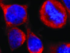
IL-6R alpha in Mouse Splenocytes.
IL-6R alpha was detected in immersion fixed mouse splenocytes using Rat Anti-Mouse IL-6R alpha Monoclonal Antibody (Catalog # MAB18301) at 15 µg/mL for 3 hours at room temperature. Cells were stained using the NorthernLights™ 557-conjugated Anti-Rat IgG Secondary Antibody (red; Catalog # NL013) and counterstained with DAPI (blue). Specific staining was localized to cytoplasm. View our protocol for Fluorescent ICC Staining of Non-adherent Cells.Applications for Mouse IL-6R alpha Antibody (255820)
Application
Recommended Usage
Immunocytochemistry
8-25 µg/mL
Sample: Immersion fixed mouse splenocytes
Sample: Immersion fixed mouse splenocytes
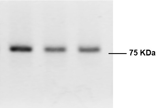
Western Blot
1 µg/mL
Sample: Recombinant Mouse IL-6R alpha (Catalog # 1830-SR)
Sample: Recombinant Mouse IL-6R alpha (Catalog # 1830-SR)
Reviewed Applications
Read 4 reviews rated 4.5 using MAB18301 in the following applications:
Formulation, Preparation, and Storage
Purification
Protein A or G purified from hybridoma culture supernatant
Reconstitution
Reconstitute at 0.5 mg/mL in sterile PBS. For liquid material, refer to CoA for concentration.
Loading...
Formulation
Lyophilized from a 0.2 μm filtered solution in PBS with Trehalose. *Small pack size (SP) is supplied either lyophilized or as a 0.2 µm filtered solution in PBS.
Shipping
Lyophilized product is shipped at ambient temperature. Liquid small pack size (-SP) is shipped with polar packs. Upon receipt, store immediately at the temperature recommended below.
Stability & Storage
Use a manual defrost freezer and avoid repeated freeze-thaw cycles.
- 12 months from date of receipt, -20 to -70 °C as supplied.
- 1 month, 2 to 8 °C under sterile conditions after reconstitution.
- 6 months, -20 to -70 °C under sterile conditions after reconstitution.
Calculators
Background: IL-6R alpha
References
- Yamasaki, K. et al. (1988) Science 241:825.
- Sugita, T. et al. (1990) J. Exp. Med. 171:2001.
- Hibi, M. et al. (1990) Cell 63:1149.
- Saito, M. et al. (1992) J. Immunol. 148:4066.
- Novick, D. et al. (1989) J. Exp. Med. 170:1409.
- Jones, S.A. et al. (2001) FASEB J. 15:43.
- Jones, S.A. and S. Rose-John (2002) Biochim. Biophys. Acta 1592:251.
Long Name
Interleukin 6 Receptor alpha
Alternate Names
CD126, IL-6 R alpha, IL-6Ra, IL6R, IL6R alpha
Gene Symbol
IL6R
UniProt
Additional IL-6R alpha Products
Product Documents for Mouse IL-6R alpha Antibody (255820)
Certificate of Analysis
To download a Certificate of Analysis, please enter a lot or batch number in the search box below.
Note: Certificate of Analysis not available for kit components.
Product Specific Notices for Mouse IL-6R alpha Antibody (255820)
For research use only
Citations for Mouse IL-6R alpha Antibody (255820)
 Powered by Bioz
Customer Reviews for Mouse IL-6R alpha Antibody (255820) (4)
4.5 out of 5
4 Customer Ratings
Have you used Mouse IL-6R alpha Antibody (255820)?
Submit a review and receive an Amazon gift card!
$25/€18/£15/$25CAN/¥2500 Yen for a review with an image
$10/€7/£6/$10CAN/¥1110 Yen for a review without an image
Submit a review
Customer Images
Showing
1
-
4 of
4 reviews
Showing All
Filter By:
-
Application: Immunocytochemistry/ImmunofluorescenceSample Tested: Cancerous basophil cellsSpecies: MouseVerified Customer | Posted 12/14/2021

-
Application: Western BlotSample Tested: Bone marrow cellsSpecies: MouseVerified Customer | Posted 05/20/2019
-
Application: Western BlotSample Tested: RAW 264.7 mouse monocyte/macrophage cell lineSpecies: MouseVerified Customer | Posted 07/15/2018
-
Application: Immunohistochemistry-FrozenSample Tested: See PMID 22295972Species: MouseVerified Customer | Posted 02/10/2015
There are no reviews that match your criteria.
Protocols
Find general support by application which include: protocols, troubleshooting, illustrated assays, videos and webinars.
- Appropriate Fixation of IHC/ICC Samples
- Cellular Response to Hypoxia Protocols
- ClariTSA™ Fluorophore Kits
- Detection & Visualization of Antibody Binding
- ICC Cell Smear Protocol for Suspension Cells
- ICC Immunocytochemistry Protocol Videos
- ICC for Adherent Cells
- Immunocytochemistry (ICC) Protocol
- Immunocytochemistry Troubleshooting
- Immunofluorescence of Organoids Embedded in Cultrex Basement Membrane Extract
- Immunohistochemistry (IHC) and Immunocytochemistry (ICC) Protocols
- Preparing Samples for IHC/ICC Experiments
- Preventing Non-Specific Staining (Non-Specific Binding)
- Primary Antibody Selection & Optimization
- Protocol for VisUCyte™ HRP Polymer Detection Reagent
- Protocol for the Fluorescent ICC Staining of Cell Smears - Graphic
- Protocol for the Fluorescent ICC Staining of Cultured Cells on Coverslips - Graphic
- Protocol for the Preparation and Fluorescent ICC Staining of Cells on Coverslips
- Protocol for the Preparation and Fluorescent ICC Staining of Non-adherent Cells
- Protocol for the Preparation and Fluorescent ICC Staining of Stem Cells on Coverslips
- Protocol for the Preparation of a Cell Smear for Non-adherent Cell ICC - Graphic
- R&D Systems Quality Control Western Blot Protocol
- TUNEL and Active Caspase-3 Detection by IHC/ICC Protocol
- The Importance of IHC/ICC Controls
- Troubleshooting Guide: Western Blot Figures
- Western Blot Conditions
- Western Blot Protocol
- Western Blot Protocol for Cell Lysates
- Western Blot Troubleshooting
- Western Blot Troubleshooting Guide
- View all Protocols, Troubleshooting, Illustrated assays and Webinars


