Mouse TRAIL R2, also called DR5, TRICK 2, TNFRSF10B, and MK is a type 1 TNF R superfamily, membrane protein which is a receptor for TRAIL (APO2 ligand). Mouse TRAIL R2 cDNA encodes a 381 amino acid (aa) precursor protein containing an extracellular cysteine-rich domain, a transmembrane domain and a cytoplasmic death domain. Human and mouse TRAIL R2 share 49% aa sequence similarity. The death domains of human TRAIL R1 and TRAIL R2 share high homology with the death domain of mouse TRAIL R2, 76% and 79%, respectively. Binding of trimeric TRAIL to TRAIL R2 induces apoptosis. The induction of apoptosis likely requires oligomerization of the receptor. Besides the death domain containing receptors TRAIL R2 and TRAIL R1/DR4, three TRAIL decoy receptors, TRAIL R3/DcR1, TRAIL R4/DcR2, and OPG, have been reported.
Mouse TRAIL R2/TNFRSF10B Antibody (118929)
R&D Systems | Catalog # MAB1540

Key Product Details
Species Reactivity
Validated:
Mouse
Cited:
Human, Mouse
Applications
Validated:
Immunohistochemistry, Western Blot
Cited:
Immunohistochemistry-Paraffin, Western Blot
Label
Unconjugated
Antibody Source
Monoclonal Rat IgG2A Clone # 118929
Loading...
Product Specifications
Immunogen
Mouse myeloma cell line NS0-derived recombinant mouse TRAIL R2/TNFRSF10B
Asn53-Ser177 (predicted)
Accession # Q9QZM4
Asn53-Ser177 (predicted)
Accession # Q9QZM4
Specificity
Detects mouse TRAIL R2/TNFRSF10B in direct ELISAs and Western blots.
Clonality
Monoclonal
Host
Rat
Isotype
IgG2A
Scientific Data Images for Mouse TRAIL R2/TNFRSF10B Antibody (118929)
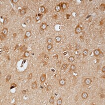
TRAIL R2/TNFRSF10B in Mouse Brain.
TRAIL R2/TNFRSF10B was detected in perfusion fixed frozen sections of mouse brain (cerebellum) using Rat Anti-Mouse TRAIL R2/TNFRSF10B Monoclonal Antibody (Catalog # MAB1540) at 25 µg/mL overnight at 4 °C. Tissue was stained using the Anti-Rat HRP-DAB Cell & Tissue Staining Kit (brown; Catalog # CTS017) and counter-stained with hematoxylin (blue). Specific staining was localized to cytoplasm of Purkinje neurons. View our protocol for Chromogenic IHC Staining of Frozen Tissue Sections.Applications for Mouse TRAIL R2/TNFRSF10B Antibody (118929)
Application
Recommended Usage
Immunohistochemistry
8-25 µg/mL
Sample: Perfusion fixed frozen sections of mouse brain (cerebellum)
Sample: Perfusion fixed frozen sections of mouse brain (cerebellum)
Western Blot
1 µg/mL
Sample: Recombinant Mouse TRAIL R2/TNFRSF10B Fc Chimera (Catalog # 721-DR)
Sample: Recombinant Mouse TRAIL R2/TNFRSF10B Fc Chimera (Catalog # 721-DR)
Reviewed Applications
Read 1 review rated 5 using MAB1540 in the following applications:
Formulation, Preparation, and Storage
Purification
Protein A or G purified from hybridoma culture supernatant
Reconstitution
Reconstitute at 0.5 mg/mL in sterile PBS. For liquid material, refer to CoA for concentration.
Loading...
Formulation
Lyophilized from a 0.2 μm filtered solution in PBS with Trehalose. *Small pack size (SP) is supplied either lyophilized or as a 0.2 µm filtered solution in PBS.
Shipping
Lyophilized product is shipped at ambient temperature. Liquid small pack size (-SP) is shipped with polar packs. Upon receipt, store immediately at the temperature recommended below.
Stability & Storage
Use a manual defrost freezer and avoid repeated freeze-thaw cycles.
- 12 months from date of receipt, -20 to -70 °C as supplied.
- 1 month, 2 to 8 °C under sterile conditions after reconstitution.
- 6 months, -20 to -70 °C under sterile conditions after reconstitution.
Calculators
Background: TRAIL R2/TNFRSF10B
References
- Chaudhary, P.M. et al. (1997) Immunity 7:821.
- Walczak, H. et al. (1997) EMBO J. 16:5386.
- Golstein, P. (1997) Curr. Biol. 7:R750.
- Wu, G.S. et al. (1999) Cancer Research 59:2770.
Long Name
TRAIL Receptor 2
Alternate Names
CD262, DR5, TNFRSF10B, TRAILR2
Gene Symbol
TNFRSF10B
UniProt
Additional TRAIL R2/TNFRSF10B Products
Product Documents for Mouse TRAIL R2/TNFRSF10B Antibody (118929)
Certificate of Analysis
To download a Certificate of Analysis, please enter a lot or batch number in the search box below.
Note: Certificate of Analysis not available for kit components.
Product Specific Notices for Mouse TRAIL R2/TNFRSF10B Antibody (118929)
For research use only
Citations for Mouse TRAIL R2/TNFRSF10B Antibody (118929)
 Powered by Bioz
Customer Reviews for Mouse TRAIL R2/TNFRSF10B Antibody (118929) (1)
5 out of 5
1 Customer Rating
Have you used Mouse TRAIL R2/TNFRSF10B Antibody (118929)?
Submit a review and receive an Amazon gift card!
$25/€18/£15/$25CAN/¥2500 Yen for a review with an image
$10/€7/£6/$10CAN/¥1110 Yen for a review without an image
Submit a review
Customer Images
Showing
1
-
1 of
1 review
Showing All
Filter By:
-
Application: ImmunohistochemistrySample Tested: Brain tissueSpecies: MouseVerified Customer | Posted 06/17/2022

There are no reviews that match your criteria.
Protocols
Find general support by application which include: protocols, troubleshooting, illustrated assays, videos and webinars.
- Antigen Retrieval Protocol (PIER)
- Antigen Retrieval for Frozen Sections Protocol
- Appropriate Fixation of IHC/ICC Samples
- Cellular Response to Hypoxia Protocols
- Chromogenic IHC Staining of Formalin-Fixed Paraffin-Embedded (FFPE) Tissue Protocol
- Chromogenic Immunohistochemistry Staining of Frozen Tissue
- ClariTSA™ Fluorophore Kits
- Detection & Visualization of Antibody Binding
- Fluorescent IHC Staining of Frozen Tissue Protocol
- Graphic Protocol for Heat-induced Epitope Retrieval
- Graphic Protocol for the Preparation and Fluorescent IHC Staining of Frozen Tissue Sections
- Graphic Protocol for the Preparation and Fluorescent IHC Staining of Paraffin-embedded Tissue Sections
- Graphic Protocol for the Preparation of Gelatin-coated Slides for Histological Tissue Sections
- IHC Sample Preparation (Frozen sections vs Paraffin)
- Immunofluorescent IHC Staining of Formalin-Fixed Paraffin-Embedded (FFPE) Tissue Protocol
- Immunohistochemistry (IHC) and Immunocytochemistry (ICC) Protocols
- Immunohistochemistry Frozen Troubleshooting
- Immunohistochemistry Paraffin Troubleshooting
- Preparing Samples for IHC/ICC Experiments
- Preventing Non-Specific Staining (Non-Specific Binding)
- Primary Antibody Selection & Optimization
- Protocol for Heat-Induced Epitope Retrieval (HIER)
- Protocol for Making a 4% Formaldehyde Solution in PBS
- Protocol for VisUCyte™ HRP Polymer Detection Reagent
- Protocol for the Preparation & Fixation of Cells on Coverslips
- Protocol for the Preparation and Chromogenic IHC Staining of Frozen Tissue Sections
- Protocol for the Preparation and Chromogenic IHC Staining of Frozen Tissue Sections - Graphic
- Protocol for the Preparation and Chromogenic IHC Staining of Paraffin-embedded Tissue Sections
- Protocol for the Preparation and Chromogenic IHC Staining of Paraffin-embedded Tissue Sections - Graphic
- Protocol for the Preparation and Fluorescent IHC Staining of Frozen Tissue Sections
- Protocol for the Preparation and Fluorescent IHC Staining of Paraffin-embedded Tissue Sections
- Protocol for the Preparation of Gelatin-coated Slides for Histological Tissue Sections
- R&D Systems Quality Control Western Blot Protocol
- TUNEL and Active Caspase-3 Detection by IHC/ICC Protocol
- The Importance of IHC/ICC Controls
- Troubleshooting Guide: Immunohistochemistry
- Troubleshooting Guide: Western Blot Figures
- Western Blot Conditions
- Western Blot Protocol
- Western Blot Protocol for Cell Lysates
- Western Blot Troubleshooting
- Western Blot Troubleshooting Guide
- View all Protocols, Troubleshooting, Illustrated assays and Webinars
Loading...
Associated Pathways

