
Key Product Details
Validated by
Biological Validation
Species Reactivity
Validated:
Mouse
Cited:
Mouse
Applications
Validated:
Western Blot, Neutralization
Cited:
Immunohistochemistry, Neutralization, Flow Cytometry, Immunocytochemistry, In vivo assay, Neutralizing
Label
Unconjugated
Antibody Source
Monoclonal Rat IgG2A Clone # 152614
Loading...
Product Specifications
Immunogen
S. frugiperda insect ovarian cell line Sf 21-derived recombinant mouse TSLP
Tyr20-Glu140
Accession # Q9JIE6
Tyr20-Glu140
Accession # Q9JIE6
Specificity
Detects mouse TSLP in Western blots.
Clonality
Monoclonal
Host
Rat
Isotype
IgG2A
Endotoxin Level
<0.10 EU per 1 μg of the antibody by the LAL method.
Scientific Data Images for Mouse TSLP Antibody (152614)
Cell Proliferation Induced by TSLP and Neutralization by Mouse TSLP Antibody.
Recombinant Mouse TSLP (Catalog # 555-TS) stimulates proliferation in the BaF3 mouse pro‑B cell line transfected with mouse IL‑7 Ra in a dose-dependent manner (orange line). Proliferation elicited by Recombinant Mouse TSLP (7.5 ng/mL) is neutralized (green line) by increasing concentrations of Mouse TSLP Monoclonal Antibody (Catalog # MAB555). The ND50 is typically 0.1-0.4 µg/mL.
Detection of Mouse TSLP by Block/Neutralize
TSLP neutralization inhibits airway structural alterations.(A) The depth of peribronchial collagen deposition was significantly reduced in anti-TSLP-treated mice, compared with HDM-exposed and IgG isotype-treated controls. (B) Epithelial goblet cells were markedly reduced in anti-TSLP-treated mice, compared to the HDM-exposed and IgG -treated controls. (C) TGF-beta 1 levels in the BALF were significantly increased in HDM-exposed mice. The blockage of TSLP with the anti-TSLP mAb reduced TGF-beta 1levels in the BALF. TGF-beta 1 protein and mRNA levels were determined in duplicate experiments. (D-G) Representative photomicrographs of Masson's trichrome-stained lung sections from mice that were exposed for 5 weeks to saline (D) or HDM (E) and pretreated with anti-TSLP mAb (F) or an IgG-treated control (G) 60 minutes prior to and during HDM exposure. (H-K) Representative photomicrographs of airway sections of PAS-stained from mice that were exposed for 5 weeks to saline (H) or HDM (I) and pretreated with anti-TSLP mAb (J) or an IgG-treated control (K) 60 minutes prior to and during the HDM exposure. Original magnification, 20×. The data shown represent the means±SEM (n = 5), **p<0.01 compared with the HDM group, ##p<0.01 compared with the IgG-treated control mice. The photomicrographs of H-K are representative of 5 independent experiments. Image collected and cropped by CiteAb from the following publication (https://dx.plos.org/10.1371/journal.pone.0051268), licensed under a CC-BY license. Not internally tested by R&D Systems.
Detection of Mouse Mouse TSLP Antibody by Flow Cytometry
TSLP neutralization inhibits surface marker expression on airway CD11c+ cells.(A) Lung cell suspensions from mice were stained with combinations of PerCP-Cy 5.5-conjugated CD11c, PE-conjugated OX40L, FITC-conjugated CD80 and APC-conjugated CD86. The dead cells and the debris were excluded based on forward scatter (FSC)/side scatter (SSC) plots. (B) A total of 1×10⁵ to 3×10⁵ viable cells were acquired using FACS, and the DCs were then sorted as CD11c+ airway cells on FSC/SSC plots. (C) Mice that were chronically exposed to HDM received an intranasal administration of anti-TSLP mAb or of a control IgG 60 minutes prior to each HDM exposure. The expression levels of OX40L, CD80 and CD86 on pulmonary DCs were then analyzed using flow cytometry. The HDM-exposed mice exhibited a significant increase in the OX40L, CD80 and CD86 surface markers on CD11c+ airway cells. However, anti-TSLP pretreatment effectively reduced OX40L, CD80 and CD86 expression on the DCs, even though the mice were continuously exposed to HDM. (D) The TSLP levels in the BALF were highly correlated with OX40L expression on CD11c+ airway cells in all of the treatment mice (n = 20, R = 0.879, p<0.01). (E) Representative data from one of the five replicates. (F) Staining for OX40L (12-4031), CD80 (11-4888), and CD86 (17-4321) from isotype-Ig-treated controls. The data represent the means±SEM (n = 5). * p<0.05 or ** p<0.01 compared with the HDM group. # p<0.05 or ## p<0.01 compared with the IgG-treated control mice. The results are derived from 4 experimental groups (5 mice/per group), and the data are representative of 5 independent experiment. Image collected and cropped by CiteAb from the following publication (https://pubmed.ncbi.nlm.nih.gov/23300949), licensed under a CC-BY license. Not internally tested by R&D Systems.Applications for Mouse TSLP Antibody (152614)
Application
Recommended Usage
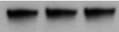
Western Blot
1 µg/mL
Sample: Recombinant Mouse TSLP (Catalog # 555-TS)
under non-reducing conditions only
Sample: Recombinant Mouse TSLP (Catalog # 555-TS)
under non-reducing conditions only
Neutralization
Measured by its ability to neutralize TSLP-induced proliferation in the BaF3 mouse pro‑B cell line transfected with mouse IL‑7 R alpha. Park, L.S. et al. (2000) J. Exp. Med. 192:659. The Neutralization Dose (ND50) is typically 0.1‑0.4 µg/mL in the presence of 7.5 ng/mL Recombinant Mouse TSLP.
Reviewed Applications
Read 2 reviews rated 3.5 using MAB555 in the following applications:
Formulation, Preparation, and Storage
Purification
Protein A or G purified from hybridoma culture supernatant
Reconstitution
Reconstitute at 0.5 mg/mL in sterile PBS. For liquid material, refer to CoA for concentration.
Loading...
Formulation
Lyophilized from a 0.2 μm filtered solution in PBS with Trehalose. *Small pack size (SP) is supplied either lyophilized or as a 0.2 µm filtered solution in PBS.
Shipping
Lyophilized product is shipped at ambient temperature. Liquid small pack size (-SP) is shipped with polar packs. Upon receipt, store immediately at the temperature recommended below.
Stability & Storage
Use a manual defrost freezer and avoid repeated freeze-thaw cycles.
- 12 months from date of receipt, -20 to -70 °C as supplied.
- 1 month, 2 to 8 °C under sterile conditions after reconstitution.
- 6 months, -20 to -70 °C under sterile conditions after reconstitution.
Calculators
Background: TSLP
Long Name
Thymic Stromal Lymphopoietin
Alternate Names
thymic stromal lymphopoietin
Gene Symbol
TSLP
UniProt
Additional TSLP Products
Product Documents for Mouse TSLP Antibody (152614)
Certificate of Analysis
To download a Certificate of Analysis, please enter a lot or batch number in the search box below.
Note: Certificate of Analysis not available for kit components.
Product Specific Notices for Mouse TSLP Antibody (152614)
For research use only
Citations for Mouse TSLP Antibody (152614)
 Powered by Bioz
Customer Reviews for Mouse TSLP Antibody (152614) (2)
3.5 out of 5
2 Customer Ratings
Have you used Mouse TSLP Antibody (152614)?
Submit a review and receive an Amazon gift card!
$25/€18/£15/$25CAN/¥2500 Yen for a review with an image
$10/€7/£6/$10CAN/¥1110 Yen for a review without an image
Submit a review
Customer Images
Showing
1
-
2 of
2 reviews
Showing All
Filter By:
-
Application: Western BlotSample Tested: Spleen tissueSpecies: MouseVerified Customer | Posted 09/27/2021

-
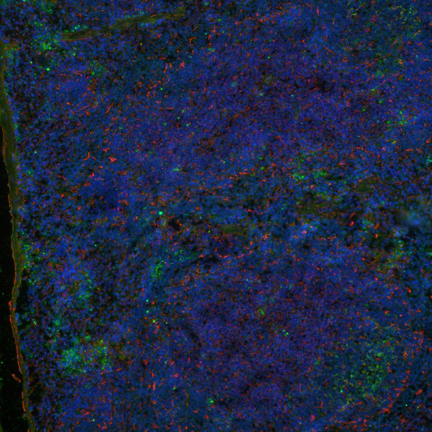
Application: Immunocytochemistry/ImmunofluorescenceSample Tested: Spleen tissueSpecies: MouseVerified Customer | Posted 02/26/2021
Bio-Techne ResponseThank you for reviewing our product. We are sorry to that that this antibody did not perform as expected. We have been in touch with the customer to resolve this issue according to our Product Guarantee and to the customer’s satisfaction. Although MAB555 has not been validated by Bio-Techne for IHC/ICC/IF application, Technical Service provided troubleshooting suggestions, which resulted in a weak signal in tissues tested. Customer observed a good signal with polyclonal antibody from Novus, catalog # NB110-55234.
There are no reviews that match your criteria.
Protocols
Find general support by application which include: protocols, troubleshooting, illustrated assays, videos and webinars.
- Cellular Response to Hypoxia Protocols
- R&D Systems Quality Control Western Blot Protocol
- Troubleshooting Guide: Western Blot Figures
- Western Blot Conditions
- Western Blot Protocol
- Western Blot Protocol for Cell Lysates
- Western Blot Troubleshooting
- Western Blot Troubleshooting Guide
- View all Protocols, Troubleshooting, Illustrated assays and Webinars
Loading...
Associated Pathways
