NCAM-1/CD56 Antibody (123A8 (same as 56C04)) - Azide and BSA Free
Novus Biologicals | Catalog # NBP2-34518
Key Product Details
Species Reactivity
Human
Applications
Validated:
Immunohistochemistry, Immunohistochemistry-Paraffin, Western Blot, Immunocytochemistry/ Immunofluorescence, Imaging Mass Cytometry
Cited:
IF/IHC
Label
Unconjugated
Antibody Source
Monoclonal Mouse IgG1 kappa Clone # 123A8 (same as 56C04)
Format
Azide and BSA Free
Loading...
Product Specifications
Immunogen
Membrane preparation of a small cell lung carcinoma
Localization
Cell surface
Marker
Neuronal Cell Marker
Specificity
This monoclonal antibody reacts with an extracellular domain (close to transmembrane) of CD56/NCAM. Three isoforms of neural cell adhesion molecule (NCAM) are produced by differential splicing of the RNA transcript from a single gene. The 135kDa isoform is the basic molecule, which is glycosylated or sialylated to produce the mature species. Anti-CD56 recognizes two proteins of the neural cell adhesion molecule, the basic molecule expressed on most neuroectodermally derived tissues and neoplasms (e.g. retinoblastoma, medulloblastomas, astrocytomas, neuroblastomas, and small cell carcinomas). It is also expressed on some mesodermally derived tumors (rhabdomyosarcoma). Anti-CD56 plays an important role in the diagnosis of nodal and nasal NK/T-cell lymphomas.
Clonality
Monoclonal
Host
Mouse
Isotype
IgG1 kappa
Description
1.0 mg/ml of antibody purified from Bioreactor Concentrate by Protein A/G. Prepared in 10mM PBS WITHOUT BSA & azide. Also available at 200 ug/ml WITH BSA & azide (NBP2-34279).
Antibody with azide - store at 2 to 8C. Antibody without azide - store at -20 to -80C.
Antibody with azide - store at 2 to 8C. Antibody without azide - store at -20 to -80C.
Scientific Data Images for NCAM-1/CD56 Antibody (123A8 (same as 56C04)) - Azide and BSA Free
![Western Blot: NCAM-1/CD56 Antibody (123A8 (same as 56C04))Azide and BSA Free [NBP2-34518]](https://resources.rndsystems.com/images/products/NCAM-1-CD56-Antibody-123A8-same-as-56C04-Azide-and-BSA-Free-Western-Blot-NBP2-34518-img0005.jpg "Western Blot: NCAM-1/CD56 Antibody (123A8 (same as 56C04))Azide and BSA Free [NBP2-34518]")
Western Blot: NCAM-1/CD56 Antibody (123A8 (same as 56C04))Azide and BSA Free [NBP2-34518]
Western Blot: NCAM-1/CD56 Antibody (123A8 (same as 56C04)) - Azide and BSA Free [NBP2-34518] - Western blot analysis of human brain tissue lysate using NCAM-1/CD56 antibody (123A8 (same as 56C04)).![Immunohistochemistry-Paraffin: NCAM-1/CD56 Antibody (123A8 (same as 56C04)) - Azide and BSA Free [NBP2-34518]](https://resources.rndsystems.com/images/products/NCAM-1-CD56-Antibody-123A8-same-as-56C04-Azide-and-BSA-Free-Immunohistochemistry-Paraffin-NBP2-34518-img0007.jpg "Immunohistochemistry-Paraffin: NCAM-1/CD56 Antibody (123A8 (same as 56C04)) - Azide and BSA Free [NBP2-34518]")
Immunohistochemistry-Paraffin: NCAM-1/CD56 Antibody (123A8 (same as 56C04)) - Azide and BSA Free [NBP2-34518]
Immunohistochemistry-Paraffin: NCAM-1/CD56 Antibody (123A8 (same as 56C04)) - Azide and BSA Free [NBP2-34518] - Formalin-fixed, paraffin-embedded human colon stained with NCAM-1/CD56 Antibody (123A8 (same as 56C04)).![Immunohistochemistry-Paraffin: NCAM-1/CD56 Antibody (123A8 (same as 56C04)) - Azide and BSA Free [NBP2-34518]](https://resources.rndsystems.com/images/products/NCAM-1-CD56-Antibody-123A8-same-as-56C04-Azide-and-BSA-Free-Immunohistochemistry-Paraffin-NBP2-34518-img0002.jpg "Immunohistochemistry-Paraffin: NCAM-1/CD56 Antibody (123A8 (same as 56C04)) - Azide and BSA Free [NBP2-34518]")
Immunohistochemistry-Paraffin: NCAM-1/CD56 Antibody (123A8 (same as 56C04)) - Azide and BSA Free [NBP2-34518]
Immunohistochemistry-Paraffin: NCAM-1/CD56 Antibody (123A8 (same as 56C04)) - Azide and BSA Free [NBP2-34518] - Formalin-fixed, paraffin-embedded human Pancreas stained with NCAM-1/CD56 Antibody (123A8 (same as 56C04))![Immunohistochemistry-Paraffin: NCAM-1/CD56 Antibody (123A8 (same as 56C04)) - Azide and BSA Free [NBP2-34518]](https://resources.rndsystems.com/images/products/NCAM-1-CD56-Antibody-123A8-same-as-56C04-Azide-and-BSA-Free-Immunohistochemistry-Paraffin-NBP2-34518-img0006.jpg "Immunohistochemistry-Paraffin: NCAM-1/CD56 Antibody (123A8 (same as 56C04)) - Azide and BSA Free [NBP2-34518]")
Immunohistochemistry-Paraffin: NCAM-1/CD56 Antibody (123A8 (same as 56C04)) - Azide and BSA Free [NBP2-34518]
Immunohistochemistry-Paraffin: NCAM-1/CD56 Antibody (123A8 (same as 56C04)) - Azide and BSA Free [NBP2-34518] - Formalin-fixed, paraffin-embedded human thyroid stained with NCAM-1/CD56 Antibody (123A8 (same as 56C04)). HIER: Tris/EDTA, pH9.0, 45min. HRP-polymer, 30min. DAB, 5min.![Imaging Mass Cytometry: NCAM-1/CD56 Antibody (123A8 (same as 56C04)) - Azide and BSA Free [NBP2-34518]](https://resources.rndsystems.com/images/products/NCAM-1-CD56-Antibody-123A8-same-as-56C04-Azide-and-BSA-Free-Imaging-Mass-Cytometry-NBP2-34518-img0004.jpg "Imaging Mass Cytometry: NCAM-1/CD56 Antibody (123A8 (same as 56C04)) - Azide and BSA Free [NBP2-34518]")
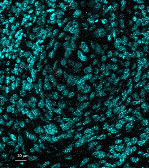
Imaging Mass Cytometry: NCAM-1/CD56 Antibody (123A8 (same as 56C04)) - Azide and BSA Free [NBP2-34518]
Imaging Mass Cytometry: NCAM-1/CD56 Antibody (123A8 (same as 56C04)) - Azide and BSA Free [NBP2-34518] - Image of human cutaneous squamous cell carcinoma tumor stained with Anti-NCAM-1/CD56 Azide/BSA Free Antibody [123A8] (in red). IMC image submitted by a verified customer review.)")
NCAM-1/CD56 Antibody (123A8 (same as 56C04))
Formalin-fixed, paraffin-embedded human thyroid stained with NCAM-1/CD56 antibody (123A8 (same as 56C04)). HIER: Tris/EDTA, pH9.0, 45min. Secondary: HRP-polymer, 30min. DAB, 5min.Applications for NCAM-1/CD56 Antibody (123A8 (same as 56C04)) - Azide and BSA Free
Application
Recommended Usage
Immunocytochemistry/ Immunofluorescence
1-2ug/ml
Immunohistochemistry-Paraffin
0.5-1ug/ml
Application Notes
Immunohistochemistry (Formalin-fixed): 1-2ug/ml for 30 minutes at RT. Staining of formalin-fixed tissues requires heating tissue sections in 10mM Tris with 1mM EDTA, pH 9.0, for 45 min at 95C followed by cooling at RT for 20 minutes.
Optimal dilution for a specific application should be determined.
Optimal dilution for a specific application should be determined.
Reviewed Applications
Read 1 review rated 4 using NBP2-34518 in the following applications:
Formulation, Preparation, and Storage
Purification
Protein A or G purified
Formulation
10 mM PBS
Format
Azide and BSA Free
Preservative
No Preservative
Concentration
1.0 mg/ml
Shipping
The product is shipped with polar packs. Upon receipt, store it immediately at the temperature recommended below.
Stability & Storage
Store at -20 to -80C. Avoid freeze-thaw cycles.
Background: NCAM-1/CD56
Long Name
Neural Cell Adhesion Molecule
Alternate Names
CD56, NCAM1
Gene Symbol
NCAM1
Additional NCAM-1/CD56 Products
Product Documents for NCAM-1/CD56 Antibody (123A8 (same as 56C04)) - Azide and BSA Free
Certificate of Analysis
To download a Certificate of Analysis, please enter a lot or batch number in the search box below.
Product Specific Notices for NCAM-1/CD56 Antibody (123A8 (same as 56C04)) - Azide and BSA Free
This product is for research use only and is not approved for use in humans or in clinical diagnosis. Primary Antibodies are guaranteed for 1 year from date of receipt.
Related Research Areas
Citations for NCAM-1/CD56 Antibody (123A8 (same as 56C04)) - Azide and BSA Free
 Powered by Bioz
Customer Reviews for NCAM-1/CD56 Antibody (123A8 (same as 56C04)) - Azide and BSA Free (1)
4 out of 5
1 Customer Rating
Have you used NCAM-1/CD56 Antibody (123A8 (same as 56C04)) - Azide and BSA Free?
Submit a review and receive an Amazon gift card!
$25/€18/£15/$25CAN/¥2500 Yen for a review with an image
$10/€7/£6/$10CAN/¥1110 Yen for a review without an image
Submit a review
Customer Images
Showing
1
-
1 of
1 review
Showing All
Filter By:
-
Application: Imaging Mass CytometrySample Tested: FFPESpecies: HumanVerified Customer | Posted 10/05/2020Mass Cytometry - Image of human cutaneous squamous cell carcinoma tumor stained with Anti-NCAM-1/CD56 Azide/BSA Free Antibody [123A8] (in red).

There are no reviews that match your criteria.
Protocols
Find general support by application which include: protocols, troubleshooting, illustrated assays, videos and webinars.
- Antigen Retrieval Protocol (PIER)
- Antigen Retrieval for Frozen Sections Protocol
- Appropriate Fixation of IHC/ICC Samples
- Cellular Response to Hypoxia Protocols
- Chromogenic IHC Staining of Formalin-Fixed Paraffin-Embedded (FFPE) Tissue Protocol
- Chromogenic Immunohistochemistry Staining of Frozen Tissue
- ClariTSA™ Fluorophore Kits
- Detection & Visualization of Antibody Binding
- Fluorescent IHC Staining of Frozen Tissue Protocol
- Graphic Protocol for Heat-induced Epitope Retrieval
- Graphic Protocol for the Preparation and Fluorescent IHC Staining of Frozen Tissue Sections
- Graphic Protocol for the Preparation and Fluorescent IHC Staining of Paraffin-embedded Tissue Sections
- Graphic Protocol for the Preparation of Gelatin-coated Slides for Histological Tissue Sections
- ICC Cell Smear Protocol for Suspension Cells
- ICC Immunocytochemistry Protocol Videos
- ICC for Adherent Cells
- IHC Sample Preparation (Frozen sections vs Paraffin)
- Immunocytochemistry (ICC) Protocol
- Immunocytochemistry Troubleshooting
- Immunofluorescence of Organoids Embedded in Cultrex Basement Membrane Extract
- Immunofluorescent IHC Staining of Formalin-Fixed Paraffin-Embedded (FFPE) Tissue Protocol
- Immunohistochemistry (IHC) and Immunocytochemistry (ICC) Protocols
- Immunohistochemistry Frozen Troubleshooting
- Immunohistochemistry Paraffin Troubleshooting
- Preparing Samples for IHC/ICC Experiments
- Preventing Non-Specific Staining (Non-Specific Binding)
- Primary Antibody Selection & Optimization
- Protocol for Heat-Induced Epitope Retrieval (HIER)
- Protocol for Making a 4% Formaldehyde Solution in PBS
- Protocol for VisUCyte™ HRP Polymer Detection Reagent
- Protocol for the Fluorescent ICC Staining of Cell Smears - Graphic
- Protocol for the Fluorescent ICC Staining of Cultured Cells on Coverslips - Graphic
- Protocol for the Preparation & Fixation of Cells on Coverslips
- Protocol for the Preparation and Chromogenic IHC Staining of Frozen Tissue Sections
- Protocol for the Preparation and Chromogenic IHC Staining of Frozen Tissue Sections - Graphic
- Protocol for the Preparation and Chromogenic IHC Staining of Paraffin-embedded Tissue Sections
- Protocol for the Preparation and Chromogenic IHC Staining of Paraffin-embedded Tissue Sections - Graphic
- Protocol for the Preparation and Fluorescent ICC Staining of Cells on Coverslips
- Protocol for the Preparation and Fluorescent ICC Staining of Non-adherent Cells
- Protocol for the Preparation and Fluorescent ICC Staining of Stem Cells on Coverslips
- Protocol for the Preparation and Fluorescent IHC Staining of Frozen Tissue Sections
- Protocol for the Preparation and Fluorescent IHC Staining of Paraffin-embedded Tissue Sections
- Protocol for the Preparation of Gelatin-coated Slides for Histological Tissue Sections
- Protocol for the Preparation of a Cell Smear for Non-adherent Cell ICC - Graphic
- R&D Systems Quality Control Western Blot Protocol
- TUNEL and Active Caspase-3 Detection by IHC/ICC Protocol
- The Importance of IHC/ICC Controls
- Troubleshooting Guide: Immunohistochemistry
- Troubleshooting Guide: Western Blot Figures
- Western Blot Conditions
- Western Blot Protocol
- Western Blot Protocol for Cell Lysates
- Western Blot Troubleshooting
- Western Blot Troubleshooting Guide
- View all Protocols, Troubleshooting, Illustrated assays and Webinars
Loading...
Associated Pathways


