Neural Stem Cell Marker Antibody Pack
Novus Biologicals | Catalog # NBP1-42826
![Knockout Validated: Neural Stem Cell Marker Antibody Pack [NBP1-42826]](https://resources.rndsystems.com/images/products/Neural-Stem-Cell-Marker-Antibody-Pack-Knockout-Validated-NBP1-42826-img0018.jpg "Western Blot: Neural Stem Cell Marker Antibody Pack [NBP1-42826]")
Loading...
Key Product Details
Species
Human, Mouse, Rat
Applications
Immunocytochemistry/ Immunofluorescence, Immunohistochemistry, Western Blot
Kit Type
Kit
Product Summary for Neural Stem Cell Marker Antibody Pack
This pack contains 1 vial each of: MAB1195 (100 ug), NB110-37235 (0.1 mL), NBP1-05197 (0.1 mL), NB100-2322 (0.1 mL), and NB300-266 (0.1 mL).
Loading...
Product Specifications
Specificity
NB100-2322: HMGB1 - Oligodendrocyte Marker
NB110-37235: SOX2 - Embryonic Stem Cell Marker
MAB1195: Neuron-specific beta-III Tubulin MAb (Clone TuJ-1)
NBP1-05197: GFAP - Astrocyte Marker
NB300-266: Nestin - Cytoskeleton-Intermediate Filaments Marker
NB110-37235: SOX2 - Embryonic Stem Cell Marker
MAB1195: Neuron-specific beta-III Tubulin MAb (Clone TuJ-1)
NBP1-05197: GFAP - Astrocyte Marker
NB300-266: Nestin - Cytoskeleton-Intermediate Filaments Marker
Application Notes
See individual datasheets of components for their validated applications. NB100-2322 - A band is seen at ~29 kDa in Western Blotting.
NB110-37235 - a band is seen at ~40 kDa in Western Blotting, representing the SOX2 protein.
NBP1-05197 - Can be used to identify astrocytic cells, their progenitors and their descendents in tissue sections and in tissue culture. The epitope recognized by 5C10 is unusually resistant to aldehyde fixation, and so is ideal of studies of formalin-fixed paraffin-embedded histological specimens. A 55 kDa band can be seen in Western blotting.
NB300-266 - a doublet band is seen at ~220-240 kDa in Western Blotting.
Reactivity Notes
See individual datasheets of components for their validated species
Scientific Data Images for Neural Stem Cell Marker Antibody Pack
Western Blot: Neural Stem Cell Marker Antibody Pack [NBP1-42826]
Western Blot: Neural Stem Cell Marker Antibody Pack [NBP1-42826] - Western blot shows lysates of HEK293T human embryonic kidney parental cell line and HMGB1 knockout (KO) HEK293T cell line. PVDF membrane was probed with 1.0 ug/ml of Rabbit Anti-Human HMGB1 Polyclonal Antibody (Catalog # NB100-2322) followed by HRP-conjugated Anti-Rabbit IgG Secondary Antibody (Catalog #HAF008). Specific band was detected for HMGB1 at approximately 30 kDa (as indicated) in the parental HEK293T cell line, but is not detectable in the knockout HEK293T cell line. This experiment was conducted under reducing conditions.![Immunohistochemistry Free-Floating: Neural Stem Cell Marker Antibody Pack [NBP1-42826]](https://resources.rndsystems.com/images/products/Neural-Stem-Cell-Marker-Antibody-Pack-Immunohistochemistry-Free-Floating-NBP1-42826-img0019.jpg "Immunohistochemistry Free-Floating: Neural Stem Cell Marker Antibody Pack [NBP1-42826]")
Immunohistochemistry Free-Floating: Neural Stem Cell Marker Antibody Pack [NBP1-42826]
Immunohistochemistry Free-Floating: Neural Stem Cell Marker Antibody Pack [NBP1-42826] - Analysis of rat cerebellum section stained with mouse GFAP mAb (NBP1-05197), dilution 1:1,000 (Green), costained with rabbit neurofilament NF-L pAb, dilution 1:2,000 (Red). Following transcardial perfusion with 4% paraformaldehyde, brain was post fixed for 24hrs, cut to 45uM, and free-floating sections were stained with antibodies. The GFAP antibody stains a network of astroglial cells, while the NF-L antibody labels neuronal cells and their processes.![Simple Western: Neural Stem Cell Marker Antibody Pack [NBP1-42826]](https://resources.rndsystems.com/images/products/Neural-Stem-Cell-Marker-Antibody-Pack-Simple-Western-NBP1-42826-img0016.jpg "Simple Western: Neural Stem Cell Marker Antibody Pack [NBP1-42826]")
Simple Western: Neural Stem Cell Marker Antibody Pack [NBP1-42826]
Simple Western: Neural Stem Cell Marker Antibody Pack [NBP1-42826] - Simple Western lane view shows lysates of rat cortical neurons, loaded at 0.2 mg/mL. A specific band was detected for beta -III Tubulin at approximately 56 kDa (as indicated) using 10 ug/mL of Mouse Anti-Neuron-specific beta -III Tubulin Monoclonal Antibody (Catalog # MAB1195). This experiment was conducted under reducing conditions and using the 12-230 kDa separation system.![Immunocytochemistry/ Immunofluorescence: Neural Stem Cell Marker Antibody Pack [NBP1-42826]](https://resources.rndsystems.com/images/products/Neural-Stem-Cell-Marker-Antibody-Pack-Immunocytochemistry-Immunofluorescence-NBP1-42826-img0013.jpg "Immunocytochemistry/ Immunofluorescence: Neural Stem Cell Marker Antibody Pack [NBP1-42826]")
Immunocytochemistry/ Immunofluorescence: Neural Stem Cell Marker Antibody Pack [NBP1-42826]
Immunocytochemistry/Immunofluorescence: Neural Stem Cell Marker Antibody Pack [NBP1-42826] - beta -III Tubulin in Differentiated Human Neural Progenitor Cells. beta -III Tubulin was detected in immersion fixed differentiated human neural progenitor cells using Neuron-specific beta -III Tubulin Monoclonal Antibody (clone TuJ-1) (Catalog # MAB1195) for 3 hours at room temperature. Cells were stained (green) and counterstained (red). View our protocol for Fluorescent ICC Staining of Cells on Coverslips.![Western Blot: Neural Stem Cell Marker Antibody Pack [NBP1-42826]](https://resources.rndsystems.com/images/products/Neural-Stem-Cell-Marker-Antibody-Pack-Western-Blot-NBP1-42826-img0003.jpg "Western Blot: Neural Stem Cell Marker Antibody Pack [NBP1-42826]")
Western Blot: Neural Stem Cell Marker Antibody Pack [NBP1-42826]
Western Blot: Neural Stem Cell Marker Antibody Pack [NBP1-42826] - Detection of SOX2 in mouse brain lysate using NB110-37235 (0.5ug/ml).![Western Blot: Neural Stem Cell Marker Antibody Pack [NBP1-42826]](https://resources.rndsystems.com/images/products/Neural-Stem-Cell-Marker-Antibody-Pack-Western-Blot-NBP1-42826-img0023.jpg "Western Blot: Neural Stem Cell Marker Antibody Pack [NBP1-42826]")
Western Blot: Neural Stem Cell Marker Antibody Pack [NBP1-42826]
Western Blot: Neural Stem Cell Marker Antibody Pack [NBP1-42826] - Western blot shows lysates of human brain (cerebellum) tissue, human brain (hypothalamus) tissue, mouse brain (cerebellum) tissue, and mouse brain (stem) tissue. PVDF membrane was probed with 0.2 ug/mL of Mouse Anti-Neuron-specific beta -III Tubulin Monoclonal Antibody (Catalog # MAB1195) followed by HRP-conjugated Anti-Mouse IgG Secondary Antibody (Catalog # HAF018). A specific band was detected for beta -III Tubulin at approximately 55 kDa (as indicated). This experiment was conducted under reducing conditions and using Immunoblot Buffer Group 1.![Simple Western: Neural Stem Cell Marker Antibody Pack [NBP1-42826]](https://resources.rndsystems.com/images/products/Neural-Stem-Cell-Marker-Antibody-Pack-Simple-Western-NBP1-42826-img0020.jpg "Simple Western: Neural Stem Cell Marker Antibody Pack [NBP1-42826]")
Simple Western: Neural Stem Cell Marker Antibody Pack [NBP1-42826]
Simple Western: Neural Stem Cell Marker Antibody Pack [NBP1-42826] - Simple Western lane view shows a specific band for GFAP in 0.05 mg/ml of Human Brain lysate using NBP1-05197. This experiment was performed under reducing conditions using the 12-230 kDa separation system.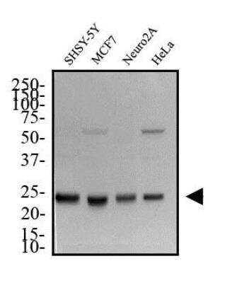
Neural Stem Cell Marker Antibody Pack [NBP1-42826] - Total protein from SHSY-5Y, MCF7, Neuro2A and HeLa was separated on a 12% gel by SDS-PAGE, transferred to PVDF membrane and blocked in 5% non-fat milk in TBST. The membrane was probed with 2.0 ug/mL anti-HMGB1 [NB100-2322] in 1% non-fat milk in TBST and detected with an anti-rabbit HRP secondary antibody using chemiluminescence.
Kit Contents for Neural Stem Cell Marker Antibody Pack
- MAB1195: Neuron-specific beta ‑III Tubulin Antibody
- NBP1-05197: GFAP Antibody (5C10) - BSA Free
- NB100-2322: HMGB1/HMG-1 Antibody - BSA Free
- HAF007: Mouse IgG Horseradish Peroxidase-conjugated Antibody
- NB300-266: Nestin Antibody (10C2) - BSA Free
- HAF008: Rabbit IgG Horseradish Peroxidase-conjugated Antibody
- NB110-37235: SOX2 Antibody - BSA Free
Formulation, Preparation, and Storage
Purification
Immunogen affinity purified
Preservative
0.02% Sodium Azide
Concentration
Concentration of individual antibodies may be found on the vial label. If unlisted please contact technical services.
Shipping
The product is shipped with polar packs. Upon receipt, store it immediately at the temperature recommended below.
Storage
Store at 4C. Do not freeze.
Background: Neural Stem Cell Marker
Additional Neural Stem Cell Marker Products
Product Documents for Neural Stem Cell Marker Antibody Pack
Certificate of Analysis
To download a Certificate of Analysis, please enter a lot or batch number in the search box below.
Product Specific Notices for Neural Stem Cell Marker Antibody Pack
This product is for research use only and is not approved for use in humans or in clinical diagnosis. Antibody Packs are guaranteed for 1 year from date of receipt.
Customer Reviews for Neural Stem Cell Marker Antibody Pack
There are currently no reviews for this product. Be the first to review Neural Stem Cell Marker Antibody Pack and earn rewards!
Have you used Neural Stem Cell Marker Antibody Pack?
Submit a review and receive an Amazon gift card!
$25/€18/£15/$25CAN/¥2500 Yen for a review with an image
$10/€7/£6/$10CAN/¥1110 Yen for a review without an image
Submit a review
Protocols
Find general support by application which include: protocols, troubleshooting, illustrated assays, videos and webinars.
- Antigen Retrieval Protocol (PIER)
- Antigen Retrieval for Frozen Sections Protocol
- Appropriate Fixation of IHC/ICC Samples
- Cellular Response to Hypoxia Protocols
- Chromogenic IHC Staining of Formalin-Fixed Paraffin-Embedded (FFPE) Tissue Protocol
- Chromogenic Immunohistochemistry Staining of Frozen Tissue
- ClariTSA™ Fluorophore Kits
- Detection & Visualization of Antibody Binding
- Fluorescent IHC Staining of Frozen Tissue Protocol
- Graphic Protocol for Heat-induced Epitope Retrieval
- Graphic Protocol for the Preparation and Fluorescent IHC Staining of Frozen Tissue Sections
- Graphic Protocol for the Preparation and Fluorescent IHC Staining of Paraffin-embedded Tissue Sections
- Graphic Protocol for the Preparation of Gelatin-coated Slides for Histological Tissue Sections
- ICC Cell Smear Protocol for Suspension Cells
- ICC Immunocytochemistry Protocol Videos
- ICC for Adherent Cells
- IHC Sample Preparation (Frozen sections vs Paraffin)
- Immunocytochemistry (ICC) Protocol
- Immunocytochemistry Troubleshooting
- Immunofluorescence of Organoids Embedded in Cultrex Basement Membrane Extract
- Immunofluorescent IHC Staining of Formalin-Fixed Paraffin-Embedded (FFPE) Tissue Protocol
- Immunohistochemistry (IHC) and Immunocytochemistry (ICC) Protocols
- Immunohistochemistry Frozen Troubleshooting
- Immunohistochemistry Paraffin Troubleshooting
- Preparing Samples for IHC/ICC Experiments
- Preventing Non-Specific Staining (Non-Specific Binding)
- Primary Antibody Selection & Optimization
- Protocol for Heat-Induced Epitope Retrieval (HIER)
- Protocol for Making a 4% Formaldehyde Solution in PBS
- Protocol for VisUCyte™ HRP Polymer Detection Reagent
- Protocol for the Fluorescent ICC Staining of Cell Smears - Graphic
- Protocol for the Fluorescent ICC Staining of Cultured Cells on Coverslips - Graphic
- Protocol for the Preparation & Fixation of Cells on Coverslips
- Protocol for the Preparation and Chromogenic IHC Staining of Frozen Tissue Sections
- Protocol for the Preparation and Chromogenic IHC Staining of Frozen Tissue Sections - Graphic
- Protocol for the Preparation and Chromogenic IHC Staining of Paraffin-embedded Tissue Sections
- Protocol for the Preparation and Chromogenic IHC Staining of Paraffin-embedded Tissue Sections - Graphic
- Protocol for the Preparation and Fluorescent ICC Staining of Cells on Coverslips
- Protocol for the Preparation and Fluorescent ICC Staining of Non-adherent Cells
- Protocol for the Preparation and Fluorescent ICC Staining of Stem Cells on Coverslips
- Protocol for the Preparation and Fluorescent IHC Staining of Frozen Tissue Sections
- Protocol for the Preparation and Fluorescent IHC Staining of Paraffin-embedded Tissue Sections
- Protocol for the Preparation of Gelatin-coated Slides for Histological Tissue Sections
- Protocol for the Preparation of a Cell Smear for Non-adherent Cell ICC - Graphic
- R&D Systems Quality Control Western Blot Protocol
- TUNEL and Active Caspase-3 Detection by IHC/ICC Protocol
- The Importance of IHC/ICC Controls
- Troubleshooting Guide: Immunohistochemistry
- Troubleshooting Guide: Western Blot Figures
- Western Blot Conditions
- Western Blot Protocol
- Western Blot Protocol for Cell Lysates
- Western Blot Troubleshooting
- Western Blot Troubleshooting Guide
- View all Protocols, Troubleshooting, Illustrated assays and Webinars
Loading...