p53 Antibody (PAb 240) - BSA Free
Novus Biologicals | Catalog # NB200-103
Key Product Details
Species Reactivity
Validated:
Human, Mouse, Rat, Xenopus (Negative), Yeast
Cited:
Human, Mouse, Rat, Yeast
Applications
Validated:
Immunohistochemistry, Immunohistochemistry-Paraffin, Immunohistochemistry-Frozen, Western Blot, ELISA, Flow Cytometry, Flow (Intracellular), Immunocytochemistry/ Immunofluorescence, Immunoprecipitation, CyTOF-ready
Cited:
Immunohistochemistry-Paraffin, Immunohistochemistry-Frozen, Western Blot, ELISA, Flow Cytometry, Immunocytochemistry/ Immunofluorescence, Immunoprecipitation, Hydrolysis Assay, IF/IHC
Label
Unconjugated
Antibody Source
Monoclonal Mouse IgG1 kappa Clone # PAb 240
Format
BSA Free
Loading...
Product Specifications
Immunogen
Gel-purified p53-beta-galactosidase fusion protein containing murine p53 from aa 14-389 (derived from pSV53C cDNA clone).
Epitope
The epitope has been mapped between amino acids 213 and 217 on human p53. The epitiope may be conformational as it reacts with mutant p53 in nondenatured form.
Reactivity Notes
Yeast reactivity reported in scientific literature (PMID: 8710879)
Localization
Nuclear/cytoplasmic
Specificity
This monoclonal recognizes both mutant forms and wild-type human p53 under denaturing conditions. It does not recognize nondenatured wild type p53. In nondenaturing conditions (IP) it only recognizes mutant p53.
Clonality
Monoclonal
Host
Mouse
Isotype
IgG1 kappa
Scientific Data Images for p53 Antibody (PAb 240) - BSA Free
![Western Blot: p53 Antibody (PAb 240)BSA Free [NB200-103]](https://resources.rndsystems.com/images/products/p53-Antibody-PAb-240-Western-Blot-NB200-103-img0001.jpg "Western Blot: p53 Antibody (PAb 240)BSA Free [NB200-103]")
Western Blot: p53 Antibody (PAb 240)BSA Free [NB200-103]
Western Blot: p53 Antibody (PAb 240) [NB200-103] - Analysis of p53 in MCF7 and HeLa lystates. Image courtesy of anonymous customer product review.![Immunocytochemistry/ Immunofluorescence: p53 Antibody (PAb 240) - BSA Free [NB200-103]](https://resources.rndsystems.com/images/products/p53-Antibody-PAb-240-Immunocytochemistry-Immunofluorescence-NB200-103-img0013.jpg "Immunocytochemistry/ Immunofluorescence: p53 Antibody (PAb 240) - BSA Free [NB200-103]")
Immunocytochemistry/ Immunofluorescence: p53 Antibody (PAb 240) - BSA Free [NB200-103]
Immunocytochemistry/Immunofluorescence: p53 Antibody (PAb 240) [NB200-103] - PC12 cells were fixed in 4% paraformaldehyde for 10 minutes and permeabilized in 0.5% Triton X-100 in PBS for 5 minutes. The cells were incubated with anti-p53 Antibody (PAb 240) NB200-103 at 2 ug/ml overnight at 4C and detected with an anti-mouse Dylight 488 (Green) at a 1:1000 dilution for 60 minutes. Nuclei were counterstained with DAPI (Blue). Cells were imaged using a 40X objective.![Immunohistochemistry-Paraffin: p53 Antibody (PAb 240) - BSA Free [NB200-103]](https://resources.rndsystems.com/images/products/p53-Antibody-PAb-240-Immunohistochemistry-Paraffin-NB200-103-img0005.jpg "Immunohistochemistry-Paraffin: p53 Antibody (PAb 240) - BSA Free [NB200-103]")
Immunohistochemistry-Paraffin: p53 Antibody (PAb 240) - BSA Free [NB200-103]
Immunohistochemistry-Paraffin: p53 Antibody (PAb 240) [NB200-103] - p53 was detected in immersion fixed paraffin-embedded sections of human prostate cancer using anti-human mouse monoclonal antibody (Catalog # NB200-103) at 1:200 dilution overnight at 4C. Tissue was stained using the VisuCyte anti-mouse HRP polymer detection reagent (Catalog # VC001) with DAB chromogen (brown) and counterstained with hematoxylin (blue). Images may not be copied, printed or otherwise disseminated without express written permission of Novus Biologicals a bio-techne brand.![Flow Cytometry: p53 Antibody (PAb 240) - BSA Free [NB200-103]](https://resources.rndsystems.com/images/products/p53-Antibody-PAb-240-Flow-Cytometry-NB200-103-img0010.jpg "Flow Cytometry: p53 Antibody (PAb 240) - BSA Free [NB200-103]")
Flow Cytometry: p53 Antibody (PAb 240) - BSA Free [NB200-103]
Flow Cytometry: p53 Antibody (PAb 240) [NB200-103] - An intracellular stain was performed on HeLa cells with p53 Antibody [PAb 240] NB200-103 (blue) and a matched isotype control (orange). Cells were fixed with 4% PFA and then permeabilized with 0.1% saponin. Cells were incubated in an antibody dilution of 1.0 ug/mL for 30 minutes at room temperature, followed by Mouse IgG (H+L) Cross-Adsorbed Secondary Antibody, Dylight 550 (35503, Thermo Fisher).![Immunocytochemistry/ Immunofluorescence: p53 Antibody (PAb 240) - BSA Free [NB200-103]](https://resources.rndsystems.com/images/products/p53-Antibody-PAb-240-Immunocytochemistry-Immunofluorescence-NB200-103-img0004.jpg "Immunocytochemistry/ Immunofluorescence: p53 Antibody (PAb 240) - BSA Free [NB200-103]")
Immunocytochemistry/ Immunofluorescence: p53 Antibody (PAb 240) - BSA Free [NB200-103]
Immunocytochemistry/Immunofluorescence: p53 Antibody (PAb 240) [NB200-103] - HeLa cells were fixed for 10 minutes using 10% formalin and then permeabilized for 5 minutes using 1X TBS + 0.5% Triton X-100. The cells were incubated with anti-p53 (PAb 240) [NB200-103] at a 1:200 dilution overnight at 4C and detected with an anti-mouse DyLight 488 (Green) at a 1:500 dilution. Actin was detected with Phalloidin 568 (Red) at a 1:200 dilution. Nuclei were counterstained with DAPI (Blue). Cells were imaged using a 40X objective.![Immunocytochemistry/ Immunofluorescence: p53 Antibody (PAb 240) - BSA Free [NB200-103]](https://resources.rndsystems.com/images/products/p53-Antibody-PAb-240-Immunocytochemistry-Immunofluorescence-NB200-103-img0011.jpg "Immunocytochemistry/ Immunofluorescence: p53 Antibody (PAb 240) - BSA Free [NB200-103]")
Immunocytochemistry/ Immunofluorescence: p53 Antibody (PAb 240) - BSA Free [NB200-103]
Immunocytochemistry/Immunofluorescence: p53 Antibody (PAb 240) [NB200-103] - HeLa cells were fixed in 4% paraformaldehyde for 10 minutes and permeabilized in 0.5% Triton X-100 in PBS for 5 minutes. The cells were incubated with anti-p53 Antibody (PAb 240) NB200-103 at 2 ug/ml overnight at 4C and detected with an anti-mouse Dylight 488 (Green) at a 1:1000 dilution for 60 minutes. Nuclei were counterstained with DAPI (Blue). Cells were imaged using a 100X objective and digitally deconvolved.![Immunocytochemistry/ Immunofluorescence: p53 Antibody (PAb 240) - BSA Free [NB200-103]](https://resources.rndsystems.com/images/products/p53-Antibody-PAb-240-Immunocytochemistry-Immunofluorescence-NB200-103-img0012.jpg "Immunocytochemistry/ Immunofluorescence: p53 Antibody (PAb 240) - BSA Free [NB200-103]")
Immunocytochemistry/ Immunofluorescence: p53 Antibody (PAb 240) - BSA Free [NB200-103]
Immunocytochemistry/Immunofluorescence: p53 Antibody (PAb 240) [NB200-103] - Neuro2a cells were fixed in 4% paraformaldehyde for 10 minutes and permeabilized in 0.5% Triton X-100 in PBS for 5 minutes. The cells were incubated with anti-p53 Antibody (PAb 240) NB200-103 at 2 ug/ml overnight at 4C and detected with an anti-mouse Dylight 488 (Green) at a 1:1000 dilution for 60 minutes. Nuclei were counterstained with DAPI (Blue). Cells were imaged using a 100X objective and digitally deconvolved.![Flow (Intracellular): p53 Antibody (PAb 240) - BSA Free [NB200-103]](https://resources.rndsystems.com/images/products/p53-Antibody-PAb-240-Flow-Intracellular-NB200-103-img0003.jpg "Flow (Intracellular): p53 Antibody (PAb 240) - BSA Free [NB200-103]")
Flow (Intracellular): p53 Antibody (PAb 240) - BSA Free [NB200-103]
Flow (Intracellular): p53 Antibody (PAb 240) [NB200-103] - An intracellular stain was performed on HeLa cells with p53 (PAb240) NB200-103 (blue) and a matched isotype control NBP2-27287 (orange). Cells were fixed with 4% PFA and then permeablized with 0.1% saponin. Cells were incubated in an antibody dilution of 2.5 ug/mL for 30 minutes at room temperature, followed by APC-conjugated anti-mouse IgG secondary antibody F0101B.![Flow Cytometry: p53 Antibody (PAb 240) - BSA Free [NB200-103]](https://resources.rndsystems.com/images/products/p53-Antibody-PAb-240-Flow-Cytometry-NB200-103-img0006.jpg "Flow Cytometry: p53 Antibody (PAb 240) - BSA Free [NB200-103]")
Flow Cytometry: p53 Antibody (PAb 240) - BSA Free [NB200-103]
Flow Cytometry: p53 Antibody (PAb 240) [NB200-103] - An intracellular stain was performed on HeLa cells with p53 (PAb240) NB200-103AF647 (blue) and a matched isotype control (orange). Cells were fixed with 4% PFA and then permeablized with 0.1% saponin. Cells were incubated in an antibody dilution of 2.5 ug/mL for 30 minutes at room temperature. Both antibodies were conjugated to Alexa Fluor 647.![Flow Cytometry: p53 Antibody (PAb 240) - BSA Free [NB200-103]](https://resources.rndsystems.com/images/products/p53-Antibody-PAb-240-Flow-Cytometry-NB200-103-img0007.jpg "Flow Cytometry: p53 Antibody (PAb 240) - BSA Free [NB200-103]")
Flow Cytometry: p53 Antibody (PAb 240) - BSA Free [NB200-103]
Flow Cytometry: p53 Antibody (PAb 240) [NB200-103] - An intracellular stain was performed on A549 cells with p53 (PAb240) NB200-103AF488 (blue) and a matched isotype control (orange). Cells were fixed with 4% PFA and then permeablized with 0.1% saponin. Cells were incubated in an antibody dilution of 10 ug/mL for 30 minutes at room temperature. Both antibodies were conjugated to Alexa Fluor 488.![Flow Cytometry: p53 Antibody (PAb 240) - BSA Free [NB200-103]](https://resources.rndsystems.com/images/products/p53-Antibody-PAb-240-Flow-Cytometry-NB200-103-img0008.jpg "Flow Cytometry: p53 Antibody (PAb 240) - BSA Free [NB200-103]")
Flow Cytometry: p53 Antibody (PAb 240) - BSA Free [NB200-103]
Flow Cytometry: p53 Antibody (PAb 240) [NB200-103] - An intracellular stain was performed on MCF7 cells with p53 [PAb 240] Antibody NB200-103AF647 (blue) and a matched isotype control (orange). Cells were fixed with 4% PFA and then permeabilized with 0.1% saponin. Cells were incubated in an antibody dilution of 2.5 ug/mL for 30 minutes at room temperature. Both antibodies were conjugated to Alexa Fluor 647.![Flow Cytometry: p53 Antibody (PAb 240) - BSA Free [NB200-103]](https://resources.rndsystems.com/images/products/p53-Antibody-PAb-240-Flow-Cytometry-NB200-103-img0009.jpg "Flow Cytometry: p53 Antibody (PAb 240) - BSA Free [NB200-103]")
Flow Cytometry: p53 Antibody (PAb 240) - BSA Free [NB200-103]
Flow Cytometry: p53 Antibody (PAb 240) [NB200-103] - An intracellular stain was performed on Neuro2a cells with p53 Antibody [PAb 240] NB200-103 (blue) and a matched isotype control (orange). Cells were fixed with 4% PFA and then permeabilized with 0.1% saponin. Cells were incubated in an antibody dilution of 1.0 ug/mL for 30 minutes at room temperature, followed by Mouse IgG (H+L) Cross-Adsorbed Secondary Antibody, Dylight 550 (35503, Thermo Fisher).Applications for p53 Antibody (PAb 240) - BSA Free
Application
Recommended Usage
ELISA
1:100-1:2000
Flow Cytometry
1:10-1:1000
Immunocytochemistry/ Immunofluorescence
5 ug/ml. Use reported in scientific literature (PMID 1394225)
Immunohistochemistry
1:250-1:500
Immunohistochemistry-Frozen
reported in multiple pieces of scientific literature
Immunohistochemistry-Paraffin
1:250-1:500
Immunoprecipitation
10ug/mg
Western Blot
1:1000-1:2000
Application Notes
Antigen retrieval with IHC-P is not essential but may optimize staining. In IP this antibody reacts with only mutant p53 protein under non-denaturing conditions. This antibody is CyTOF ready.
Reviewed Applications
Read 3 reviews rated 4.3 using NB200-103 in the following applications:
Flow Cytometry Panel Builder
Bio-Techne Knows Flow Cytometry
Save time and reduce costly mistakes by quickly finding compatible reagents using the Panel Builder Tool.
Advanced Features
- Spectra Viewer - Custom analysis of spectra from multiple fluorochromes
- Spillover Popups - Visualize the spectra of individual fluorochromes
- Antigen Density Selector - Match fluorochrome brightness with antigen density

Formulation, Preparation, and Storage
Purification
Protein G purified
Formulation
PBS
Format
BSA Free
Preservative
0.02% Sodium Azide
Concentration
1.0 mg/ml
Shipping
The product is shipped with polar packs. Upon receipt, store it immediately at the temperature recommended below.
Stability & Storage
Store at 4C short term. Aliquot and store at -20C long term. Avoid freeze-thaw cycles.
Background: p53
Alternate Names
BCC7, LFS1, TP53, TRP53
Gene Symbol
TP53
Additional p53 Products
Product Documents for p53 Antibody (PAb 240) - BSA Free
Certificate of Analysis
To download a Certificate of Analysis, please enter a lot or batch number in the search box below.
Product Specific Notices for p53 Antibody (PAb 240) - BSA Free
This product is for research use only and is not approved for use in humans or in clinical diagnosis. Primary Antibodies are guaranteed for 1 year from date of receipt.
Citations for p53 Antibody (PAb 240) - BSA Free
 Powered by Bioz
Customer Reviews for p53 Antibody (PAb 240) - BSA Free (3)
4.3 out of 5
3 Customer Ratings
Have you used p53 Antibody (PAb 240) - BSA Free?
Submit a review and receive an Amazon gift card!
$25/€18/£15/$25CAN/¥2500 Yen for a review with an image
$10/€7/£6/$10CAN/¥1110 Yen for a review without an image
Submit a review
Customer Images
Showing
1
-
3 of
3 reviews
Showing All
Filter By:
-
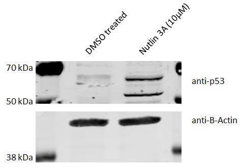
Application: Western BlotSample Tested: C166 mouse cell lineSpecies: MouseVerified Customer | Posted 09/14/201825µg of C166 whole cell lysate. Right lane Nutlin 3A treated sample which leads to induction of p53. Left lane DMSO treated control.

-
Application: Western BlotSample Tested:Species: RatVerified Customer | Posted 03/26/2014
-
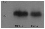
Application: Western BlotSample Tested: Whole Cell Lysate of MCF-7 and HeLa cells, Sample Amount: 10 ulSpecies: HumanVerified Customer | Posted 10/13/2010
There are no reviews that match your criteria.
Protocols
Find general support by application which include: protocols, troubleshooting, illustrated assays, videos and webinars.
- 7-Amino Actinomycin D (7-AAD) Cell Viability Flow Cytometry Protocol
- Antigen Retrieval Protocol (PIER)
- Antigen Retrieval for Frozen Sections Protocol
- Appropriate Fixation of IHC/ICC Samples
- Cellular Response to Hypoxia Protocols
- Chromogenic IHC Staining of Formalin-Fixed Paraffin-Embedded (FFPE) Tissue Protocol
- Chromogenic Immunohistochemistry Staining of Frozen Tissue
- ClariTSA™ Fluorophore Kits
- Detection & Visualization of Antibody Binding
- ELISA Sample Preparation & Collection Guide
- ELISA Troubleshooting Guide
- Extracellular Membrane Flow Cytometry Protocol
- Flow Cytometry Protocol for Cell Surface Markers
- Flow Cytometry Protocol for Staining Membrane Associated Proteins
- Flow Cytometry Staining Protocols
- Flow Cytometry Troubleshooting Guide
- Fluorescent IHC Staining of Frozen Tissue Protocol
- Graphic Protocol for Heat-induced Epitope Retrieval
- Graphic Protocol for the Preparation and Fluorescent IHC Staining of Frozen Tissue Sections
- Graphic Protocol for the Preparation and Fluorescent IHC Staining of Paraffin-embedded Tissue Sections
- Graphic Protocol for the Preparation of Gelatin-coated Slides for Histological Tissue Sections
- How to Run an R&D Systems DuoSet ELISA
- How to Run an R&D Systems Quantikine ELISA
- How to Run an R&D Systems Quantikine™ QuicKit™ ELISA
- ICC Cell Smear Protocol for Suspension Cells
- ICC Immunocytochemistry Protocol Videos
- ICC for Adherent Cells
- IHC Sample Preparation (Frozen sections vs Paraffin)
- Immunocytochemistry (ICC) Protocol
- Immunocytochemistry Troubleshooting
- Immunofluorescence of Organoids Embedded in Cultrex Basement Membrane Extract
- Immunofluorescent IHC Staining of Formalin-Fixed Paraffin-Embedded (FFPE) Tissue Protocol
- Immunohistochemistry (IHC) and Immunocytochemistry (ICC) Protocols
- Immunohistochemistry Frozen Troubleshooting
- Immunohistochemistry Paraffin Troubleshooting
- Immunoprecipitation Protocol
- Intracellular Flow Cytometry Protocol Using Alcohol (Methanol)
- Intracellular Flow Cytometry Protocol Using Detergents
- Intracellular Nuclear Staining Flow Cytometry Protocol Using Detergents
- Intracellular Staining Flow Cytometry Protocol Using Alcohol Permeabilization
- Intracellular Staining Flow Cytometry Protocol Using Detergents to Permeabilize Cells
- Preparing Samples for IHC/ICC Experiments
- Preventing Non-Specific Staining (Non-Specific Binding)
- Primary Antibody Selection & Optimization
- Propidium Iodide Cell Viability Flow Cytometry Protocol
- Protocol for Heat-Induced Epitope Retrieval (HIER)
- Protocol for Liperfluo
- Protocol for Making a 4% Formaldehyde Solution in PBS
- Protocol for VisUCyte™ HRP Polymer Detection Reagent
- Protocol for the Characterization of Human Th22 Cells
- Protocol for the Characterization of Human Th9 Cells
- Protocol for the Fluorescent ICC Staining of Cell Smears - Graphic
- Protocol for the Fluorescent ICC Staining of Cultured Cells on Coverslips - Graphic
- Protocol for the Preparation & Fixation of Cells on Coverslips
- Protocol for the Preparation and Chromogenic IHC Staining of Frozen Tissue Sections
- Protocol for the Preparation and Chromogenic IHC Staining of Frozen Tissue Sections - Graphic
- Protocol for the Preparation and Chromogenic IHC Staining of Paraffin-embedded Tissue Sections
- Protocol for the Preparation and Chromogenic IHC Staining of Paraffin-embedded Tissue Sections - Graphic
- Protocol for the Preparation and Fluorescent ICC Staining of Cells on Coverslips
- Protocol for the Preparation and Fluorescent ICC Staining of Non-adherent Cells
- Protocol for the Preparation and Fluorescent ICC Staining of Stem Cells on Coverslips
- Protocol for the Preparation and Fluorescent IHC Staining of Frozen Tissue Sections
- Protocol for the Preparation and Fluorescent IHC Staining of Paraffin-embedded Tissue Sections
- Protocol for the Preparation of Gelatin-coated Slides for Histological Tissue Sections
- Protocol for the Preparation of a Cell Smear for Non-adherent Cell ICC - Graphic
- Protocol: Annexin V and PI Staining by Flow Cytometry
- Protocol: Annexin V and PI Staining for Apoptosis by Flow Cytometry
- Quantikine HS ELISA Kit Assay Principle, Alkaline Phosphatase
- Quantikine HS ELISA Kit Principle, Streptavidin-HRP Polymer
- R&D Systems Quality Control Western Blot Protocol
- Sandwich ELISA (Colorimetric) – Biotin/Streptavidin Detection Protocol
- Sandwich ELISA (Colorimetric) – Direct Detection Protocol
- TUNEL and Active Caspase-3 Detection by IHC/ICC Protocol
- The Importance of IHC/ICC Controls
- Troubleshooting Guide: ELISA
- Troubleshooting Guide: Fluorokine Flow Cytometry Kits
- Troubleshooting Guide: Immunohistochemistry
- Troubleshooting Guide: Western Blot Figures
- Western Blot Conditions
- Western Blot Protocol
- Western Blot Protocol for Cell Lysates
- Western Blot Troubleshooting
- Western Blot Troubleshooting Guide
- View all Protocols, Troubleshooting, Illustrated assays and Webinars
Loading...
Associated Pathways


