RelA/NFkB p65 Antibody (112A1021) - BSA Free
Novus Biologicals | Catalog # NB100-56712
Key Product Details
Species Reactivity
Validated:
Cited:
Applications
Validated:
Cited:
Label
Antibody Source
Format
Product Specifications
Immunogen
Reactivity Notes
Clonality
Host
Isotype
Scientific Data Images for RelA/NFkB p65 Antibody (112A1021) - BSA Free
![Western Blot: RelA/NFkB p65 Antibody (112A1021)BSA Free [NB100-56712]](https://resources.rndsystems.com/images/products/RelA-NFkB-p65-Antibody-112A1021-Western-Blot-NB100-56712-img0013.jpg "Western Blot: RelA/NFkB p65 Antibody (112A1021)BSA Free [NB100-56712]")
Western Blot: RelA/NFkB p65 Antibody (112A1021)BSA Free [NB100-56712]
Western Blot: RelA/NFkB p65 Antibody (112A1021) [NB100-56712] - Analysis of P65 using p65 antibody at 2 ug/mL in 30 ug of A) Ramos, B) Daudi, C) HeLa and D) mouse NIH 3T3 cell lysate.![Simple Western: RelA/NFkB p65 Antibody (112A1021)BSA Free [NB100-56712]](https://resources.rndsystems.com/images/products/RelA-NFkB-p65-Antibody-112A1021-Simple-Western-NB100-56712-img0006.jpg "Simple Western: RelA/NFkB p65 Antibody (112A1021)BSA Free [NB100-56712]")
Simple Western: RelA/NFkB p65 Antibody (112A1021)BSA Free [NB100-56712]
Simple Western: RelA/NFkB p65 Antibody (112A1021) [NB100-56712] - Lane view shows a specific band for NFkB p65 in 1.0 mg/mL of HeLa lysate. This experiment was performed under reducing conditions using the 12-230 kDa separation system.![Western Blot: RelA/NFkB p65 Antibody (112A1021)BSA Free [NB100-56712]](https://resources.rndsystems.com/images/products/RelA-NFkB-p65-Antibody-112A1021-Western-Blot-NB100-56712-img0016.jpg "Western Blot: RelA/NFkB p65 Antibody (112A1021)BSA Free [NB100-56712]")
Western Blot: RelA/NFkB p65 Antibody (112A1021)BSA Free [NB100-56712]
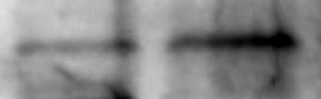
Western Blot: RelA/NFkB p65 Antibody (112A1021) [NB100-56712] - Lysates of HeLa human cervical epithelial carcinoma cell line and Daudi human Burkitt's lymphoma cell line. PVDF membrane was probed with 1 ug/mL mouse anti-RelA/NFkB p65 monoclonal (NB100-56712, Novus Biologicals), followed by 1:2000 dilution of the appropriate HRP-conjugated secondary antibody, donkey anti-mouse IgG (HAF018).![Immunohistochemistry-Paraffin: RelA/NFkB p65 Antibody (112A1021) - BSA Free [NB100-56712]](https://resources.rndsystems.com/images/products/RelA-NFkB-p65-Antibody-112A1021-Immunohistochemistry-Paraffin-NB100-56712-img0009.jpg "Immunohistochemistry-Paraffin: RelA/NFkB p65 Antibody (112A1021) - BSA Free [NB100-56712]")
Immunohistochemistry-Paraffin: RelA/NFkB p65 Antibody (112A1021) - BSA Free [NB100-56712]
Immunohistochemistry-Paraffin: RelA/NFkB p65 Antibody (112A1021) [NB100-56712] - Ovarian cystadenocarcinoma probed with p65 antibody at 5 ug/mL. Human tissue TMA was used for this test. Staining of formalin-fixed tissues is enhanced by boiling tissue sections in 10 mM sodium citrate buffer, pH 6.0 for 10-20 min followed by cooling at RT for 20 min.![Flow Cytometry: RelA/NFkB p65 Antibody (112A1021) - BSA Free [NB100-56712]](https://resources.rndsystems.com/images/products/RelA-NFkB-p65-Antibody-112A1021-Flow-Cytometry-NB100-56712-img0004.jpg "Flow Cytometry: RelA/NFkB p65 Antibody (112A1021) - BSA Free [NB100-56712]")
Flow Cytometry: RelA/NFkB p65 Antibody (112A1021) - BSA Free [NB100-56712]
Flow Cytometry: RelA/NFkB p65 Antibody (112A1021) [NB100-56712] - Intracellular staining of 293 HEK cells using 0.5 ug of p65 antibody. Green histogram represents the isotype control (p65) antibody. This NFkB p65 antibody was used for this test with an anti-mouse IgG PE conjugated secondary antibody.![Simple Western: RelA/NFkB p65 Antibody (112A1021)BSA Free [NB100-56712]](https://resources.rndsystems.com/images/products/RelA-NFkB-p65-Antibody-112A1021-Simple-Western-NB100-56712-img0017.jpg "Simple Western: RelA/NFkB p65 Antibody (112A1021)BSA Free [NB100-56712]")
Simple Western: RelA/NFkB p65 Antibody (112A1021)BSA Free [NB100-56712]
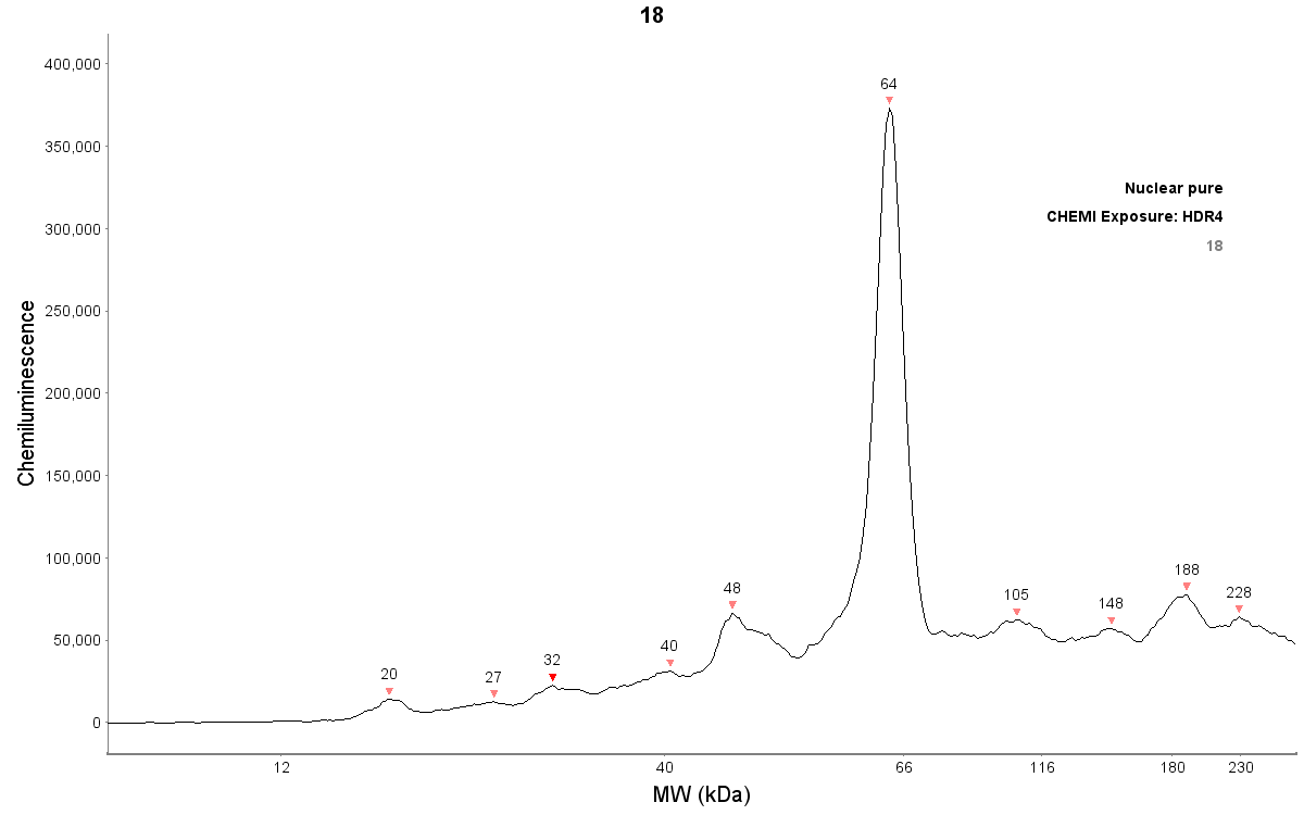
Simple Western: RelA/NFkB p65 Antibody (112A1021) [NB100-56712] - Detection of p65 by Simple Western (JESS) in chemiluminescence in nuclear extract of reconstructed human epidermis lysate (about 600 ug/mL protein concentration).Antibody at 1:20. Simple Western image submitted by a verified customer review.Applications for RelA/NFkB p65 Antibody (112A1021) - BSA Free
Flow (Intracellular)
Flow Cytometry
Immunohistochemistry
Immunohistochemistry-Paraffin
Immunoprecipitation
Simple Western
Western Blot
See Simple Western Antibody Database for Simple Western validation: Tested in HeLa lysate 1.0 mg/mL, separated by Size, antibody dilution of 1:25, apparent MW was 69 kDa. Separated by Size-Wes, Sally Sue/Peggy Sue. This antibody is CyTOF ready.
Reviewed Applications
Read 2 reviews rated 4.5 using NB100-56712 in the following applications:
Flow Cytometry Panel Builder
Bio-Techne Knows Flow Cytometry
Save time and reduce costly mistakes by quickly finding compatible reagents using the Panel Builder Tool.
Advanced Features
- Spectra Viewer - Custom analysis of spectra from multiple fluorochromes
- Spillover Popups - Visualize the spectra of individual fluorochromes
- Antigen Density Selector - Match fluorochrome brightness with antigen density

Formulation, Preparation, and Storage
Purification
Formulation
Format
Preservative
Concentration
Shipping
Stability & Storage
Background: RelA/NFkB p65
Long Name
Alternate Names
Gene Symbol
Additional RelA/NFkB p65 Products
Product Documents for RelA/NFkB p65 Antibody (112A1021) - BSA Free
Certificate of Analysis
To download a Certificate of Analysis, please enter a lot or batch number in the search box below.
Product Specific Notices for RelA/NFkB p65 Antibody (112A1021) - BSA Free
This product is for research use only and is not approved for use in humans or in clinical diagnosis. Primary Antibodies are guaranteed for 1 year from date of receipt.
Related Research Areas
Citations for RelA/NFkB p65 Antibody (112A1021) - BSA Free
 Powered by Bioz
Customer Reviews for RelA/NFkB p65 Antibody (112A1021) - BSA Free (2)
Have you used RelA/NFkB p65 Antibody (112A1021) - BSA Free?
Submit a review and receive an Amazon gift card!
$25/€18/£15/$25CAN/¥2500 Yen for a review with an image
$10/€7/£6/$10CAN/¥1110 Yen for a review without an image
Submit a review
Customer Images
-
Application: ImmunoprecipitationSample Tested: A549 human alveolar adenocarcinoma cell lineSpecies: HumanVerified Customer | Posted 02/07/2020Acetylated Rela following treatment with MS275 Class 1 HDAC inhibitor500 ug of whole cell lysates from cells either left untreated Left or treated with 10 uM MS275 selective Class 1 HDAC inhibitor, Right were immunoprecipitated using 5 ug of RelA/NFkB p65 Antibody 112A1021 conjugated to 50 uL of protein G Dynabeads. Antibody was crosslinked to beads to reduce background using BS3 reagent. following precipitation/washing bound proteins were eluted and ran on a 4-15% gel. Total acetylation was visualized using rabbit anti-acetylated-lysine. bands pictured visualized at ~65 kDA.

-
Application: Simple WesternSample Tested: Human foreskin, adult ski, engineered human skin, keratinocytes, HaCaT cellsSpecies: HumanVerified Customer | Posted 06/17/2019Detection of p65 by Simple Western (JESS) in chemiluminescence in nuclear extract of reconstructed human epidermis lysate (about 600 ug/mL protein concentration). Antibody dilution: 1/20JESS system
There are no reviews that match your criteria.
Protocols
Find general support by application which include: protocols, troubleshooting, illustrated assays, videos and webinars.
- 7-Amino Actinomycin D (7-AAD) Cell Viability Flow Cytometry Protocol
- Antigen Retrieval Protocol (PIER)
- Antigen Retrieval for Frozen Sections Protocol
- Appropriate Fixation of IHC/ICC Samples
- Cellular Response to Hypoxia Protocols
- Chromogenic IHC Staining of Formalin-Fixed Paraffin-Embedded (FFPE) Tissue Protocol
- Chromogenic Immunohistochemistry Staining of Frozen Tissue
- ClariTSA™ Fluorophore Kits
- Detection & Visualization of Antibody Binding
- Extracellular Membrane Flow Cytometry Protocol
- Flow Cytometry Protocol for Cell Surface Markers
- Flow Cytometry Protocol for Staining Membrane Associated Proteins
- Flow Cytometry Staining Protocols
- Flow Cytometry Troubleshooting Guide
- Fluorescent IHC Staining of Frozen Tissue Protocol
- Graphic Protocol for Heat-induced Epitope Retrieval
- Graphic Protocol for the Preparation and Fluorescent IHC Staining of Frozen Tissue Sections
- Graphic Protocol for the Preparation and Fluorescent IHC Staining of Paraffin-embedded Tissue Sections
- Graphic Protocol for the Preparation of Gelatin-coated Slides for Histological Tissue Sections
- IHC Sample Preparation (Frozen sections vs Paraffin)
- Immunofluorescent IHC Staining of Formalin-Fixed Paraffin-Embedded (FFPE) Tissue Protocol
- Immunohistochemistry (IHC) and Immunocytochemistry (ICC) Protocols
- Immunohistochemistry Frozen Troubleshooting
- Immunohistochemistry Paraffin Troubleshooting
- Immunoprecipitation Protocol
- Intracellular Flow Cytometry Protocol Using Alcohol (Methanol)
- Intracellular Flow Cytometry Protocol Using Detergents
- Intracellular Nuclear Staining Flow Cytometry Protocol Using Detergents
- Intracellular Staining Flow Cytometry Protocol Using Alcohol Permeabilization
- Intracellular Staining Flow Cytometry Protocol Using Detergents to Permeabilize Cells
- Preparing Samples for IHC/ICC Experiments
- Preventing Non-Specific Staining (Non-Specific Binding)
- Primary Antibody Selection & Optimization
- Propidium Iodide Cell Viability Flow Cytometry Protocol
- Protocol for Heat-Induced Epitope Retrieval (HIER)
- Protocol for Liperfluo
- Protocol for Making a 4% Formaldehyde Solution in PBS
- Protocol for VisUCyte™ HRP Polymer Detection Reagent
- Protocol for the Characterization of Human Th22 Cells
- Protocol for the Characterization of Human Th9 Cells
- Protocol for the Preparation & Fixation of Cells on Coverslips
- Protocol for the Preparation and Chromogenic IHC Staining of Frozen Tissue Sections
- Protocol for the Preparation and Chromogenic IHC Staining of Frozen Tissue Sections - Graphic
- Protocol for the Preparation and Chromogenic IHC Staining of Paraffin-embedded Tissue Sections
- Protocol for the Preparation and Chromogenic IHC Staining of Paraffin-embedded Tissue Sections - Graphic
- Protocol for the Preparation and Fluorescent IHC Staining of Frozen Tissue Sections
- Protocol for the Preparation and Fluorescent IHC Staining of Paraffin-embedded Tissue Sections
- Protocol for the Preparation of Gelatin-coated Slides for Histological Tissue Sections
- Protocol: Annexin V and PI Staining by Flow Cytometry
- Protocol: Annexin V and PI Staining for Apoptosis by Flow Cytometry
- R&D Systems Quality Control Western Blot Protocol
- TUNEL and Active Caspase-3 Detection by IHC/ICC Protocol
- The Importance of IHC/ICC Controls
- Troubleshooting Guide: Fluorokine Flow Cytometry Kits
- Troubleshooting Guide: Immunohistochemistry
- Troubleshooting Guide: Western Blot Figures
- Western Blot Conditions
- Western Blot Protocol
- Western Blot Protocol for Cell Lysates
- Western Blot Troubleshooting
- Western Blot Troubleshooting Guide
- View all Protocols, Troubleshooting, Illustrated assays and Webinars
FAQs for RelA/NFkB p65 Antibody (112A1021) - BSA Free
-
Q: Are the following monoclonals purified from ascites or from tissue culture supernatant? NB100-56541, NB100-56705, NB600-1298, NB100-56534, NB100-56505, NBP2-24873, NB600-1107, NBP2-24917, NB100-56524, NB100-56712
A: These antibodies are all purified from tissue culture supernatant.
-
Q: We are interested in your item, NB100-56712 and I found this description, Predicted to react with Cow, Gorilla, New World Monkey and Pig, on your datasheet. I want to blast their identity(100%) by myself but I could not find RelA/NFkB p65 protein sequence for Gorilla. Would you please kindly provide us the gene ID or protein ID for Gorilla RelA?
A:
The gorilla RelA protein that was blasted against the immunogen for our NB100-56712 can be found on the following link: Ultiprot
-
Q: Are the following monoclonals purified from ascites or from tissue culture supernatant? NB100-56541, NB100-56705, NB600-1298, NB100-56534, NB100-56505, NBP2-24873, NB600-1107, NBP2-24917, NB100-56524, NB100-56712
A: These antibodies are all purified from tissue culture supernatant.
-
Q: We are interested in your item, NB100-56712 and I found this description, Predicted to react with Cow, Gorilla, New World Monkey and Pig, on your datasheet. I want to blast their identity(100%) by myself but I could not find RelA/NFkB p65 protein sequence for Gorilla. Would you please kindly provide us the gene ID or protein ID for Gorilla RelA?
A:
The gorilla RelA protein that was blasted against the immunogen for our NB100-56712 can be found on the following link: Ultiprot


