RelA/NFkB p65 Antibody (112A1021) [Biotin]
Novus Biologicals | Catalog # NB100-56055
Key Product Details
Species Reactivity
Validated:
Human, Mouse, Rat
Cited:
Rat
Applications
Validated:
Immunohistochemistry, Immunohistochemistry-Paraffin, Western Blot, Flow Cytometry, Immunoprecipitation
Cited:
Immunohistochemistry-Paraffin
Label
Biotin
Antibody Source
Monoclonal Mouse IgG1 kappa Clone # 112A1021
Loading...
Product Specifications
Immunogen
This NfkB p65 antibody was raised against a synthetic peptide corresponding to amino acids 526-539 of human NF-kB (p65) protein.
Reactivity Notes
Predicted to react with Bovine, Gorilla, New World Monkey and Porcine.
Clonality
Monoclonal
Host
Mouse
Isotype
IgG1 kappa
Scientific Data Images for RelA/NFkB p65 Antibody (112A1021) [Biotin]
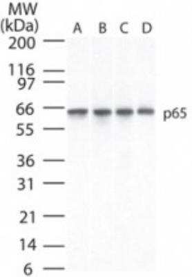
Western Blot: RelA/NFkB p65 Antibody (112A1021) [Biotin] [NB100-56055] - Analysis ofRelA/NFkB p65 Antibody (112A1021) [Biotin]using NB100-56055 at 2 ug/ml in 30 ug of A) Ramos, B) Daudi, C) HeLa and D) mouse NIH 3T3 cell lysate.
Applications for RelA/NFkB p65 Antibody (112A1021) [Biotin]
Application
Recommended Usage
Flow Cytometry
1ul/1 million cells
Immunohistochemistry-Paraffin
1:10-1:500
Immunoprecipitation
1:10-1:500
Western Blot
1:100-1:2000
Application Notes
Optimal dilution of this antibody should be experimentally determined.
Reviewed Applications
Read 1 review rated 5 using NB100-56055 in the following applications:
Flow Cytometry Panel Builder
Bio-Techne Knows Flow Cytometry
Save time and reduce costly mistakes by quickly finding compatible reagents using the Panel Builder Tool.
Advanced Features
- Spectra Viewer - Custom analysis of spectra from multiple fluorochromes
- Spillover Popups - Visualize the spectra of individual fluorochromes
- Antigen Density Selector - Match fluorochrome brightness with antigen density

Formulation, Preparation, and Storage
Purification
Protein G purified
Formulation
PBS
Preservative
0.05% Sodium Azide
Concentration
Please see the vial label for concentration. If unlisted please contact technical services.
Shipping
The product is shipped with polar packs. Upon receipt, store it immediately at the temperature recommended below.
Stability & Storage
Store at 4C. Do not freeze.
Background: RelA/NFkB p65
Long Name
v-rel Reticuloendotheliosis Viral Oncogene Homolog A
Alternate Names
NFkB p65, NFKB3, p65RelA
Gene Symbol
RELA
Additional RelA/NFkB p65 Products
Product Documents for RelA/NFkB p65 Antibody (112A1021) [Biotin]
Certificate of Analysis
To download a Certificate of Analysis, please enter a lot or batch number in the search box below.
Product Specific Notices for RelA/NFkB p65 Antibody (112A1021) [Biotin]
This product is for research use only and is not approved for use in humans or in clinical diagnosis. Primary Antibodies are guaranteed for 1 year from date of receipt.
Related Research Areas
Citations for RelA/NFkB p65 Antibody (112A1021) [Biotin]
 Powered by Bioz
Customer Reviews for RelA/NFkB p65 Antibody (112A1021) [Biotin] (1)
5 out of 5
1 Customer Rating
Have you used RelA/NFkB p65 Antibody (112A1021) [Biotin]?
Submit a review and receive an Amazon gift card!
$25/€18/£15/$25CAN/¥2500 Yen for a review with an image
$10/€7/£6/$10CAN/¥1110 Yen for a review without an image
Submit a review
Customer Images
![RelA/NFkB p65 Antibody (112A1021) [Biotin] NB100-56055](https://resources.rndsystems.com/images/reviews/23567_1_0_0_0_0_0_0_0_0_0.jpg)
Showing
1
-
1 of
1 review
Showing All
Filter By:
-
Application: Western BlotSample Tested: NIH 3T3 cell lysateSpecies: MouseVerified Customer | Posted 09/14/2021Western blot in NIH 3T3 cell lysate. RelA/NFkB p65 Antibody.
![RelA/NFkB p65 Antibody (112A1021) [Biotin] NB100-56055](data:image/png;base64,R0lGODlhAQABAAD/ACwAAAAAAQABAAACADs=)
There are no reviews that match your criteria.
Protocols
Find general support by application which include: protocols, troubleshooting, illustrated assays, videos and webinars.
- 7-Amino Actinomycin D (7-AAD) Cell Viability Flow Cytometry Protocol
- Antigen Retrieval Protocol (PIER)
- Antigen Retrieval for Frozen Sections Protocol
- Appropriate Fixation of IHC/ICC Samples
- Cellular Response to Hypoxia Protocols
- Chromogenic IHC Staining of Formalin-Fixed Paraffin-Embedded (FFPE) Tissue Protocol
- Chromogenic Immunohistochemistry Staining of Frozen Tissue
- ClariTSA™ Fluorophore Kits
- Detection & Visualization of Antibody Binding
- Extracellular Membrane Flow Cytometry Protocol
- Flow Cytometry Protocol for Cell Surface Markers
- Flow Cytometry Protocol for Staining Membrane Associated Proteins
- Flow Cytometry Staining Protocols
- Flow Cytometry Troubleshooting Guide
- Fluorescent IHC Staining of Frozen Tissue Protocol
- Graphic Protocol for Heat-induced Epitope Retrieval
- Graphic Protocol for the Preparation and Fluorescent IHC Staining of Frozen Tissue Sections
- Graphic Protocol for the Preparation and Fluorescent IHC Staining of Paraffin-embedded Tissue Sections
- Graphic Protocol for the Preparation of Gelatin-coated Slides for Histological Tissue Sections
- IHC Sample Preparation (Frozen sections vs Paraffin)
- Immunofluorescent IHC Staining of Formalin-Fixed Paraffin-Embedded (FFPE) Tissue Protocol
- Immunohistochemistry (IHC) and Immunocytochemistry (ICC) Protocols
- Immunohistochemistry Frozen Troubleshooting
- Immunohistochemistry Paraffin Troubleshooting
- Immunoprecipitation Protocol
- Intracellular Flow Cytometry Protocol Using Alcohol (Methanol)
- Intracellular Flow Cytometry Protocol Using Detergents
- Intracellular Nuclear Staining Flow Cytometry Protocol Using Detergents
- Intracellular Staining Flow Cytometry Protocol Using Alcohol Permeabilization
- Intracellular Staining Flow Cytometry Protocol Using Detergents to Permeabilize Cells
- Preparing Samples for IHC/ICC Experiments
- Preventing Non-Specific Staining (Non-Specific Binding)
- Primary Antibody Selection & Optimization
- Propidium Iodide Cell Viability Flow Cytometry Protocol
- Protocol for Heat-Induced Epitope Retrieval (HIER)
- Protocol for Liperfluo
- Protocol for Making a 4% Formaldehyde Solution in PBS
- Protocol for VisUCyte™ HRP Polymer Detection Reagent
- Protocol for the Characterization of Human Th22 Cells
- Protocol for the Characterization of Human Th9 Cells
- Protocol for the Preparation & Fixation of Cells on Coverslips
- Protocol for the Preparation and Chromogenic IHC Staining of Frozen Tissue Sections
- Protocol for the Preparation and Chromogenic IHC Staining of Frozen Tissue Sections - Graphic
- Protocol for the Preparation and Chromogenic IHC Staining of Paraffin-embedded Tissue Sections
- Protocol for the Preparation and Chromogenic IHC Staining of Paraffin-embedded Tissue Sections - Graphic
- Protocol for the Preparation and Fluorescent IHC Staining of Frozen Tissue Sections
- Protocol for the Preparation and Fluorescent IHC Staining of Paraffin-embedded Tissue Sections
- Protocol for the Preparation of Gelatin-coated Slides for Histological Tissue Sections
- Protocol: Annexin V and PI Staining by Flow Cytometry
- Protocol: Annexin V and PI Staining for Apoptosis by Flow Cytometry
- R&D Systems Quality Control Western Blot Protocol
- TUNEL and Active Caspase-3 Detection by IHC/ICC Protocol
- The Importance of IHC/ICC Controls
- Troubleshooting Guide: Fluorokine Flow Cytometry Kits
- Troubleshooting Guide: Immunohistochemistry
- Troubleshooting Guide: Western Blot Figures
- Western Blot Conditions
- Western Blot Protocol
- Western Blot Protocol for Cell Lysates
- Western Blot Troubleshooting
- Western Blot Troubleshooting Guide
- View all Protocols, Troubleshooting, Illustrated assays and Webinars


