RelA/NFkB p65 Antibody - BSA Free
Novus Biologicals | Catalog # NB100-2176
![Western Blot: RelA/NFkB p65 Antibody [NB100-2176]](https://resources.rndsystems.com/images/products/RelA-NFkB-p65-Antibody-Western-Blot-NB100-2176-img0006.jpg "Western Blot: RelA/NFkB p65 Antibody [NB100-2176]")
Key Product Details
Validated by
Species Reactivity
Validated:
Cited:
Applications
Validated:
Cited:
Label
Antibody Source
Format
Product Specifications
Immunogen
Localization
Specificity
Clonality
Host
Isotype
Theoretical MW
Disclaimer note: The observed molecular weight of the protein may vary from the listed predicted molecular weight due to post translational modifications, post translation cleavages, relative charges, and other experimental factors.
Scientific Data Images for RelA/NFkB p65 Antibody - BSA Free
![Immunohistochemistry-Paraffin: RelA/NFkB p65 Antibody [NB100-2176]](https://resources.rndsystems.com/images/products/RelA-NFkB-p65-Antibody-Immunohistochemistry-Paraffin-NB100-2176-img0004.jpg "Immunohistochemistry-Paraffin: RelA/NFkB p65 Antibody [NB100-2176]")
Immunohistochemistry-Paraffin: RelA/NFkB p65 Antibody [NB100-2176]
Immunohistochemistry-Paraffin: RelA/NFkB p65 Antibody [NB100-2176] - Staining of formalin-fixed, paraffin-embedded human DLBCL showing nuclear expression of NFkB p65 in the tumor cells.![Western Blot: RelA/NFkB p65 Antibody [NB100-2176]](https://resources.rndsystems.com/images/products/RelA-NFkB-p65-Antibody-Western-Blot-NB100-2176-img0005.jpg "Western Blot: RelA/NFkB p65 Antibody [NB100-2176]")
Western Blot: RelA/NFkB p65 Antibody [NB100-2176]
Western Blot: RelA/NFkB p65 Antibody [NB100-2176] - Analysis of NFkB p65 expression in A431 (A) and HeLa (B) whole cell lysates.
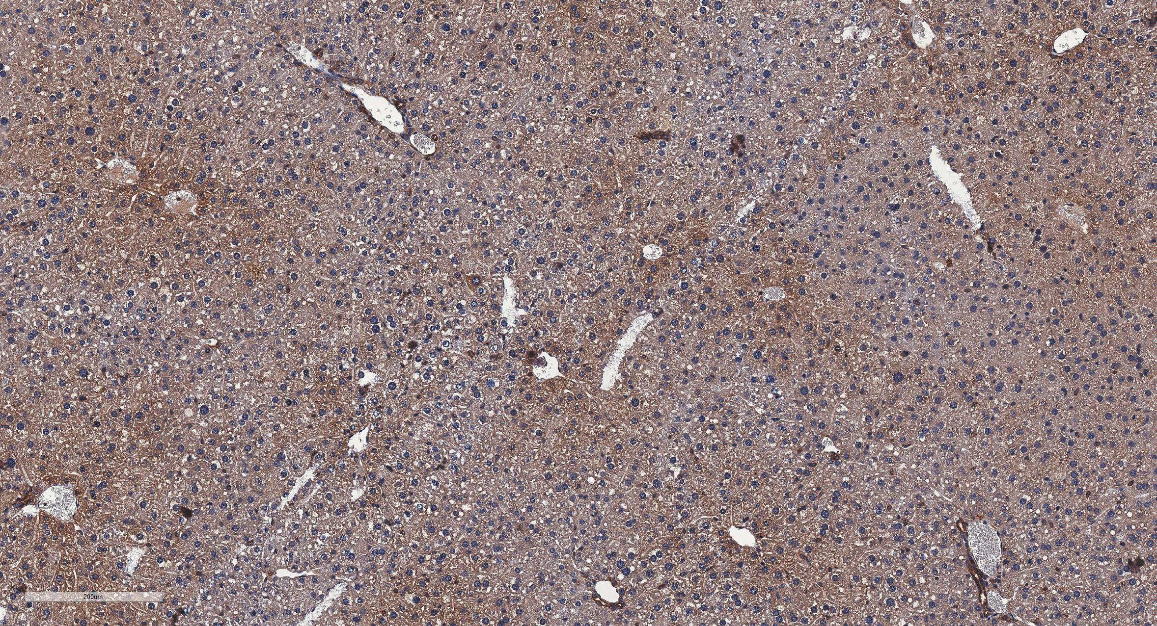
Immunohistochemistry-Paraffin: Rabbit Polyclonal RelA/NFkB p65 Antibody [NB100-2176]
Immunohistochemistry-Paraffin: Rabbit Polyclonal RelA/NFkB p65 Antibody [NB100-2176] - RelA/NFkB p65 IHC (brown) in the mice liver tissue; bars, 200 μm. Image from a verified customer review.
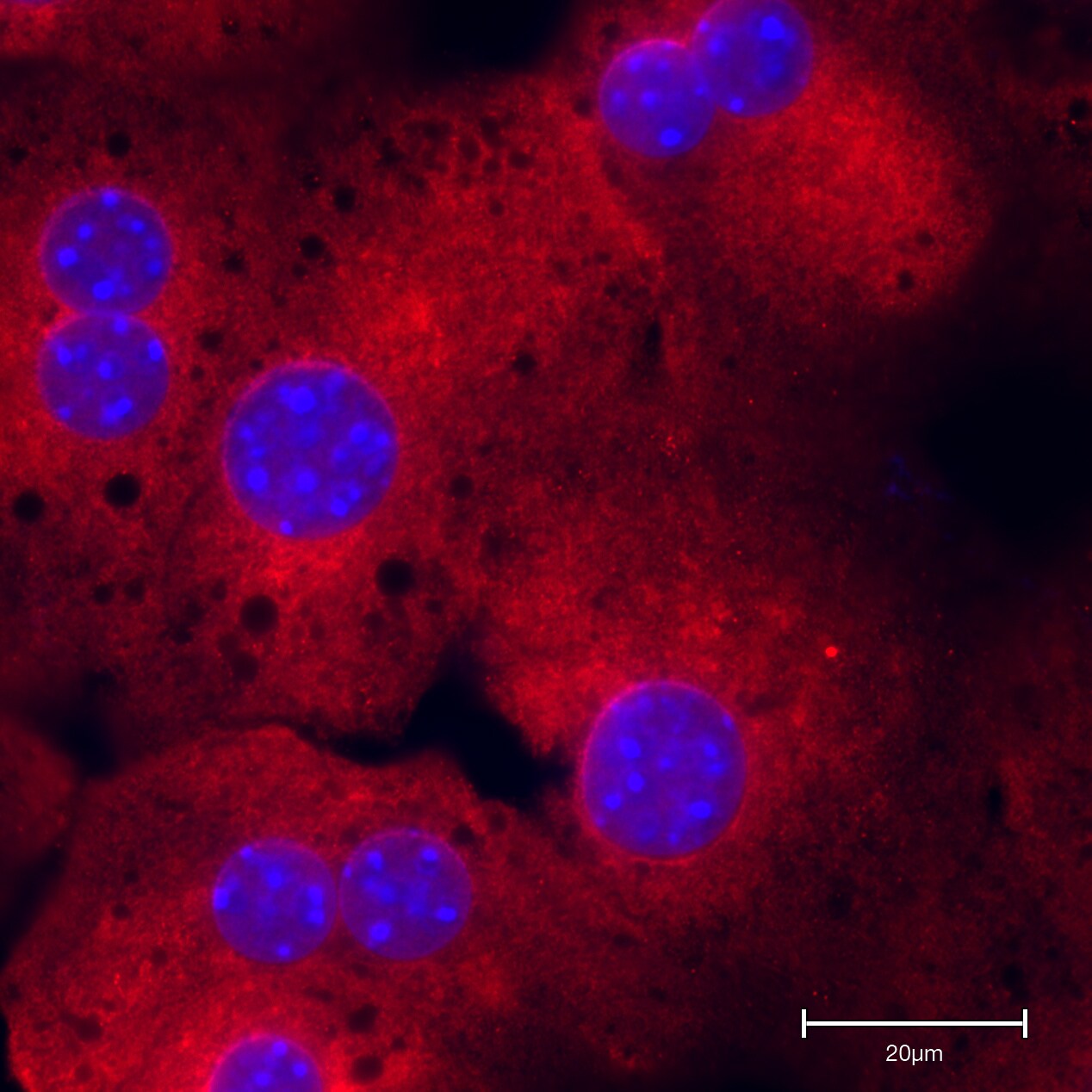
Immunocytochemistry/Immunofluorescence: Rabbit Polyclonal RelA/NFkB p65 Antibody [NB100-2176]
Immunocytochemistry/Immunofluorescence: Rabbit Polyclonal RelA/NFkB p65 Antibody [NB100-2176] - Mice hepatocytes stained for RelA/NFkB p65. Image from a verified customer review.Applications for RelA/NFkB p65 Antibody - BSA Free
Immunohistochemistry
Immunohistochemistry-Paraffin
Immunoprecipitation
Western Blot
Reviewed Applications
Read 3 reviews rated 5 using NB100-2176 in the following applications:
Formulation, Preparation, and Storage
Purification
Formulation
Format
Preservative
Concentration
Shipping
Stability & Storage
Background: RelA/NFkB p65
Long Name
Alternate Names
Gene Symbol
UniProt
Additional RelA/NFkB p65 Products
Product Documents for RelA/NFkB p65 Antibody - BSA Free
Certificate of Analysis
To download a Certificate of Analysis, please enter a lot or batch number in the search box below.
Product Specific Notices for RelA/NFkB p65 Antibody - BSA Free
This product is for research use only and is not approved for use in humans or in clinical diagnosis. Primary Antibodies are guaranteed for 1 year from date of receipt.
Related Research Areas
Citations for RelA/NFkB p65 Antibody - BSA Free
 Powered by Bioz
Customer Reviews for RelA/NFkB p65 Antibody - BSA Free (3)
Have you used RelA/NFkB p65 Antibody - BSA Free?
Submit a review and receive an Amazon gift card!
$25/€18/£15/$25CAN/¥2500 Yen for a review with an image
$10/€7/£6/$10CAN/¥1110 Yen for a review without an image
Submit a review
Customer Images
-
Application: ImmunofluorescenceSample Tested: Primary mouse hepatocytesSpecies: MouseVerified Customer | Posted 11/19/2024Mice hepatocytes stained for the NB100-2176

-
Application: Immunohistochemistry-ParaffinSample Tested: Prostate tissueSpecies: MouseVerified Customer | Posted 11/07/2024NFkB p65 IHC (brown) in the mice liver tissue; bars, 200 μm.
-
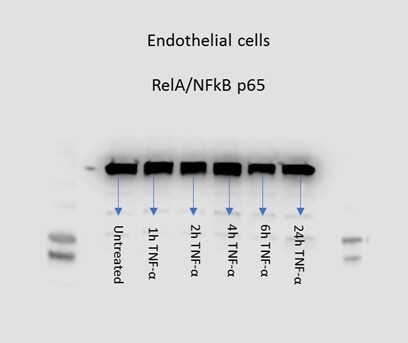
Application: Western BlotSample Tested: Endothelial and Whole cell lysatesSpecies: HumanVerified Customer | Posted 08/31/2017Human glomerular endothelial cell lysates, time-response treatment with 10ng/ml TNF-alpha.
There are no reviews that match your criteria.
Protocols
Find general support by application which include: protocols, troubleshooting, illustrated assays, videos and webinars.
- Antigen Retrieval Protocol (PIER)
- Antigen Retrieval for Frozen Sections Protocol
- Appropriate Fixation of IHC/ICC Samples
- Cellular Response to Hypoxia Protocols
- ChIP Protocol Video
- Chromatin Immunoprecipitation (ChIP) Protocol
- Chromatin Immunoprecipitation Protocol
- Chromogenic IHC Staining of Formalin-Fixed Paraffin-Embedded (FFPE) Tissue Protocol
- Chromogenic Immunohistochemistry Staining of Frozen Tissue
- ClariTSA™ Fluorophore Kits
- Detection & Visualization of Antibody Binding
- Fluorescent IHC Staining of Frozen Tissue Protocol
- Graphic Protocol for Heat-induced Epitope Retrieval
- Graphic Protocol for the Preparation and Fluorescent IHC Staining of Frozen Tissue Sections
- Graphic Protocol for the Preparation and Fluorescent IHC Staining of Paraffin-embedded Tissue Sections
- Graphic Protocol for the Preparation of Gelatin-coated Slides for Histological Tissue Sections
- ICC Cell Smear Protocol for Suspension Cells
- ICC Immunocytochemistry Protocol Videos
- ICC for Adherent Cells
- IHC Sample Preparation (Frozen sections vs Paraffin)
- Immunocytochemistry (ICC) Protocol
- Immunocytochemistry Troubleshooting
- Immunofluorescence of Organoids Embedded in Cultrex Basement Membrane Extract
- Immunofluorescent IHC Staining of Formalin-Fixed Paraffin-Embedded (FFPE) Tissue Protocol
- Immunohistochemistry (IHC) and Immunocytochemistry (ICC) Protocols
- Immunohistochemistry Frozen Troubleshooting
- Immunohistochemistry Paraffin Troubleshooting
- Immunoprecipitation Protocol
- Preparing Samples for IHC/ICC Experiments
- Preventing Non-Specific Staining (Non-Specific Binding)
- Primary Antibody Selection & Optimization
- Protocol for Heat-Induced Epitope Retrieval (HIER)
- Protocol for Making a 4% Formaldehyde Solution in PBS
- Protocol for VisUCyte™ HRP Polymer Detection Reagent
- Protocol for the Fluorescent ICC Staining of Cell Smears - Graphic
- Protocol for the Fluorescent ICC Staining of Cultured Cells on Coverslips - Graphic
- Protocol for the Preparation & Fixation of Cells on Coverslips
- Protocol for the Preparation and Chromogenic IHC Staining of Frozen Tissue Sections
- Protocol for the Preparation and Chromogenic IHC Staining of Frozen Tissue Sections - Graphic
- Protocol for the Preparation and Chromogenic IHC Staining of Paraffin-embedded Tissue Sections
- Protocol for the Preparation and Chromogenic IHC Staining of Paraffin-embedded Tissue Sections - Graphic
- Protocol for the Preparation and Fluorescent ICC Staining of Cells on Coverslips
- Protocol for the Preparation and Fluorescent ICC Staining of Non-adherent Cells
- Protocol for the Preparation and Fluorescent ICC Staining of Stem Cells on Coverslips
- Protocol for the Preparation and Fluorescent IHC Staining of Frozen Tissue Sections
- Protocol for the Preparation and Fluorescent IHC Staining of Paraffin-embedded Tissue Sections
- Protocol for the Preparation of Gelatin-coated Slides for Histological Tissue Sections
- Protocol for the Preparation of a Cell Smear for Non-adherent Cell ICC - Graphic
- R&D Systems Quality Control Western Blot Protocol
- TUNEL and Active Caspase-3 Detection by IHC/ICC Protocol
- The Importance of IHC/ICC Controls
- Troubleshooting Guide: Immunohistochemistry
- Troubleshooting Guide: Western Blot Figures
- Western Blot Conditions
- Western Blot Protocol
- Western Blot Protocol for Cell Lysates
- Western Blot Troubleshooting
- Western Blot Troubleshooting Guide
- View all Protocols, Troubleshooting, Illustrated assays and Webinars
FAQs for RelA/NFkB p65 Antibody - BSA Free
-
Q: I performed Western blot with NFkB p65 Antibody (NB100-2176) using brain and liver tissues extracts. My question: is it possible that NF-kB-Ikb complex could be detected by this antibody? Because I found 2 bands in the Western blot results, one is located at 65 and the other above 100 kD.
A: It possible that NB100-2176 could detect the entire NFkB complex in addition to just the p65 protein. If your samples were not completely reduced and denatured, they could have remained bound in your gel. The band above 100 kDa could also be an aggregate of NFkB. You may consider probing that same membrane for a different NFkB complex protein that you would expect to remain bound under these conditions. A negative signal in this case would tell you that it's an aggregate of NFkB, or maybe that it's a non-specific band. Did you run a negative control?


