STAT5A Antibody (OTI9F7)
Novus Biologicals | Catalog # NBP2-00622
Key Product Details
Species Reactivity
Validated:
Human, Mouse, Rat, Canine, Monkey, Primate
Cited:
Human
Applications
Validated:
Immunohistochemistry, Immunohistochemistry-Paraffin, Western Blot, Flow Cytometry, Immunocytochemistry/ Immunofluorescence
Cited:
IF/IHC
Label
Unconjugated
Antibody Source
Monoclonal Mouse IgG1 Clone # OTI9F7
Loading...
Product Specifications
Immunogen
Full length human recombinant protein of human STAT5A (NP_003143) produced in HEK293T cell.
Reactivity Notes
Please note that this antibody is reactive to Mouse and derived from the same host, Mouse. Mouse-On-Mouse blocking reagent may be needed for IHC and ICC experiments to reduce high background signal. You can find these reagents under catalog numbers PK-2200-NB and MP-2400-NB. Please contact Technical Support if you have any questions.
Clonality
Monoclonal
Host
Mouse
Isotype
IgG1
Theoretical MW
90.5 kDa.
Disclaimer note: The observed molecular weight of the protein may vary from the listed predicted molecular weight due to post translational modifications, post translation cleavages, relative charges, and other experimental factors.
Disclaimer note: The observed molecular weight of the protein may vary from the listed predicted molecular weight due to post translational modifications, post translation cleavages, relative charges, and other experimental factors.
Scientific Data Images for STAT5A Antibody (OTI9F7)
![Western Blot: STAT5A Antibody (OTI9F7) [NBP2-00622]](https://resources.rndsystems.com/images/products/STAT5A-Antibody-OTI9F7-Western-Blot-NBP2-00622-img0011.jpg "Western Blot: STAT5A Antibody (OTI9F7) [NBP2-00622]")
Western Blot: STAT5A Antibody (OTI9F7) [NBP2-00622]
Western Blot: STAT5A Antibody (OTI9F7) [NBP2-00622] - Analysis of extracts (35ug) from 9 different cell lines by using anti-STAT5A monoclonal antibody.![Immunohistochemistry-Paraffin: STAT5A Antibody (OTI9F7) [NBP2-00622]](https://resources.rndsystems.com/images/products/STAT5A-Antibody-OTI9F7-Immunohistochemistry-Paraffin-NBP2-00622-img0010.jpg "Immunohistochemistry-Paraffin: STAT5A Antibody (OTI9F7) [NBP2-00622]")
Immunohistochemistry-Paraffin: STAT5A Antibody (OTI9F7) [NBP2-00622]
Immunohistochemistry-Paraffin: STAT5A Antibody (OTI9F7) [NBP2-00622] - Staining of paraffin-embedded Human prostate tissue using anti-STAT5A mouse monoclonal antibody.![Flow Cytometry: STAT5A Antibody (OTI9F7) [NBP2-00622]](https://resources.rndsystems.com/images/products/STAT5A-Antibody-OTI9F7-Flow-Cytometry-NBP2-00622-img0002.jpg "Flow Cytometry: STAT5A Antibody (OTI9F7) [NBP2-00622]")
Flow Cytometry: STAT5A Antibody (OTI9F7) [NBP2-00622]
Flow Cytometry: STAT5A Antibody (OTI9F7) [NBP2-00622] - Analysis of Jurkat cells, using anti-STAT5A antibody, (Red), compared to a nonspecific negative control antibody (Blue).![Western Blot: STAT5A Antibody (OTI9F7) [NBP2-00622]](https://resources.rndsystems.com/images/products/STAT5A-Antibody-OTI9F7-Western-Blot-NBP2-00622-img0003.jpg "Western Blot: STAT5A Antibody (OTI9F7) [NBP2-00622]")
Western Blot: STAT5A Antibody (OTI9F7) [NBP2-00622]
Western Blot: STAT5A Antibody (OTI9F7) [NBP2-00622] - HEK293T cells were transfected with the pCMV6-ENTRY control (Left lane) or pCMV6-ENTRY STAT5A (Right lane) cDNA for 48 hrs and lysed. Equivalent amounts of cell lysates (5 ug per lane) were separated by SDS-PAGE and immunoblotted with anti-STAT5A.![Immunohistochemistry-Paraffin: STAT5A Antibody (OTI9F7) [NBP2-00622]](https://resources.rndsystems.com/images/products/STAT5A-Antibody-OTI9F7-Immunohistochemistry-Paraffin-NBP2-00622-img0004.jpg "Immunohistochemistry-Paraffin: STAT5A Antibody (OTI9F7) [NBP2-00622]")
Immunohistochemistry-Paraffin: STAT5A Antibody (OTI9F7) [NBP2-00622]
Immunohistochemistry-Paraffin: STAT5A Antibody (OTI9F7) [NBP2-00622] - Staining of paraffin-embedded Carcinoma of Human bladder tissue using anti-STAT5A mouse monoclonal antibody.![Immunohistochemistry-Paraffin: STAT5A Antibody (OTI9F7) [NBP2-00622]](https://resources.rndsystems.com/images/products/STAT5A-Antibody-OTI9F7-Immunohistochemistry-Paraffin-NBP2-00622-img0005.jpg "Immunohistochemistry-Paraffin: STAT5A Antibody (OTI9F7) [NBP2-00622]")
Immunohistochemistry-Paraffin: STAT5A Antibody (OTI9F7) [NBP2-00622]
Immunohistochemistry-Paraffin: STAT5A Antibody (OTI9F7) [NBP2-00622] - Staining of paraffin-embedded Carcinoma of Human pancreas tissue using anti-STAT5A mouse monoclonal antibody.![Immunohistochemistry-Paraffin: STAT5A Antibody (OTI9F7) [NBP2-00622]](https://resources.rndsystems.com/images/products/STAT5A-Antibody-OTI9F7-Immunohistochemistry-Paraffin-NBP2-00622-img0006.jpg "Immunohistochemistry-Paraffin: STAT5A Antibody (OTI9F7) [NBP2-00622]")
Immunohistochemistry-Paraffin: STAT5A Antibody (OTI9F7) [NBP2-00622]
Immunohistochemistry-Paraffin: STAT5A Antibody (OTI9F7) [NBP2-00622] - Staining of paraffin-embedded Carcinoma of Human prostate tissue using anti-STAT5A mouse monoclonal antibody.![Immunohistochemistry-Paraffin: STAT5A Antibody (OTI9F7) [NBP2-00622]](https://resources.rndsystems.com/images/products/STAT5A-Antibody-OTI9F7-Immunohistochemistry-Paraffin-NBP2-00622-img0007.jpg "Immunohistochemistry-Paraffin: STAT5A Antibody (OTI9F7) [NBP2-00622]")
Immunohistochemistry-Paraffin: STAT5A Antibody (OTI9F7) [NBP2-00622]
Immunohistochemistry-Paraffin: STAT5A Antibody (OTI9F7) [NBP2-00622] - Staining of paraffin-embedded Human Kidney tissue using anti-STAT5A mouse monoclonal antibody.![Immunohistochemistry-Paraffin: STAT5A Antibody (OTI9F7) [NBP2-00622]](https://resources.rndsystems.com/images/products/STAT5A-Antibody-OTI9F7-Immunohistochemistry-Paraffin-NBP2-00622-img0008.jpg "Immunohistochemistry-Paraffin: STAT5A Antibody (OTI9F7) [NBP2-00622]")
Immunohistochemistry-Paraffin: STAT5A Antibody (OTI9F7) [NBP2-00622]
Immunohistochemistry-Paraffin: STAT5A Antibody (OTI9F7) [NBP2-00622] - Staining of paraffin-embedded Human lymph node tissue using anti-STAT5A mouse monoclonal antibody.![Immunohistochemistry-Paraffin: STAT5A Antibody (OTI9F7) [NBP2-00622]](https://resources.rndsystems.com/images/products/STAT5A-Antibody-OTI9F7-Immunohistochemistry-Paraffin-NBP2-00622-img0009.jpg "Immunohistochemistry-Paraffin: STAT5A Antibody (OTI9F7) [NBP2-00622]")
Immunohistochemistry-Paraffin: STAT5A Antibody (OTI9F7) [NBP2-00622]
Immunohistochemistry-Paraffin: STAT5A Antibody (OTI9F7) [NBP2-00622] - Staining of paraffin-embedded Human lymphoma tissue using anti-STAT5A mouse monoclonal antibody.![Flow Cytometry: STAT5A Antibody (OTI9F7) [NBP2-00622]](https://resources.rndsystems.com/images/products/STAT5A-Antibody-OTI9F7-Flow-Cytometry-NBP2-00622-img0001.jpg "Flow Cytometry: STAT5A Antibody (OTI9F7) [NBP2-00622]")
Flow Cytometry: STAT5A Antibody (OTI9F7) [NBP2-00622]
Flow Cytometry: STAT5A Antibody (OTI9F7) [NBP2-00622] - Analysis of Hela cells, using anti-STAT5A antibody, (Red), compared to a nonspecific negative control antibody (Blue).Applications for STAT5A Antibody (OTI9F7)
Application
Recommended Usage
Flow Cytometry
1:100
Immunocytochemistry/ Immunofluorescence
1:100
Immunohistochemistry
1:150
Immunohistochemistry-Paraffin
1:150
Western Blot
1:500-2000
Reviewed Applications
Read 2 reviews rated 4 using NBP2-00622 in the following applications:
Flow Cytometry Panel Builder
Bio-Techne Knows Flow Cytometry
Save time and reduce costly mistakes by quickly finding compatible reagents using the Panel Builder Tool.
Advanced Features
- Spectra Viewer - Custom analysis of spectra from multiple fluorochromes
- Spillover Popups - Visualize the spectra of individual fluorochromes
- Antigen Density Selector - Match fluorochrome brightness with antigen density

Formulation, Preparation, and Storage
Purification
Immunogen affinity purified
Formulation
PBS (pH 7.3), 1.0% BSA and 50% Glycerol
Preservative
0.02% Sodium Azide
Concentration
0.73 mg/ml
Shipping
The product is shipped with polar packs. Upon receipt, store it immediately at the temperature recommended below.
Stability & Storage
Store at -20C. Avoid freeze-thaw cycles.
Background: STAT5a
Long Name
Signal Transducer and Activator of Transcription 5
Alternate Names
MGF, signal transducer and activator of transcription 5A, STAT5
Entrez Gene IDs
6776 (Human)
Gene Symbol
STAT5A
Additional STAT5a Products
Product Documents for STAT5A Antibody (OTI9F7)
Certificate of Analysis
To download a Certificate of Analysis, please enter a lot or batch number in the search box below.
Product Specific Notices for STAT5A Antibody (OTI9F7)
This product is for research use only and is not approved for use in humans or in clinical diagnosis. Primary Antibodies are guaranteed for 1 year from date of receipt.
Citations for STAT5A Antibody (OTI9F7)
 Powered by Bioz
Customer Reviews for STAT5A Antibody (OTI9F7) (2)
4 out of 5
2 Customer Ratings
Have you used STAT5A Antibody (OTI9F7)?
Submit a review and receive an Amazon gift card!
$25/€18/£15/$25CAN/¥2500 Yen for a review with an image
$10/€7/£6/$10CAN/¥1110 Yen for a review without an image
Submit a review
Customer Images
Showing
1
-
2 of
2 reviews
Showing All
Filter By:
-
Application: Simple WesternSample Tested: K562 human chronic myelogenous leukemia cell lineSpecies: HumanVerified Customer | Posted 03/22/2017

-
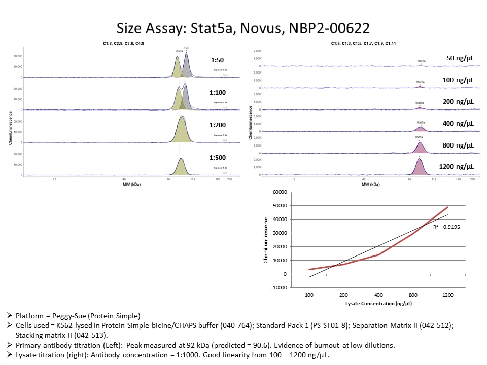
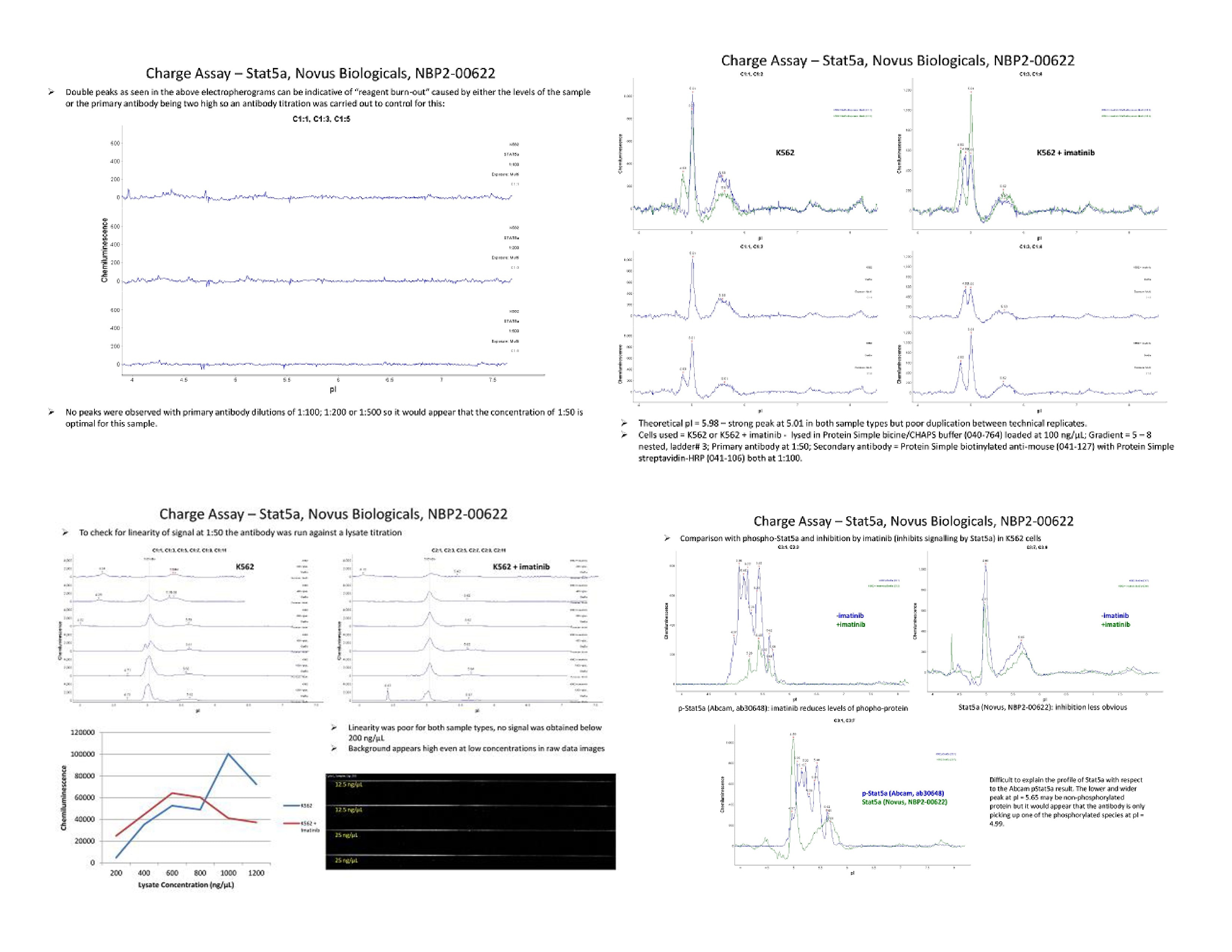
Application: Simple WesternSample Tested: K562 human chronic myelogenous leukemia cell lineSpecies: HumanVerified Customer | Posted 02/06/2017Sta5aCells used = K562 or K562 + imatinib - lysed in Protein Simple bicine/CHAPS buffer (040-764) loaded at 100 ng/µL; Gradient = 5 – 8 nested, ladder# 3; Primary antibody at 1:50; Secondary antibody = Protein Simple biotinylated anti-mouse (041-127) with Protein Simple streptavidin-HRP (041-106) both at 1:100.
There are no reviews that match your criteria.
Protocols
Find general support by application which include: protocols, troubleshooting, illustrated assays, videos and webinars.
- 7-Amino Actinomycin D (7-AAD) Cell Viability Flow Cytometry Protocol
- Antigen Retrieval Protocol (PIER)
- Antigen Retrieval for Frozen Sections Protocol
- Appropriate Fixation of IHC/ICC Samples
- Cellular Response to Hypoxia Protocols
- Chromogenic IHC Staining of Formalin-Fixed Paraffin-Embedded (FFPE) Tissue Protocol
- Chromogenic Immunohistochemistry Staining of Frozen Tissue
- ClariTSA™ Fluorophore Kits
- Detection & Visualization of Antibody Binding
- Extracellular Membrane Flow Cytometry Protocol
- Flow Cytometry Protocol for Cell Surface Markers
- Flow Cytometry Protocol for Staining Membrane Associated Proteins
- Flow Cytometry Staining Protocols
- Flow Cytometry Troubleshooting Guide
- Fluorescent IHC Staining of Frozen Tissue Protocol
- Graphic Protocol for Heat-induced Epitope Retrieval
- Graphic Protocol for the Preparation and Fluorescent IHC Staining of Frozen Tissue Sections
- Graphic Protocol for the Preparation and Fluorescent IHC Staining of Paraffin-embedded Tissue Sections
- Graphic Protocol for the Preparation of Gelatin-coated Slides for Histological Tissue Sections
- ICC Cell Smear Protocol for Suspension Cells
- ICC Immunocytochemistry Protocol Videos
- ICC for Adherent Cells
- IHC Sample Preparation (Frozen sections vs Paraffin)
- Immunocytochemistry (ICC) Protocol
- Immunocytochemistry Troubleshooting
- Immunofluorescence of Organoids Embedded in Cultrex Basement Membrane Extract
- Immunofluorescent IHC Staining of Formalin-Fixed Paraffin-Embedded (FFPE) Tissue Protocol
- Immunohistochemistry (IHC) and Immunocytochemistry (ICC) Protocols
- Immunohistochemistry Frozen Troubleshooting
- Immunohistochemistry Paraffin Troubleshooting
- Intracellular Flow Cytometry Protocol Using Alcohol (Methanol)
- Intracellular Flow Cytometry Protocol Using Detergents
- Intracellular Nuclear Staining Flow Cytometry Protocol Using Detergents
- Intracellular Staining Flow Cytometry Protocol Using Alcohol Permeabilization
- Intracellular Staining Flow Cytometry Protocol Using Detergents to Permeabilize Cells
- Preparing Samples for IHC/ICC Experiments
- Preventing Non-Specific Staining (Non-Specific Binding)
- Primary Antibody Selection & Optimization
- Propidium Iodide Cell Viability Flow Cytometry Protocol
- Protocol for Heat-Induced Epitope Retrieval (HIER)
- Protocol for Liperfluo
- Protocol for Making a 4% Formaldehyde Solution in PBS
- Protocol for VisUCyte™ HRP Polymer Detection Reagent
- Protocol for the Characterization of Human Th22 Cells
- Protocol for the Characterization of Human Th9 Cells
- Protocol for the Fluorescent ICC Staining of Cell Smears - Graphic
- Protocol for the Fluorescent ICC Staining of Cultured Cells on Coverslips - Graphic
- Protocol for the Preparation & Fixation of Cells on Coverslips
- Protocol for the Preparation and Chromogenic IHC Staining of Frozen Tissue Sections
- Protocol for the Preparation and Chromogenic IHC Staining of Frozen Tissue Sections - Graphic
- Protocol for the Preparation and Chromogenic IHC Staining of Paraffin-embedded Tissue Sections
- Protocol for the Preparation and Chromogenic IHC Staining of Paraffin-embedded Tissue Sections - Graphic
- Protocol for the Preparation and Fluorescent ICC Staining of Cells on Coverslips
- Protocol for the Preparation and Fluorescent ICC Staining of Non-adherent Cells
- Protocol for the Preparation and Fluorescent ICC Staining of Stem Cells on Coverslips
- Protocol for the Preparation and Fluorescent IHC Staining of Frozen Tissue Sections
- Protocol for the Preparation and Fluorescent IHC Staining of Paraffin-embedded Tissue Sections
- Protocol for the Preparation of Gelatin-coated Slides for Histological Tissue Sections
- Protocol for the Preparation of a Cell Smear for Non-adherent Cell ICC - Graphic
- Protocol: Annexin V and PI Staining by Flow Cytometry
- Protocol: Annexin V and PI Staining for Apoptosis by Flow Cytometry
- R&D Systems Quality Control Western Blot Protocol
- TUNEL and Active Caspase-3 Detection by IHC/ICC Protocol
- The Importance of IHC/ICC Controls
- Troubleshooting Guide: Fluorokine Flow Cytometry Kits
- Troubleshooting Guide: Immunohistochemistry
- Troubleshooting Guide: Western Blot Figures
- Western Blot Conditions
- Western Blot Protocol
- Western Blot Protocol for Cell Lysates
- Western Blot Troubleshooting
- Western Blot Troubleshooting Guide
- View all Protocols, Troubleshooting, Illustrated assays and Webinars


