TGF-beta 1, 2, 3 Antibody (1D11)
R&D Systems | Catalog # MAB1835

Key Product Details
Validated by
Species Reactivity
Validated:
Cited:
Applications
Validated:
Cited:
Label
Antibody Source
Product Specifications
Immunogen
Specificity
Clonality
Host
Isotype
Endotoxin Level
Scientific Data Images for TGF-beta 1, 2, 3 Antibody (1D11)
TGF‑ beta 1 Inhibition of IL‑4-dependent Cell Proliferation and Neutralization by TGF‑ beta 1, 2, 3 Antibody.
Recombinant Human TGF-beta 1 (Catalog # 240-B) inhibits Recombinant Mouse IL-4 (Catalog # 404-ML) induced proliferation in the HT-2 mouse T cell line in a dose-dependent manner (orange line). Inhibition of Recombinant Mouse IL-4 (7.5 ng/mL) activity elicited by Recombinant Human TGF-beta 1 (1 ng/mL) is neutralized (green line) by increasing concentrations of Mouse Anti-TGF-beta 1, 2, 3 Monoclonal Antibody (Catalog # MAB1835). The ND50 is typically 0.25-1.25 µg/mL.
Detection of Mouse TGF beta 1/2/3 by In vivo assay
Apoptotic cell-induced immunomodulation in mice with collagen-induced arthritis (CIA) is dependent on transforming growth factor (TGF)-beta. CIA mice that received or did not receive apoptotic cells, with or without anti-TGF-beta blocking antibody were scored daily (a). Data are shown as mean ± SEM of five mice per group from one of two representative experiments; *p < 0.05, **p < 0.01 (Friedman test analysis of variance (ANOVA) with Dunn's multiple comparisons test). Anti-collagen IgG2a antibodies (Ab) were quantified in plasma (b). Data from 5 to 16 mice from two to three independent experiments (no difference, Kruskal-Wallis test ANOVA with Dunn’s multiple comparisons test). Lymph node cells were collected, cultured, and T cell proliferation in response to collagen protein or CD3-specific Ab stimulations was evaluated (c). Data are shown as mean ± SEM from one of two representative experiments; *p < 0.05, **p < 0.01 (nonparametric unpaired t test). Percent and absolute number of Foxp3+ T regulatory cells (Treg) were evaluated in the spleen (d) and suppressive assays were performed by isolating and adding Treg at a different ratio into collagen-specific cultures and responder T cell proliferation was assessed (e). Data are shown as mean ± SEM from one experiment (d Kruskal-Wallis test ANOVA with Dunn’s multiple comparisons test; e Friedman test ANOVA with Dunn’s multiple comparisons test). Plasmacytoid dendritic cells (pDC), conventional dendritic cells (cDC) and macrophages (macro) were isolated and cultured with naïve allogenic T cells to determine Treg polarization (f), and their response to TLR ligand was assessed through evaluation of CD40 costimulatory molecule expression (g). Data from individual mouse plus mean (black bar) from one experiment representative of four are shown; *p < 0.05, ***p < 0.001 (f ordinary one-way ANOVA with Tukey’s multiple comparisons test; g Kruskal-Wallis test ANOVA with Dunn’s multiple comparisons test). BrDU 5-Bromo-2’-deoxyuridine Im
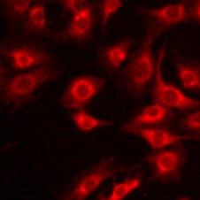
Detection of Human TGF beta 1/2/3 by Immunocytochemistry/Immunofluorescence
TGF beta 1 stimulates the expression of ECM components in human and rabbit primary cells. (A) Selected ECM mRNA transcripts were measured using qRT-PCR in human RPTEC or human IPF134 following 48 hr stimulation with TGF beta 1 (10 ng/mL). Data show transcript abundance relative to the unstimulated control (indicated by the dashed line) and are shown as mean ± SD of four (RPTEC) or seven (IPF134) independent experiments. ****P < 0.0001; ****P < 0.001; **P < 0.01; Student’s t-test. (B) The accumulation of ECM in human RPTEC (i) or human IPF (ii) cells stimulated with 10ng/mL TGF beta 1 was evaluated via measurement of the incorporation of 14C-labelled amino acids into the deposited ECM. Data presented are the mean ± SD of three independent experiments. **P < 0.01; Student’s t-test. (C) Increased mature ECM accumulation following treatment of cells with a pro-fibrotic stimulus can be visualised following in-situ fluorescent staining using the Flamingo dye. Cells from three different systems (primary human RPTEC; primary human IPF134; co-culture of primary rabbit RPTEC with primary rabbit renal fibroblasts) were cultured in the absence (top) or presence (bottom) of 10 ng/ml TGF beta 1 before decellularised matrix was fixed and stained in situ with Flamingo fluorescent dye. Images show a single field. Image collected and cropped by CiteAb from the following publication (https://pubmed.ncbi.nlm.nih.gov/28855577), licensed under a CC-BY license. Not internally tested by R&D Systems.
Detection of Mouse TGF-beta 1, 2, 3 Antibody by Flow Cytometry
CBirTox induced expression of Foxp3 is dependent on TGF-beta but independent of RA signaling.(A) CD11c+ DCs were isolated from the spleen, MLN or LP and pulsed with 1 μg/ml of CBirTox or CBir1 peptide for 2 hours prior to co-culture with CD4+ CBir1 Tg T cells for 4 days before flow cytometry analysis. Results represent 2–3 independent experiments (B-C) CD11c+ DCs were isolated from the spleen and pulsed with 1 μg/ml of CBirTox for 1 hour before co-culture with CD4+ CBir1 Tg T cells in the presence of 1 μM of the RA inhibitor LE135 or RA for 4 days. Representative flow plots (B) of 3 independent experiments (C) are shown. (D-E) Splenic CD19+ B cells were pulsed with 2 μg of CBirTox for 2 hours before co-culture with with CD4+ CBir1 Tg T cells with or without the presence of 10 μg/ml anti-TGF-beta (1,2,3) for 4 days prior to flow cytometry analysis. Representative flow plots (D) of 4–5 independent experiments (E) are shown. Results of all experiments are expressed as the mean ± SEM. ***p<0.0005, NS, not significant. Groups of three or more were analyzed using one-way ANOVA with Bonferroni’s post test. Image collected and cropped by CiteAb from the following publication (https://pubmed.ncbi.nlm.nih.gov/28750075), licensed under a CC-BY license. Not internally tested by R&D Systems.
Detection of Zebrafish TGF beta 1/2/3 by Immunohistochemistry
Expression of genes involved in fibrosis at 6 months.(A) IHC and ISH (inlets) showing MMP9 expression in proliferating myofibroblasts and ductular cells. (B) IHC for TGF beta 1. Contrary to MMP9, TGF beta 1 is expressed only in proliferating ductular cells which are also positive for cytokeratin. (C, D) ISH for PDGFAa and IL1b. Transcripts of both genes are detected in a small subset of proliferating myofibroblasts and ductular cells. (A–C, red arrowheads, ductular cells). Microscopic images are 400×. Bars, 50 µm. Image collected and cropped by CiteAb from the following open publication (https://pubmed.ncbi.nlm.nih.gov/22164219), licensed under a CC-BY license. Not internally tested by R&D Systems.
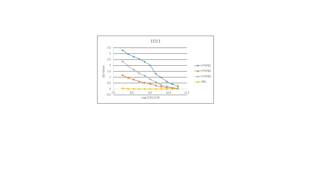
Human TGF-beta 1 ELISA Standard Curve
Recombinant Human TGF‑ beta 1 (Catalog # 240-B) was serially diluted and captured by Mouse Anti-TGF-beta 1, 2, 3 Monoclonal Antibody (Catalog # MAB1835) coated on a Clear Polystyrene Microplate (Catalog # DY990). Chicken Anti-TGF‑ beta 1 Biotinylated Antigen Affinity-purified Polyclonal Antibody (Catalog # ) was incubated with the protein captured on the plate. Detection of the standard curve was achieved by incubating Streptavidin-HRP (Catalog # DY998)Applications for TGF-beta 1, 2, 3 Antibody (1D11)
Western Blot
Sample: Recombinant Human TGF-beta 1 (Catalog # 240-B) under non-reducing conditions only
Neutralization
Binding Inhibition
Human TGF-beta 1 Sandwich Immunoassay
Reviewed Applications
Read 12 reviews rated 4.4 using MAB1835 in the following applications:
Formulation, Preparation, and Storage
Purification
Reconstitution
Reconstitute at 0.5 mg/mL in sterile PBS. For liquid material, refer to CoA for concentration.
Formulation
Shipping
Stability & Storage
- 12 months from date of receipt, -20 to -70 °C as supplied.
- 1 month, 2 to 8 °C under sterile conditions after reconstitution.
- 6 months, -20 to -70 °C under sterile conditions after reconstitution.
Calculators
Background: TGF-beta 1, 2, 3
References
- Ayala A. et al. (1992) FASEB J. 6:A1604.
- Roberts A.B. and Sporn M.B., eds. (1990) Peptide Growth Factors and Their Receptors I, Springer-Verlag, 419.
- Dasch J.R. et al. (1989) J. Immunol. 142:1536.
Long Name
Additional TGF-beta 1, 2, 3 Products
Product Documents for TGF-beta 1, 2, 3 Antibody (1D11)
Certificate of Analysis
To download a Certificate of Analysis, please enter a lot or batch number in the search box below.
Note: Certificate of Analysis not available for kit components.
Product Specific Notices for TGF-beta 1, 2, 3 Antibody (1D11)
For research use only
Citations for TGF-beta 1, 2, 3 Antibody (1D11)
 Powered by Bioz
Customer Reviews for TGF-beta 1, 2, 3 Antibody (1D11) (12)
Have you used TGF-beta 1, 2, 3 Antibody (1D11)?
Submit a review and receive an Amazon gift card!
$25/€18/£15/$25CAN/¥2500 Yen for a review with an image
$10/€7/£6/$10CAN/¥1110 Yen for a review without an image
Submit a review
Customer Images
-
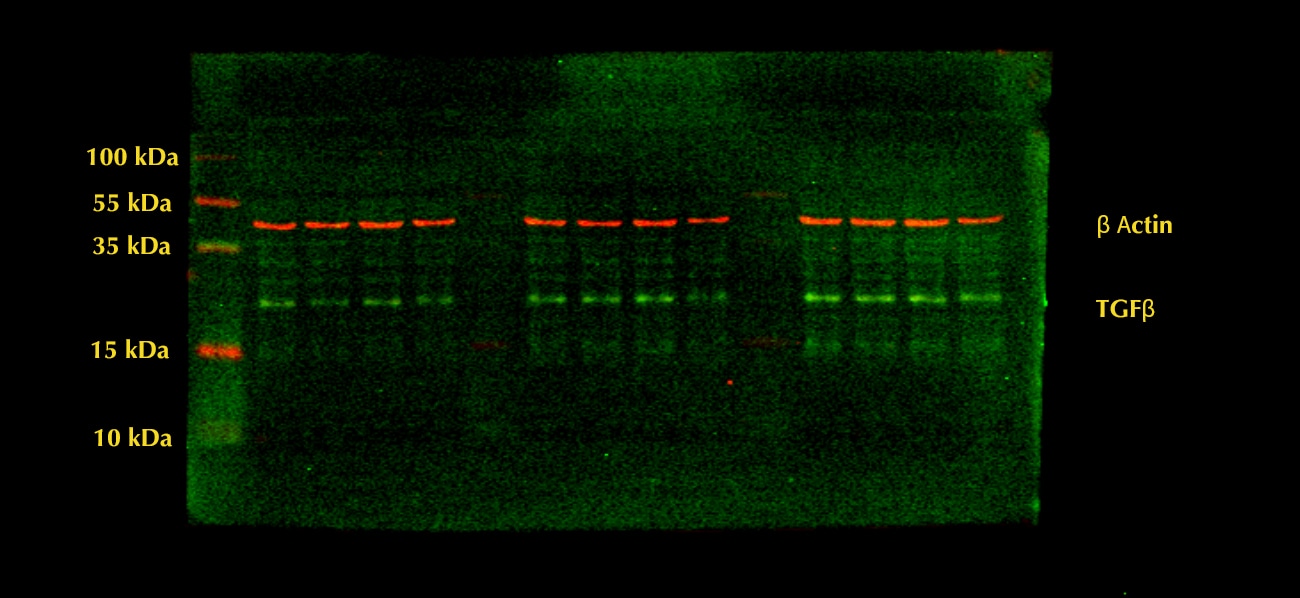
Application: Western BlotSample Tested: HepG2 human hepatocellular carcinoma cell lineSpecies: HumanVerified Customer | Posted 12/06/2022

-
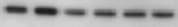
Application: Western BlotSample Tested: Dermal fibroblastsSpecies: MouseVerified Customer | Posted 08/05/2021
-
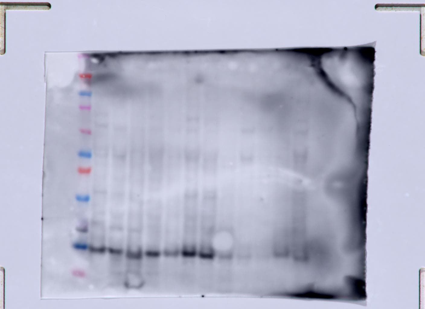
Application: Western BlotSample Tested: Adult pancreasSpecies: MouseVerified Customer | Posted 10/27/2020
-
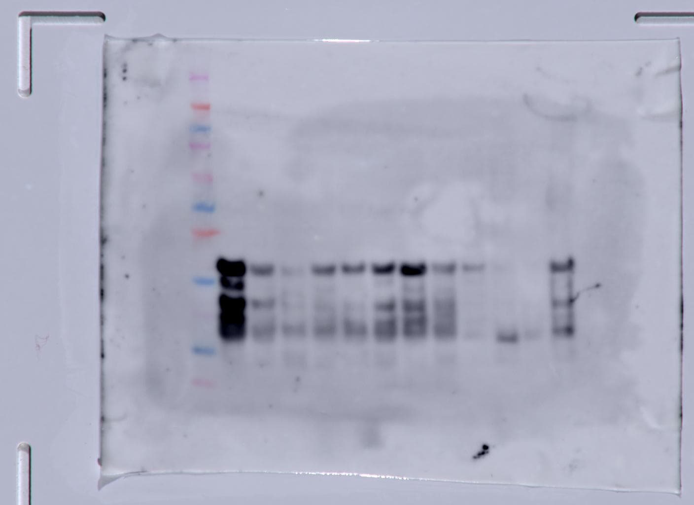
Application: Western BlotSample Tested: Pancreas tissueSpecies: MouseVerified Customer | Posted 10/13/2020
-
Application: Block/NeutralizeSample Tested: 3T3-L1 mouse embryonic fibroblast adipose-like cell lineSpecies: MouseVerified Customer | Posted 07/09/2019
-
Application: Western BlotSample Tested: Muscle tissueSpecies: MouseVerified Customer | Posted 04/30/2019
-
Application: ELISASample Tested: Recombinant proteinSpecies: HumanVerified Customer | Posted 03/28/2019
-
Application: Western BlotSample Tested: Skin cancer tissueSpecies: MouseVerified Customer | Posted 01/28/2019
-
Application: ImmunohistochemistrySample Tested: Tonsil tissueSpecies: HumanVerified Customer | Posted 05/22/2018
-
Application: ImmunohistochemistrySample Tested: Skin cancer tissueSpecies: MouseVerified Customer | Posted 04/24/2018
-
Application: Immunocytochemistry/ImmunofluorescenceSample Tested: A549 human lung carcinoma cell lineSpecies: HumanVerified Customer | Posted 07/21/2017
-
Application: Block/NeutralizeSample Tested: Peritoneal macrophagesSpecies: MouseVerified Customer | Posted 05/16/2017
There are no reviews that match your criteria.
Protocols
Find general support by application which include: protocols, troubleshooting, illustrated assays, videos and webinars.
- Cellular Response to Hypoxia Protocols
- R&D Systems Quality Control Western Blot Protocol
- Troubleshooting Guide: Western Blot Figures
- Western Blot Conditions
- Western Blot Protocol
- Western Blot Protocol for Cell Lysates
- Western Blot Troubleshooting
- Western Blot Troubleshooting Guide
- View all Protocols, Troubleshooting, Illustrated assays and Webinars
FAQs for TGF-beta 1, 2, 3 Antibody (1D11)
-
Q: Can the TGF-beta 1, 2, 3 Antibody, Cat# MAB1835, neutralize the activity of all three TGF-beta proteins?
A: Yes, this antibody can neutralize the activity of TGF-beta 1, 2, and 3 proteins.
Associated Pathways


