TLR5 Antibody (19D759.2) - BSA Free
Novus Biologicals | Catalog # NBP2-24787
Key Product Details
Validated by
Biological Validation
Species Reactivity
Validated:
Human, Mouse, Canine
Cited:
Human, Mouse
Applications
Validated:
Immunohistochemistry, Immunohistochemistry-Paraffin, Immunohistochemistry-Frozen, Western Blot, Flow Cytometry, Flow (Cell Surface), Flow (Intracellular), Immunocytochemistry/ Immunofluorescence, Immunoprecipitation, Dot Blot
Cited:
Immunohistochemistry-Paraffin, Western Blot, Block/Neutralize, Flow Cytometry, Flow (Cell Surface), Immunocytochemistry/ Immunofluorescence, Cytometric Bead Assay Standard, Flow Cytometry Control, IF/IHC
Label
Unconjugated
Antibody Source
Monoclonal Mouse IgG2a Kappa Clone # 19D759.2
Format
BSA Free
Loading...
Product Specifications
Immunogen
This antibody was developed against KLH-conjugated synthetic peptide corresponding to a portion of human TLR5 between amino acids 700-800. The antibody also reacts with mouse TLR5.
Clonality
Monoclonal
Host
Mouse
Isotype
IgG2a Kappa
Scientific Data Images for TLR5 Antibody (19D759.2) - BSA Free
![Immunohistochemistry-Paraffin: TLR5 Antibody (19D759.2) - BSA Free [NBP2-24787]](https://resources.rndsystems.com/images/products/TLR5-Antibody-19D759-2-Immunohistochemistry-Paraffin-NBP2-24787-img0005.jpg "Immunohistochemistry-Paraffin: TLR5 Antibody (19D759.2) - BSA Free [NBP2-24787]")
Immunohistochemistry-Paraffin: TLR5 Antibody (19D759.2) - BSA Free [NBP2-24787]
Immunohistochemistry-Paraffin: TLR5 Antibody (19D759.2) [NBP2-24787] - Analysis of FFPE tissue section of normal human skin using 5 ug/ml concentration of TLR5 antibody (clone 19D759.2). Intense membrane staining of TLR5 was observed specifically in the pigmented basel cells of epidermal layer [10X Magnification]![Flow Cytometry: TLR5 Antibody (19D759.2) - BSA Free [NBP2-24787]](https://resources.rndsystems.com/images/products/TLR5-Antibody-19D759-2-Flow-Cytometry-NBP2-24787-img0011.jpg "Flow Cytometry: TLR5 Antibody (19D759.2) - BSA Free [NBP2-24787]")
Flow Cytometry: TLR5 Antibody (19D759.2) - BSA Free [NBP2-24787]
Flow Cytometry: TLR5 Antibody (19D759.2) [NBP2-24787] - Analysis using the PE conjugate of NBP2-24787. Staining of CD14-/CD83+/TLR5+ diffferentiated myeloid dendritic cells (mDCs). PBMCs were stimulated 5 days with GM-CSF+IL4 prior to the staining. The cells were stained for surface markers CD14 and CD83 FITC, and TLR5 PE. CD14- cells were gated and stained with PE-conjugated isotype control (mouse IgG2a), and FITC-conjugated CD83 (left). CD14- cells were gated and stained with 1 ug PE-conjugated TLR5, and FITC-conjugated CD83 (right).![Western Blot: TLR5 Antibody (19D759.2)BSA Free [NBP2-24787]](https://resources.rndsystems.com/images/products/TLR5-Antibody-19D759-2-Western-Blot-NBP2-24787-img0006.jpg "Western Blot: TLR5 Antibody (19D759.2)BSA Free [NBP2-24787]")
Western Blot: TLR5 Antibody (19D759.2)BSA Free [NBP2-24787]
Western Blot: TLR5 Antibody (19D759.2) [NBP2-24787] - BEAS-2B cells whole cell lysate. Image from verified customer review.![Immunocytochemistry/ Immunofluorescence: TLR5 Antibody (19D759.2) - BSA Free [NBP2-24787]](https://resources.rndsystems.com/images/products/TLR5-Antibody-19D759-2-Immunocytochemistry-Immunofluorescence-NBP2-24787-img0012.jpg "Immunocytochemistry/ Immunofluorescence: TLR5 Antibody (19D759.2) - BSA Free [NBP2-24787]")
Immunocytochemistry/ Immunofluorescence: TLR5 Antibody (19D759.2) - BSA Free [NBP2-24787]
Immunocytochemistry/Immunofluorescence: TLR5 Antibody (19D759.2) [NBP2-24787] - Staining using in-vitro cultured fixed non-permeabilized MCF7 breast cancer cells (ATCC). EpCAM (cell membrane) staining is represented in green (FITC filter), nuclei staining is represented in blue (DAPI filter). Image from a verified customer review using the Alexa Fluor 488 conjugated format of this antibody.![Immunohistochemistry: TLR5 Antibody (19D759.2) - BSA Free [NBP2-24787]](https://resources.rndsystems.com/images/products/TLR5-Antibody-19D759-2-Immunohistochemistry-NBP2-24787-img0013.jpg "Immunohistochemistry: TLR5 Antibody (19D759.2) - BSA Free [NBP2-24787]")
Immunohistochemistry: TLR5 Antibody (19D759.2) - BSA Free [NBP2-24787]
TLR5-Antibody-19D759-2-Immunohistochemistry-NBP2-24787-img0013.jpg![Flow Cytometry: TLR5 Antibody (19D759.2) - BSA Free [NBP2-24787]](https://resources.rndsystems.com/images/products/TLR5-Antibody-19D759-2-BSA-Free-Flow-Cytometry-NBP2-24787-img0014.jpg "Flow Cytometry: TLR5 Antibody (19D759.2) - BSA Free [NBP2-24787]")
Flow Cytometry: TLR5 Antibody (19D759.2) - BSA Free [NBP2-24787]
Flow Cytometry: TLR5 Antibody (19D759.2) - BSA Free [NBP2-24787] - Flow analysis of TLR5 in mouse MC38 cells. Image from verified customer review.![Immunohistochemistry-Paraffin: TLR5 Antibody (19D759.2) - BSA Free [NBP2-24787]](https://resources.rndsystems.com/images/products/TLR5-Antibody-19D759-2-Immunohistochemistry-Paraffin-NBP2-24787-img0004.jpg "Immunohistochemistry-Paraffin: TLR5 Antibody (19D759.2) - BSA Free [NBP2-24787]")
Immunohistochemistry-Paraffin: TLR5 Antibody (19D759.2) - BSA Free [NBP2-24787]
Immunohistochemistry-Paraffin: TLR5 Antibody (19D759.2) [NBP2-24787] - Human skin stained with TLR5 antibody (5 ug/ml), peroxidase-conjugate and DAB chromogen.![Immunohistochemistry-Paraffin: TLR5 Antibody (19D759.2) - BSA Free [NBP2-24787]](https://resources.rndsystems.com/images/products/TLR5-Antibody-19D759-2-Immunohistochemistry-Paraffin-NBP2-24787-img0002.jpg "Immunohistochemistry-Paraffin: TLR5 Antibody (19D759.2) - BSA Free [NBP2-24787]")
Immunohistochemistry-Paraffin: TLR5 Antibody (19D759.2) - BSA Free [NBP2-24787]
Immunohistochemistry-Paraffin: TLR5 Antibody (19D759.2) [NBP2-24787] - Analysis of TLR5 in FFPE mouse brain tissue using TLR5 antibody at 5 ug/ml.![Flow Cytometry: TLR5 Antibody (19D759.2) - BSA Free [NBP2-24787]](https://resources.rndsystems.com/images/products/TLR5-Antibody-19D759-2-Flow-Cytometry-NBP2-24787-img0003.jpg "Flow Cytometry: TLR5 Antibody (19D759.2) - BSA Free [NBP2-24787]")
Flow Cytometry: TLR5 Antibody (19D759.2) - BSA Free [NBP2-24787]
Flow Cytometry: TLR5 Antibody (19D759.2) [NBP2-24787] - Intracellular flow analysis of TLR5 in human monocytes using 0.5 ug of TLR5 antibody. Shaded histogram represents cells without antibody; green represents isotype control; red represents TLR5 antibody. this antibody secondary antibody was used in this test.Applications for TLR5 Antibody (19D759.2) - BSA Free
Application
Recommended Usage
Dot Blot
reported in scientific literature (PMID 27248820)
Flow (Cell Surface)
reported in scientific literature (PMID 24412598)
Flow (Intracellular)
0.5-1 ug/10^6 cells. Use reported in scientific literature (PMID 24412598)
Flow Cytometry
0.5-1 ug/10^6 cells
Immunocytochemistry/ Immunofluorescence
5 ug/ml. Use reported by customer review
Immunohistochemistry
1:10-1:500. Use reported in scientific literature (PMID 27392931)
Immunohistochemistry-Frozen
10 ug/ml
Immunohistochemistry-Paraffin
5 ug/ml
Western Blot
1-3 ug/ml
Application Notes
Staining of formalin-fixed tissues is enhanced by boiling tissue sections in 10 mM sodium citrate buffer, pH 6.0 for 10-20 min followed by cooling at RT for 20 min.
Reviewed Applications
Read 1 review rated 4 using NBP2-24787 in the following applications:
Flow Cytometry Panel Builder
Bio-Techne Knows Flow Cytometry
Save time and reduce costly mistakes by quickly finding compatible reagents using the Panel Builder Tool.
Advanced Features
- Spectra Viewer - Custom analysis of spectra from multiple fluorochromes
- Spillover Popups - Visualize the spectra of individual fluorochromes
- Antigen Density Selector - Match fluorochrome brightness with antigen density

Formulation, Preparation, and Storage
Purification
Protein G purified
Formulation
PBS
Format
BSA Free
Preservative
0.05% Sodium Azide
Concentration
1.0 mg/ml
Shipping
The product is shipped with polar packs. Upon receipt, store it immediately at the temperature recommended below.
Stability & Storage
Store at -20C. Avoid freeze-thaw cycles.
Background: TLR5
Long Name
Toll-like Receptor 5
Alternate Names
TIL-3
Gene Symbol
TLR5
Additional TLR5 Products
Product Documents for TLR5 Antibody (19D759.2) - BSA Free
Certificate of Analysis
To download a Certificate of Analysis, please enter a lot or batch number in the search box below.
Product Specific Notices for TLR5 Antibody (19D759.2) - BSA Free
This product is for research use only and is not approved for use in humans or in clinical diagnosis. Primary Antibodies are guaranteed for 1 year from date of receipt.
Citations for TLR5 Antibody (19D759.2) - BSA Free
 Powered by Bioz
Customer Reviews for TLR5 Antibody (19D759.2) - BSA Free (1)
4 out of 5
1 Customer Rating
Have you used TLR5 Antibody (19D759.2) - BSA Free?
Submit a review and receive an Amazon gift card!
$25/€18/£15/$25CAN/¥2500 Yen for a review with an image
$10/€7/£6/$10CAN/¥1110 Yen for a review without an image
Submit a review
Customer Images
Showing
1
-
1 of
1 review
Showing All
Filter By:
-
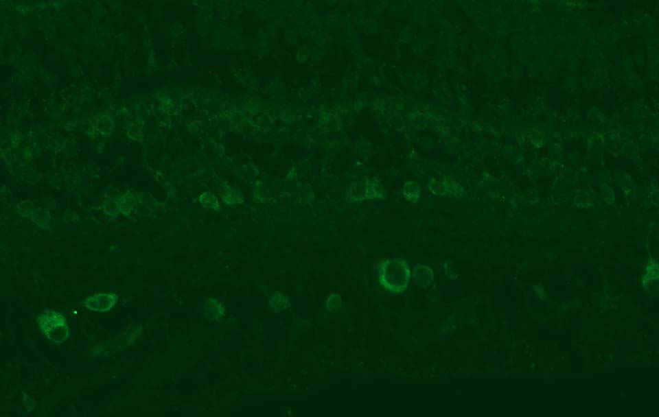
Application: Immunohistochemistry-ParaffinSample Tested: formalin fixed paraffin embedded macaque retina tissueSpecies: maccaca mulattaVerified Customer | Posted 04/06/2018TLR5 antibody stains retinal ganglion cells and round cells in the inner nuclear layer of paraffin embedded rhesus macaque retina tissueheat mediated antigen retrieval (pH 6 citrate buffer) 1:50 dilution of antibody, overnight at 4C goat anti-mouse AlexaFluor 488 secondary antibody (1:400, 1 hr RT)

There are no reviews that match your criteria.
Protocols
Find general support by application which include: protocols, troubleshooting, illustrated assays, videos and webinars.
- 7-Amino Actinomycin D (7-AAD) Cell Viability Flow Cytometry Protocol
- Antigen Retrieval Protocol (PIER)
- Antigen Retrieval for Frozen Sections Protocol
- Appropriate Fixation of IHC/ICC Samples
- Cellular Response to Hypoxia Protocols
- Chromogenic IHC Staining of Formalin-Fixed Paraffin-Embedded (FFPE) Tissue Protocol
- Chromogenic Immunohistochemistry Staining of Frozen Tissue
- ClariTSA™ Fluorophore Kits
- Detection & Visualization of Antibody Binding
- Extracellular Membrane Flow Cytometry Protocol
- Flow Cytometry Protocol for Cell Surface Markers
- Flow Cytometry Protocol for Staining Membrane Associated Proteins
- Flow Cytometry Staining Protocols
- Flow Cytometry Troubleshooting Guide
- Fluorescent IHC Staining of Frozen Tissue Protocol
- Graphic Protocol for Heat-induced Epitope Retrieval
- Graphic Protocol for the Preparation and Fluorescent IHC Staining of Frozen Tissue Sections
- Graphic Protocol for the Preparation and Fluorescent IHC Staining of Paraffin-embedded Tissue Sections
- Graphic Protocol for the Preparation of Gelatin-coated Slides for Histological Tissue Sections
- ICC Cell Smear Protocol for Suspension Cells
- ICC Immunocytochemistry Protocol Videos
- ICC for Adherent Cells
- IHC Sample Preparation (Frozen sections vs Paraffin)
- Immunocytochemistry (ICC) Protocol
- Immunocytochemistry Troubleshooting
- Immunofluorescence of Organoids Embedded in Cultrex Basement Membrane Extract
- Immunofluorescent IHC Staining of Formalin-Fixed Paraffin-Embedded (FFPE) Tissue Protocol
- Immunohistochemistry (IHC) and Immunocytochemistry (ICC) Protocols
- Immunohistochemistry Frozen Troubleshooting
- Immunohistochemistry Paraffin Troubleshooting
- Immunoprecipitation Protocol
- Intracellular Flow Cytometry Protocol Using Alcohol (Methanol)
- Intracellular Flow Cytometry Protocol Using Detergents
- Intracellular Nuclear Staining Flow Cytometry Protocol Using Detergents
- Intracellular Staining Flow Cytometry Protocol Using Alcohol Permeabilization
- Intracellular Staining Flow Cytometry Protocol Using Detergents to Permeabilize Cells
- Preparing Samples for IHC/ICC Experiments
- Preventing Non-Specific Staining (Non-Specific Binding)
- Primary Antibody Selection & Optimization
- Propidium Iodide Cell Viability Flow Cytometry Protocol
- Protocol for Heat-Induced Epitope Retrieval (HIER)
- Protocol for Liperfluo
- Protocol for Making a 4% Formaldehyde Solution in PBS
- Protocol for VisUCyte™ HRP Polymer Detection Reagent
- Protocol for the Characterization of Human Th22 Cells
- Protocol for the Characterization of Human Th9 Cells
- Protocol for the Fluorescent ICC Staining of Cell Smears - Graphic
- Protocol for the Fluorescent ICC Staining of Cultured Cells on Coverslips - Graphic
- Protocol for the Preparation & Fixation of Cells on Coverslips
- Protocol for the Preparation and Chromogenic IHC Staining of Frozen Tissue Sections
- Protocol for the Preparation and Chromogenic IHC Staining of Frozen Tissue Sections - Graphic
- Protocol for the Preparation and Chromogenic IHC Staining of Paraffin-embedded Tissue Sections
- Protocol for the Preparation and Chromogenic IHC Staining of Paraffin-embedded Tissue Sections - Graphic
- Protocol for the Preparation and Fluorescent ICC Staining of Cells on Coverslips
- Protocol for the Preparation and Fluorescent ICC Staining of Non-adherent Cells
- Protocol for the Preparation and Fluorescent ICC Staining of Stem Cells on Coverslips
- Protocol for the Preparation and Fluorescent IHC Staining of Frozen Tissue Sections
- Protocol for the Preparation and Fluorescent IHC Staining of Paraffin-embedded Tissue Sections
- Protocol for the Preparation of Gelatin-coated Slides for Histological Tissue Sections
- Protocol for the Preparation of a Cell Smear for Non-adherent Cell ICC - Graphic
- Protocol: Annexin V and PI Staining by Flow Cytometry
- Protocol: Annexin V and PI Staining for Apoptosis by Flow Cytometry
- R&D Systems Quality Control Western Blot Protocol
- TUNEL and Active Caspase-3 Detection by IHC/ICC Protocol
- The Importance of IHC/ICC Controls
- Troubleshooting Guide: Fluorokine Flow Cytometry Kits
- Troubleshooting Guide: Immunohistochemistry
- Troubleshooting Guide: Western Blot Figures
- Western Blot Conditions
- Western Blot Protocol
- Western Blot Protocol for Cell Lysates
- Western Blot Troubleshooting
- Western Blot Troubleshooting Guide
- View all Protocols, Troubleshooting, Illustrated assays and Webinars


