Tyrosine Hydroxylase [p Ser40] Antibody - Azide Free
Novus Biologicals | Catalog # NB300-173
Loading...
Key Product Details
Validated by
Independent Antibodies
Species Reactivity
Validated:
Mouse, Rat, Mammal
Cited:
Mouse, Rat
Applications
Validated:
Immunohistochemistry, Immunohistochemistry-Frozen, Western Blot, Immunocytochemistry/ Immunofluorescence
Cited:
Immunohistochemistry-Frozen, Western Blot, Immunocytochemistry/ Immunofluorescence, IF/IHC
Label
Unconjugated
Antibody Source
Polyclonal Rabbit IgG
Format
Azide Free
Loading...
Product Specifications
Immunogen
Synthetic phospho-peptide corresponding to amino acid residues surrounding Tyrosine Hydroxylase conjugated to KLH. Accession # P04177
Reactivity Notes
Reactivity assumed based on sequence identity to a wide variety of mammalian and non-mammalian species.
Modification
p Ser40
Marker
Neuronal Marker
Specificity
Specific for the ~60 kDa tyrosine hydroxylase protein phosphorylated at Ser40. Some higher molecular weight bands may be detected by the antibody depending upon the brain region being studied, protein loads and the detection methods used. The antibody has three orders of magnitude selectivity over dephospho TH.
Clonality
Polyclonal
Host
Rabbit
Isotype
IgG
Theoretical MW
60 kDa.
Disclaimer note: The observed molecular weight of the protein may vary from the listed predicted molecular weight due to post translational modifications, post translation cleavages, relative charges, and other experimental factors.
Disclaimer note: The observed molecular weight of the protein may vary from the listed predicted molecular weight due to post translational modifications, post translation cleavages, relative charges, and other experimental factors.
Scientific Data Images for Tyrosine Hydroxylase [p Ser40] Antibody - Azide Free
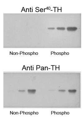
Tyrosine Hydroxylase [p Ser40] Antibody [NB300-173] - Western blot of recombinant phospho-TH and non-phospho-TH showing selective immunolabeling by the phosphospecific antibody of the ~60 kDa TH phosphorylated at Ser40. The pan-specific antibody (anti-pan-TH) recognized both the phospho- and non-phospho-TH; while most importantly, the phospho-specific antibody (anti-Ser40 TH) recognized only phospho-TH.
![Immunohistochemistry: Tyrosine Hydroxylase [p Ser40] Antibody [NB300-173]](https://resources.rndsystems.com/images/products/Tyrosine-Hydroxylase-[p-Ser40]-Antibody-Immunohistochemistry-NB300-173-img0004.jpg "Immunohistochemistry: Tyrosine Hydroxylase [p Ser40] Antibody [NB300-173]")
Immunohistochemistry: Tyrosine Hydroxylase [p Ser40] Antibody [NB300-173]
Immunohistochemistry: Tyrosine Hydroxylase [p Ser40] Antibody [NB300-173] - Immunohistochemical staining of retina with the pan-tyrosine hydroxylase (pan-TH) and phospho-specific tyrosine hydroxylase (phospho-TH) antibodies. The pan-TH antibody shows extensive labeling in this photomicrograph of the retina. In contrast, the phospho-TH antibody selectively labels only the two amacrine cells in this light-stimulated retina example.Applications for Tyrosine Hydroxylase [p Ser40] Antibody - Azide Free
Application
Recommended Usage
Immunocytochemistry/ Immunofluorescence
1:1000
Immunohistochemistry
1:1000
Immunohistochemistry-Frozen
1:1000
Western Blot
1:1000
Reviewed Applications
Read 1 review rated 4 using NB300-173 in the following applications:
Formulation, Preparation, and Storage
Purification
Antigen Affinity-purified
Formulation
10 mM HEPES (pH 7.5), 0.15 M NaCl, 0.1 mg/mL BSA, 50% Glycerol
Format
Azide Free
Preservative
No Preservative
Concentration
Please see the vial label for concentration. If unlisted please contact technical services.
Shipping
The product is shipped with polar packs. Upon receipt, store it immediately at the temperature recommended below.
Stability & Storage
Store at -20C. Avoid freeze-thaw cycles.
Background: Tyrosine Hydroxylase
Two transcription factor binding sites in the proximal region of the TH gene, the TPA-responsive element (TRE) and the c-AMP responsive element (CRE), have been implicated in the complex regulation of the TH gene. Dysregulation of breakdown for the amino acid, tyrosine, by TH is a result of a genetic disorder that results in Tyrosinemia (high levels of tyrosine in the blood, tissue and organs).
Tyrosine hydroxylase deficiency is a disorder that primarily affects movement, where individuals display symptoms that include lack of coordination when walking, postural tremors and unusual body positioning. TH deficient dopamine-responsive dystonia (DRD), also known as Segawa syndrome, is a rare genetic disorder that is associated with low levels of TH and is diagnosed during childhood with characteristic symptoms including increased muscle tone (dystonia) and signs of Parkinsonism like bradykinesia, tremors, rigidity and postural instability (2). Correspondingly, TH is also linked to Parkinson's disease in older adults, where low dopamine levels are a consistent neurochemical abnormality. Functional polymorphisms of the TH gene may be involved in the pathogenesis of neuropsychiatric diseases such as schizophrenia and other affective disorders where dopamine is often dysregulated (3).
References
1. Hamanaka, Y., & Mizunami, M. (2019). Tyrosine hydroxylase-immunoreactive neurons in the mushroom body of the field cricket, Gryllus bimaculatus. Cell Tissue Res, 376(1), 97-111. doi:10.1007/s00441-018-2969-9
2. Li, L., & Zhou, F. M. (2013). Parallel dopamine D1 receptor activity dependence of l-Dopa-induced normal movement and dyskinesia in mice. Neuroscience, 236, 66-76. doi:10.1016/j.neuroscience.2012.12.065
3. Borkar, C. D., Bharne, A. P., Nagalakshmi, B., Sakharkar, A. J., Subhedar, N. K., & Kokare, D. M. (2018). Cocaine- and Amphetamine-Regulated Transcript Peptide (CART) Alleviates MK-801-Induced Schizophrenic Dementia-Like Symptoms. Neuroscience, 375, 94-107. doi:10.1016/j.neuroscience.2018.01.056
Alternate Names
TH, TYH
Gene Symbol
TH
UniProt
Additional Tyrosine Hydroxylase Products
Product Documents for Tyrosine Hydroxylase [p Ser40] Antibody - Azide Free
Certificate of Analysis
To download a Certificate of Analysis, please enter a lot or batch number in the search box below.
Product Specific Notices for Tyrosine Hydroxylase [p Ser40] Antibody - Azide Free
This product is for research use only and is not approved for use in humans or in clinical diagnosis. Primary Antibodies are guaranteed for 1 year from date of receipt.
Citations for Tyrosine Hydroxylase [p Ser40] Antibody - Azide Free
 Powered by Bioz
Customer Reviews for Tyrosine Hydroxylase [p Ser40] Antibody - Azide Free (1)
4 out of 5
1 Customer Rating
Have you used Tyrosine Hydroxylase [p Ser40] Antibody - Azide Free?
Submit a review and receive an Amazon gift card!
$25/€18/£15/$25CAN/¥2500 Yen for a review with an image
$10/€7/£6/$10CAN/¥1110 Yen for a review without an image
Submit a review
Showing
1
-
1 of
1 review
Showing All
Filter By:
-
Application: Immunohistochemistry-FrozenSample Tested: mouse brainSpecies: MouseVerified Customer | Posted 03/23/2010
There are no reviews that match your criteria.
Protocols
Find general support by application which include: protocols, troubleshooting, illustrated assays, videos and webinars.
- Antigen Retrieval Protocol (PIER)
- Antigen Retrieval for Frozen Sections Protocol
- Appropriate Fixation of IHC/ICC Samples
- Cellular Response to Hypoxia Protocols
- Chromogenic IHC Staining of Formalin-Fixed Paraffin-Embedded (FFPE) Tissue Protocol
- Chromogenic Immunohistochemistry Staining of Frozen Tissue
- ClariTSA™ Fluorophore Kits
- Detection & Visualization of Antibody Binding
- Fluorescent IHC Staining of Frozen Tissue Protocol
- Graphic Protocol for Heat-induced Epitope Retrieval
- Graphic Protocol for the Preparation and Fluorescent IHC Staining of Frozen Tissue Sections
- Graphic Protocol for the Preparation and Fluorescent IHC Staining of Paraffin-embedded Tissue Sections
- Graphic Protocol for the Preparation of Gelatin-coated Slides for Histological Tissue Sections
- ICC Cell Smear Protocol for Suspension Cells
- ICC Immunocytochemistry Protocol Videos
- ICC for Adherent Cells
- IHC Sample Preparation (Frozen sections vs Paraffin)
- Immunocytochemistry (ICC) Protocol
- Immunocytochemistry Troubleshooting
- Immunofluorescence of Organoids Embedded in Cultrex Basement Membrane Extract
- Immunofluorescent IHC Staining of Formalin-Fixed Paraffin-Embedded (FFPE) Tissue Protocol
- Immunohistochemistry (IHC) and Immunocytochemistry (ICC) Protocols
- Immunohistochemistry Frozen Troubleshooting
- Immunohistochemistry Paraffin Troubleshooting
- Preparing Samples for IHC/ICC Experiments
- Preventing Non-Specific Staining (Non-Specific Binding)
- Primary Antibody Selection & Optimization
- Protocol for Heat-Induced Epitope Retrieval (HIER)
- Protocol for Making a 4% Formaldehyde Solution in PBS
- Protocol for VisUCyte™ HRP Polymer Detection Reagent
- Protocol for the Fluorescent ICC Staining of Cell Smears - Graphic
- Protocol for the Fluorescent ICC Staining of Cultured Cells on Coverslips - Graphic
- Protocol for the Preparation & Fixation of Cells on Coverslips
- Protocol for the Preparation and Chromogenic IHC Staining of Frozen Tissue Sections
- Protocol for the Preparation and Chromogenic IHC Staining of Frozen Tissue Sections - Graphic
- Protocol for the Preparation and Chromogenic IHC Staining of Paraffin-embedded Tissue Sections
- Protocol for the Preparation and Chromogenic IHC Staining of Paraffin-embedded Tissue Sections - Graphic
- Protocol for the Preparation and Fluorescent ICC Staining of Cells on Coverslips
- Protocol for the Preparation and Fluorescent ICC Staining of Non-adherent Cells
- Protocol for the Preparation and Fluorescent ICC Staining of Stem Cells on Coverslips
- Protocol for the Preparation and Fluorescent IHC Staining of Frozen Tissue Sections
- Protocol for the Preparation and Fluorescent IHC Staining of Paraffin-embedded Tissue Sections
- Protocol for the Preparation of Gelatin-coated Slides for Histological Tissue Sections
- Protocol for the Preparation of a Cell Smear for Non-adherent Cell ICC - Graphic
- R&D Systems Quality Control Western Blot Protocol
- TUNEL and Active Caspase-3 Detection by IHC/ICC Protocol
- The Importance of IHC/ICC Controls
- Troubleshooting Guide: Immunohistochemistry
- Troubleshooting Guide: Western Blot Figures
- Western Blot Conditions
- Western Blot Protocol
- Western Blot Protocol for Cell Lysates
- Western Blot Troubleshooting
- Western Blot Troubleshooting Guide
- View all Protocols, Troubleshooting, Illustrated assays and Webinars
Loading...
Associated Pathways
