Vimentin Antibody (812) - Azide and BSA Free
Novus Biologicals | Catalog # NBP2-43560
![Knockout Validated: Vimentin Antibody (812) [NBP2-43560]](https://resources.rndsystems.com/images/products/Vimentin-Antibody-812-Western-Blot-NBP2-43560-img0012.jpg "Western Blot: Vimentin Antibody (812) [NBP2-43560]")
Loading...
Key Product Details
Validated by
Knockout/Knockdown
Species Reactivity
Validated:
Human, Mouse, Rat, Feline
Cited:
Mouse
Predicted:
Bovine (98%), Canine (98%), Chicken (92%), Chimpanzee (99%), Guinea Pig (97%). Backed by our 100% Guarantee.
Applications
Validated:
Knockout Validated, Immunohistochemistry, Immunohistochemistry-Paraffin, Immunohistochemistry-Frozen, Western Blot, Sandwich ELISA, Flow Cytometry, Immunocytochemistry/ Immunofluorescence, Immunoprecipitation, Knockdown Validated
Cited:
Immunohistochemistry-Frozen, Flow Cytometry, Immunocytochemistry/ Immunofluorescence
Label
Unconjugated
Antibody Source
Monoclonal Mouse IgG2B Clone # 812
Format
Azide and BSA Free
Loading...
Product Specifications
Immunogen
Recombinant protein encompassing a sequence within the center region of human Vimentin. The exact sequence is proprietary.
Reactivity Notes
Cat (100%), Xenopus laevis (84%).
Clonality
Monoclonal
Host
Mouse
Isotype
IgG2B
Theoretical MW
54 kDa.
Disclaimer note: The observed molecular weight of the protein may vary from the listed predicted molecular weight due to post translational modifications, post translation cleavages, relative charges, and other experimental factors.
Disclaimer note: The observed molecular weight of the protein may vary from the listed predicted molecular weight due to post translational modifications, post translation cleavages, relative charges, and other experimental factors.
Scientific Data Images for Vimentin Antibody (812) - Azide and BSA Free
Western Blot: Vimentin Antibody (812) [NBP2-43560]
Western Blot: Vimentin Antibody (812) [NBP2-43560] - Wild-type (WT) and Vimentin knockout (KO) 293T cell extracts (30 ug) were separated by 10% SDS-PAGE, and the membrane was blotted with Vimentin antibody (812) diluted at 1:5000. HRP-conjugated anti-mouse IgG antibody was used to detect the primary antibody.![Immunohistochemistry-Frozen: Vimentin Antibody (812) [NBP2-43560]](https://resources.rndsystems.com/images/products/Vimentin-Antibody-812-Immunohistochemistry-Frozen-NBP2-43560-img0006.jpg "Immunohistochemistry-Frozen: Vimentin Antibody (812) [NBP2-43560]")
Immunohistochemistry-Frozen: Vimentin Antibody (812) [NBP2-43560]
Immunohistochemistry-Frozen: Vimentin Antibody (812) [NBP2-43560] - Frozen sectioned E13.5 Rat brain. Green: Vimentin protein stained by Vimentin antibody [812] diluted at 1:250.Red: beta Tubulin 3/ TUJ1, a mature neuron marker, stained by beta Tubulin 3/ TUJ1 antibody diluted at 1:250. Blue: Fluoroshield with DAPI.![Immunohistochemistry-Frozen: Vimentin Antibody (812) [NBP2-43560]](https://resources.rndsystems.com/images/products/Vimentin-Antibody-812-Immunohistochemistry-Frozen-NBP2-43560-img0001.jpg "Immunohistochemistry-Frozen: Vimentin Antibody (812) [NBP2-43560]")
Immunohistochemistry-Frozen: Vimentin Antibody (812) [NBP2-43560]
Immunohistochemistry-Frozen: Vimentin Antibody (812) [NBP2-43560] - Analysis of Frozen section of embryonic mouse brain (mE18.5). Red: Vimentin antibody [812] diluted at 1:250. Blue: DAPI.![Western Blot: Vimentin Antibody (812) [NBP2-43560]](https://resources.rndsystems.com/images/products/Vimentin-Antibody-812-Western-Blot-NBP2-43560-img0002.jpg "Western Blot: Vimentin Antibody (812) [NBP2-43560]")
Western Blot: Vimentin Antibody (812) [NBP2-43560]
Western Blot: Vimentin Antibody (812) [NBP2-43560] - Analysis of A. 30 ug 293T whole cell lysate/extract B. 30 ug HeLa whole cell lysate/extract 10 % SDS-PAGE Vimentin antibody [812] dilution: 1:1000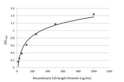
Sandwich ELISA: Vimentin Antibody (812) [NBP2-43560] - Sandwich ELISA detection of recombinant full-length Vimentin protein using Vimentin Antibody (812) as capture antibody at concentration of 5 ug/mL. HRP-conjugated rabbit IgG antibody was diluted at 1:10000 and used to detect the primary antibody.
 [NBP2-43560] -")
Western Blot: Vimentin Antibody (812) [NBP2-43560] -
Western Blot: Vimentin Antibody (812) [NBP2-43560] - Non-transfected (-) and transfected (+) 293T whole cell extracts (10 ug) were separated by 10% SDS-PAGE, and the membrane was blotted with Vimentin antibody [GT812] (NBP2-43560) diluted at 1:10000. The HRP-conjugated anti-mouse IgG antibody was used to detect the primary antibody. [NBP2-43560] -")
ELISA: Vimentin Antibody (812) [NBP2-43560] -
ELISA: Vimentin Antibody (812) [NBP2-43560] - Sandwich ELISA detection of recombinant full-length Vimentin protein using NBP2-43560 as capture antibody at concentration of 5 ug/mL and detection antibody at concentration of 1 ug/mL. Rabbit IgG antibody (HRP) was diluted at 1:10000 and used to detect the primary antibody. [NBP2-43560] -")
Western Blot: Vimentin Antibody (812) [NBP2-43560] -
Western Blot: Vimentin Antibody (812) [NBP2-43560] - Whole cell extract (30 ug) was separated by 10% SDS-PAGE, and the membrane was blotted with Vimentin antibody (NBP2-43560) diluted at 1:500000. The HRP-conjugated anti-mouse IgG antibody was used to detect the primary antibody, and the signal was developed with Trident ECL plus-Enhanced. [NBP2-43560] -")
Western Blot: Vimentin Antibody (812) [NBP2-43560] -
Various whole cell extracts (30 ug) were separated by 10% SDS-PAGE, and the membrane was blotted with Vimentin antibody [GT812] (NBP2-43560) diluted at 1:3000. [NBP2-43560] -")
 [NBP2-43560] -")
Western Blot: Vimentin Antibody (812) [NBP2-43560] -
Various whole cell extracts (30 ug) were separated by 10% SDS-PAGE, and the membrane was blotted with Vimentin antibody [GT812] (NBP2-43560) diluted at 1:5000. [NBP2-43560] -")
Western Blot: Vimentin Antibody (812) [NBP2-43560] -
Whole cell extract (30 ug) was separated by 10% SDS-PAGE, and the membrane was blotted with Vimentin antibody [GT812] (NBP2-43560) diluted at 1:75000.Applications for Vimentin Antibody (812) - Azide and BSA Free
Application
Recommended Usage
Flow Cytometry
Assay-dependent dilution
Immunocytochemistry/ Immunofluorescence
Assay dependent
Immunohistochemistry
1:100-1:1000
Immunohistochemistry-Frozen
1:100-1:1000
Immunohistochemistry-Paraffin
1:100-1:1000
Western Blot
1:500-1:75000
Flow Cytometry Panel Builder
Bio-Techne Knows Flow Cytometry
Save time and reduce costly mistakes by quickly finding compatible reagents using the Panel Builder Tool.
Advanced Features
- Spectra Viewer - Custom analysis of spectra from multiple fluorochromes
- Spillover Popups - Visualize the spectra of individual fluorochromes
- Antigen Density Selector - Match fluorochrome brightness with antigen density

Formulation, Preparation, and Storage
Purification
Protein G purified
Formulation
PBS
Format
Azide and BSA Free
Preservative
No Preservative
Concentration
Concentrations vary lot to lot. See vial label for concentration. If unlisted please contact technical services.
Shipping
The product is shipped with polar packs. Upon receipt, store it immediately at the temperature recommended below.
Stability & Storage
Aliquot and store at -20C or -80C. Avoid freeze-thaw cycles.
Background: Vimentin
Activated macrophages have been shown to secrete phosphorylated vimentin which can be stimulated by a variety of pathophysiological factors including oxidized low-density lipoproteins and TNF-alpha or inhibited by IL-10 (1). The vimentin protein is often expressed at the cell surface playing a role in cell-cell interactions, tissue damage and repair, immune response, and pathogen recognition (1). Vimentin functions in many cytoskeletal processes including cell migration, which is highlighted by its upregulation during epithelial-to-mesenchymal transition (EMT) (4,5). Vimentin is a commonly used marker for EMT and is expressed by many tumor types (5). For example, high metastasis of oral squamous cell carcinomas also showed high vimentin positive expression in immunohistochemical staining analysis (5). A number of vimentin targeting compounds are in cancer-related clinical trials, however, given the multifunctional role of vimentin, the effect of inhibition on non-malignant cells needs to be thoroughly examined (5).
References
1. Ramos, I., Stamatakis, K., Oeste, C. L., & Perez-Sala, D. (2020). Vimentin as a Multifaceted Player and Potential Therapeutic Target in Viral Infections. International Journal of Molecular Sciences. https://doi.org/10.3390/ijms21134675
2. Uniprot (P08670)
3. Morrow, C. S., & Moore, D. L. (2020). Vimentin's side gig: Regulating cellular proteostasis in mammalian systems. Cytoskeleton (Hoboken, N.J.). https://doi.org/10.1002/cm.21645
4. van Bodegraven, E. J., & Etienne-Manneville, S. (2020). Intermediate filaments against actomyosin: the david and goliath of cell migration. Current Opinion in Cell Biology. https://doi.org/10.1016/j.ceb.2020.05.006
5. Strouhalova, K., Prechova, M., Gandalovicova, A., Brabek, J., Gregor, M., & Rosel, D. (2020). Vimentin Intermediate Filaments as Potential Target for Cancer Treatment. Cancers. https://doi.org/10.3390/cancers12010184
Alternate Names
VIM
Gene Symbol
VIM
Additional Vimentin Products
Product Documents for Vimentin Antibody (812) - Azide and BSA Free
Certificate of Analysis
To download a Certificate of Analysis, please enter a lot or batch number in the search box below.
Product Specific Notices for Vimentin Antibody (812) - Azide and BSA Free
This product is for research use only and is not approved for use in humans or in clinical diagnosis. Primary Antibodies are guaranteed for 1 year from date of receipt.
Citations for Vimentin Antibody (812) - Azide and BSA Free
 Powered by Bioz
Customer Reviews for Vimentin Antibody (812) - Azide and BSA Free
There are currently no reviews for this product. Be the first to review Vimentin Antibody (812) - Azide and BSA Free and earn rewards!
Have you used Vimentin Antibody (812) - Azide and BSA Free?
Submit a review and receive an Amazon gift card!
$25/€18/£15/$25CAN/¥2500 Yen for a review with an image
$10/€7/£6/$10CAN/¥1110 Yen for a review without an image
Submit a review
Protocols
Find general support by application which include: protocols, troubleshooting, illustrated assays, videos and webinars.
- 7-Amino Actinomycin D (7-AAD) Cell Viability Flow Cytometry Protocol
- Antigen Retrieval Protocol (PIER)
- Antigen Retrieval for Frozen Sections Protocol
- Appropriate Fixation of IHC/ICC Samples
- Cellular Response to Hypoxia Protocols
- Chromogenic IHC Staining of Formalin-Fixed Paraffin-Embedded (FFPE) Tissue Protocol
- Chromogenic Immunohistochemistry Staining of Frozen Tissue
- ClariTSA™ Fluorophore Kits
- Detection & Visualization of Antibody Binding
- ELISA Sample Preparation & Collection Guide
- ELISA Troubleshooting Guide
- Extracellular Membrane Flow Cytometry Protocol
- Flow Cytometry Protocol for Cell Surface Markers
- Flow Cytometry Protocol for Staining Membrane Associated Proteins
- Flow Cytometry Staining Protocols
- Flow Cytometry Troubleshooting Guide
- Fluorescent IHC Staining of Frozen Tissue Protocol
- Graphic Protocol for Heat-induced Epitope Retrieval
- Graphic Protocol for the Preparation and Fluorescent IHC Staining of Frozen Tissue Sections
- Graphic Protocol for the Preparation and Fluorescent IHC Staining of Paraffin-embedded Tissue Sections
- Graphic Protocol for the Preparation of Gelatin-coated Slides for Histological Tissue Sections
- How to Run an R&D Systems DuoSet ELISA
- How to Run an R&D Systems Quantikine ELISA
- How to Run an R&D Systems Quantikine™ QuicKit™ ELISA
- ICC Cell Smear Protocol for Suspension Cells
- ICC Immunocytochemistry Protocol Videos
- ICC for Adherent Cells
- IHC Sample Preparation (Frozen sections vs Paraffin)
- Immunocytochemistry (ICC) Protocol
- Immunocytochemistry Troubleshooting
- Immunofluorescence of Organoids Embedded in Cultrex Basement Membrane Extract
- Immunofluorescent IHC Staining of Formalin-Fixed Paraffin-Embedded (FFPE) Tissue Protocol
- Immunohistochemistry (IHC) and Immunocytochemistry (ICC) Protocols
- Immunohistochemistry Frozen Troubleshooting
- Immunohistochemistry Paraffin Troubleshooting
- Immunoprecipitation Protocol
- Intracellular Flow Cytometry Protocol Using Alcohol (Methanol)
- Intracellular Flow Cytometry Protocol Using Detergents
- Intracellular Nuclear Staining Flow Cytometry Protocol Using Detergents
- Intracellular Staining Flow Cytometry Protocol Using Alcohol Permeabilization
- Intracellular Staining Flow Cytometry Protocol Using Detergents to Permeabilize Cells
- Preparing Samples for IHC/ICC Experiments
- Preventing Non-Specific Staining (Non-Specific Binding)
- Primary Antibody Selection & Optimization
- Propidium Iodide Cell Viability Flow Cytometry Protocol
- Protocol for Heat-Induced Epitope Retrieval (HIER)
- Protocol for Liperfluo
- Protocol for Making a 4% Formaldehyde Solution in PBS
- Protocol for VisUCyte™ HRP Polymer Detection Reagent
- Protocol for the Characterization of Human Th22 Cells
- Protocol for the Characterization of Human Th9 Cells
- Protocol for the Fluorescent ICC Staining of Cell Smears - Graphic
- Protocol for the Fluorescent ICC Staining of Cultured Cells on Coverslips - Graphic
- Protocol for the Preparation & Fixation of Cells on Coverslips
- Protocol for the Preparation and Chromogenic IHC Staining of Frozen Tissue Sections
- Protocol for the Preparation and Chromogenic IHC Staining of Frozen Tissue Sections - Graphic
- Protocol for the Preparation and Chromogenic IHC Staining of Paraffin-embedded Tissue Sections
- Protocol for the Preparation and Chromogenic IHC Staining of Paraffin-embedded Tissue Sections - Graphic
- Protocol for the Preparation and Fluorescent ICC Staining of Cells on Coverslips
- Protocol for the Preparation and Fluorescent ICC Staining of Non-adherent Cells
- Protocol for the Preparation and Fluorescent ICC Staining of Stem Cells on Coverslips
- Protocol for the Preparation and Fluorescent IHC Staining of Frozen Tissue Sections
- Protocol for the Preparation and Fluorescent IHC Staining of Paraffin-embedded Tissue Sections
- Protocol for the Preparation of Gelatin-coated Slides for Histological Tissue Sections
- Protocol for the Preparation of a Cell Smear for Non-adherent Cell ICC - Graphic
- Protocol: Annexin V and PI Staining by Flow Cytometry
- Protocol: Annexin V and PI Staining for Apoptosis by Flow Cytometry
- Quantikine HS ELISA Kit Assay Principle, Alkaline Phosphatase
- Quantikine HS ELISA Kit Principle, Streptavidin-HRP Polymer
- R&D Systems Quality Control Western Blot Protocol
- Sandwich ELISA (Colorimetric) – Biotin/Streptavidin Detection Protocol
- Sandwich ELISA (Colorimetric) – Direct Detection Protocol
- TUNEL and Active Caspase-3 Detection by IHC/ICC Protocol
- The Importance of IHC/ICC Controls
- Troubleshooting Guide: ELISA
- Troubleshooting Guide: Fluorokine Flow Cytometry Kits
- Troubleshooting Guide: Immunohistochemistry
- Troubleshooting Guide: Western Blot Figures
- Western Blot Conditions
- Western Blot Protocol
- Western Blot Protocol for Cell Lysates
- Western Blot Troubleshooting
- Western Blot Troubleshooting Guide
- View all Protocols, Troubleshooting, Illustrated assays and Webinars

