Interleukin 17 (also known as CTLA-8) is a T cell-expressed pleiotropic cytokine that exhibits a high degree of homology to a protein encoded by the ORF13 gene of herpesvirus Saimiri. cDNA clones encoding IL-17 have been isolated from activated rat, mouse and human T cells. Human IL-17 cDNA encodes a 155 amino acid (aa) residue precursor protein with a 19 amino acid residue signal peptide that is cleaved to yield the 136 aa residue mature IL-17 containing one potential N-linked glycosylation site. Both recombinant and natural IL-17 have been shown to exist as disulfide linked homodimers. At the amino acid level, human IL-17 shows 72% and 63% sequence identity with herpesvirus and rat IL-17, respectively. An IL-17 specific mouse cell surface receptor (IL-17 R) has recently been cloned. While the expression of IL-17 mRNA is restricted to activated T cells, the expression of mIL-17 R mRNA has been detected in virtually all cells and tissues tested. IL-17 exhibits multiple biological activities on a variety of cells including the induction of IL-6 and IL-8 production in fibroblasts, the enhancement of surface expression of ICAM-1 in fibroblasts, activation of NF-kappa B and costimulation of T cell proliferation.

Key Product Details
Validated by
Biological Validation
Species Reactivity
Validated:
Human
Cited:
Human, Mouse, Canine
Applications
Validated:
Immunohistochemistry, Neutralization, Intracellular Staining by Flow Cytometry, Dual RNAscope ISH-IHC Compatible, Immunocytochemistry, CyTOF-ready
Cited:
Immunohistochemistry, Immunohistochemistry-Paraffin, Immunohistochemistry-Frozen, Western Blot, Neutralization, Flow Cytometry, Immunocytochemistry, Cell Culture, ELISA Detection, ELISA Development, Mass Cytometry, Imaging Mass Cytometry, Tissue Culture
Label
Unconjugated
Antibody Source
Polyclonal Goat IgG
Loading...
Product Specifications
Immunogen
E. coli-derived recombinant human IL-17
Ile20-Ala155
Accession # Q16552
Ile20-Ala155
Accession # Q16552
Specificity
Detects human IL-17 in direct ELISAs. In direct ELISAs, approximately 30% cross-reactivity with recombinant canine IL-17, approximately 10% cross-reactivity with recombinant human IL-17F and less than 5% cross-reactivity with recombinant mouse IL‑17 is observed.
Clonality
Polyclonal
Host
Goat
Isotype
IgG
Endotoxin Level
<0.10 EU per 1 μg of the antibody by the LAL method.
Scientific Data Images for Human IL-17/IL-17A Antibody
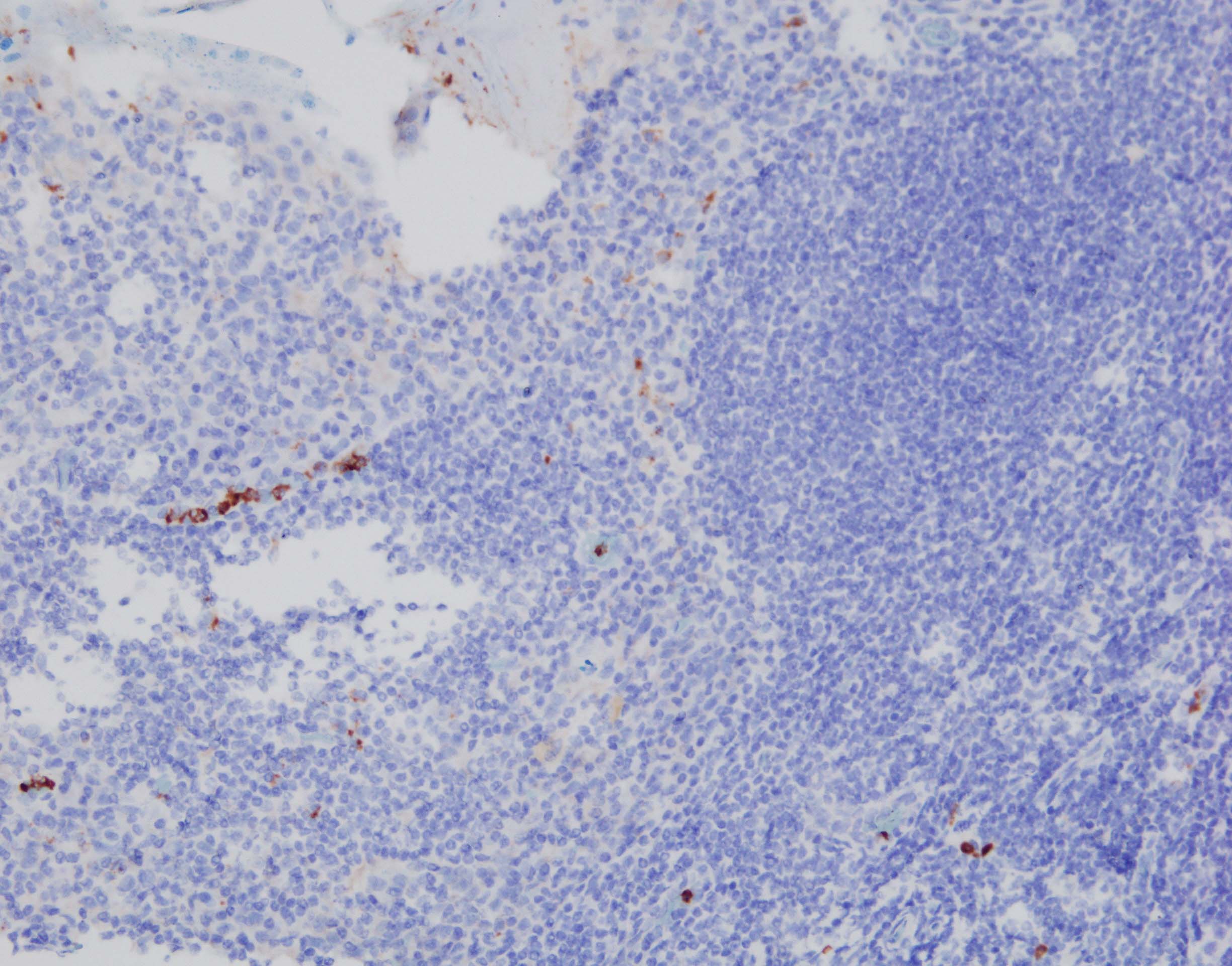
IL‑17/IL‑17A in Human Tonsil.
IL-17/IL-17A was detected in immersion fixed paraffin-embedded sections of human tonsil using Goat Anti-Human IL-17/IL-17A Antigen Affinity-purified Polyclonal Antibody (Catalog # AF-317-NA) at 1 µg/mL for 1 hour at room temperature followed by incubation with the Anti-Goat IgG VisUCyte™ HRP Polymer Antibody (VC004). Tissue was stained using DAB (brown) and counterstained with hematoxylin (blue). Specific staining was localized to lymphocytes. View our protocol for IHC Staining with VisUCyte HRP Polymer Detection Reagents.
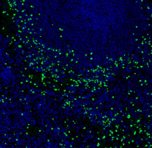
Detection of IL‑17/IL‑17A in Human Crohn's Intestine
IL-17A mRNA (red) and IL17A protein (green) were detected in formalin-fixed paraffin-embedded tissue sections of human Crohn’s Disease Intestine. ACD’s Integrated Co-Detection Workflow was performed using ACD RNAScope Probe Hs-IL-17A (Catalog # 310931) and goat anti-human IL-17/IL-17A polyclonal antibody (R&D Systems Catalog # AF-317-NA) at 1ug/mL. Tissue was stained using RNAscope® 2.5 HD Detection Kit-RED (Catalog # 322360) and RNAscope® 2.5 LS Green Accessory Pack (Catalog # 322550). Tissue was counterstained with 50% hematoxylin (blue).
Detection of IL‑17 in Human PBMCs by Flow Cytometry.
Human peripheral blood mononuclear cells were unstimulated (light orange filled histogram) or treated with 50 ng/mL PMA and 250 ng/mL Ca 2+ionomycin for 16 hours, then stained with Goat Anti-Human IL-17 Antigen Affinity-purified Polyclonal Antibody (Catalog # AF-317-NA, dark orange filled histogram) or isotype control antibody (AB-108-C, open histogram), followed by Allophycocyanin-conjugated Anti-Goat IgG Secondary Antibody (F0108). To facilitate intracellular staining, cells were fixed with paraformaldehyde and permeabilized with saponin.
IL‑6 Secretion Induced by IL‑17 and Neutralization by Human IL‑17 Antibody.
Recombinant Human IL-17 (317-ILB) stimulates IL-6 secretion in NIH/3T3 mouse embryonic fibroblasts in a dose-dependent manner (orange line), as measured by the mouse IL-6 Quantikine ELISA Kit (Catalog # M6000B). IL-6 secretion elicited by Recombinant Human IL-17 (15 ng/mL) is neutralized (green line) by increasing concentrations of Goat Anti-Human IL-17 Antigen Affinity-purified Polyclonal Antibody (Catalog # AF-317-NA). The ND50 is typically 0.02‑0.12 µg/mL.
Detection of Human IL-17/IL-17A by Immunohistochemistry
Expression of psoriasis‐related cytokines in paradoxical psoriasiform reactions. Immunohistochemistry analysis performed on paradoxical skin lesions obtained from patients 1 (Pt1) and 2 (Pt2) shows similar values of IL‐17A+ cells, a reduction of dermal IFN‐ gamma + cells and an increase of IL‐22+ or IL‐36 gamma + cells, when compared with psoriatic skin lesions. LS and NLS skin of the same psoriatic patient (n = 3) was analyzed. Graphs show the mean of number of positive cells + SD per three sections. One out of three representative stainings is shown. *p < 0.01, **p < 0.05, versus classical psoriasis. Scale bars, 200 μm. Image collected and cropped by CiteAb from the following publication (https://pubmed.ncbi.nlm.nih.gov/31577850), licensed under a CC-BY license. Not internally tested by R&D Systems.
Detection of Human IL-17/IL-17A by Immunohistochemistry
Inflammatory cell infiltrates of early acne lesions in immunohistochemistry.Representative immunohistochemical staining showing IL-17A+ cells in early acne lesion (a). Staining of IL-17A+ and CD3+ cells shows that IL-17A (in green) and CD3 (in red) were detected in acne vulgaris lesions. Cell nuclei were counterstained in blue. Arrows indicate IL-17A and CD3 double positive cells (b). T-bet+ cells (c) were more numerous than IL-17A+ cells or Foxp3+ cells (d) in the lesional acne skin. Large number of CD4+ cells (e), mostly lymphocytes, was seen around pilosebaceous unit and perivascularly. CD8+ cells (f) were fewer in number than CD4+ cells. Large number of CD68+ macrophages (g) and a few CD83+ cells (h), which are mature dendritic cells, were detected around sebaceous follicles. (Bar = 200 µm; in the insets bar = 50 µm). Image collected and cropped by CiteAb from the following publication (https://pubmed.ncbi.nlm.nih.gov/25153527), licensed under a CC-BY license. Not internally tested by R&D Systems.
Detection of IL-17/IL-17A by Western Blot
IL-17 upregulates FAP expression by activating the p-STAT3 signaling pathway.A Protein levels of FAP, pSTAT3 (Tyr705), pSTAT3 (Ser727), and STAT3 after 20 ng/ml IL-17a stimulation or PBS treatment. B Protein levels of FAP, IL-17, ACTA2, and VIMENTIN in IL-17-OE cells, FAP-OE cells, and the corresponding negative control cell lines. C Protein levels of pSTAT3 (Tyr705), pSTAT3 (Ser727), STAT3, pP65, and P65 in IL-17-OE cells, FAP-OE cells, and the corresponding negative control cell lines. D The binding ability of pSTAT3 (Tyr705) and STAT3 to the FAP promoter region in FAP-OE and FAP-NC cells was evaluated by a CUT&RUN assay. E The binding ability of pSTAT3 (Tyr705) and STAT3 to the FAP promoter region in IL-17-OE and IL-17-NC cells was evaluated by a CUT&RUN assay. F The mRNA expression of FAP of LX2 cell line after 24 h treatment of 50 μg/mL Colivelin. G The expression of FAP, STAT3, and p-STAT3 in LX2 cell line after 24 h treatment of 50 μg/mL Colivelin. H The FAP, STAT3, p-STAT3 level in FAP-OE and FAP-NC cell lines after 2 μM HJC0152 treatment. Image collected and cropped by CiteAb from the following open publication (https://www.nature.com/articles/s41420-024-01995-4), licensed under a CC-BY license. Not internally tested by R&D Systems.
Human IL-17 / IL-17A ELISA Standard Curve
Recombinant Human IL‑17/IL‑17A (Catalog # 317-ILB) was serially diluted and captured by Mouse Anti-Human/Primate IL‑17/IL‑17A Monoclonal Antibody (Catalog # MAB317) coated on a Clear Polystyrene Microplate (Catalog # DY990). Goat Anti-Human IL‑17/IL‑17A Antigen Affinity-purified Polyclonal Antibody (Catalog # AF-317-NA) was biotinylated and incubated with the protein captured on the plate. Detection of the standard curve was achieved by incubating Streptavidin-HRP (Catalog # DY998)Applications for Human IL-17/IL-17A Antibody
Application
Recommended Usage
CyTOF-ready
Ready to be labeled using established conjugation methods. No BSA or other carrier proteins that could interfere with conjugation.
Dual RNAscope ISH-IHC Compatible
0.5 - 5 µg/mL
Sample: Immersion fixed paraffin-embedded sections of human Crohn's intestine
Sample: Immersion fixed paraffin-embedded sections of human Crohn's intestine
Immunocytochemistry
5-15 µg/mL
Sample: Immersion fixed human peripheral blood mononuclear cells treated with PHA
Sample: Immersion fixed human peripheral blood mononuclear cells treated with PHA
Immunohistochemistry
1-15 µg/mL
Sample: Immersion fixed paraffin-embedded sections of human tonsil
Sample: Immersion fixed paraffin-embedded sections of human tonsil
Intracellular Staining by Flow Cytometry
2.5 µg/106 cells
Sample: Human peripheral blood mononuclear cells treated with PMA and Ca2+ ionomycin, fixed with paraformaldehyde, and permeabilized with saponin
Sample: Human peripheral blood mononuclear cells treated with PMA and Ca2+ ionomycin, fixed with paraformaldehyde, and permeabilized with saponin
Neutralization
Measured by its ability to neutralize IL‑17-induced IL‑6 secretion in NIH/3T3 mouse embryonic fibroblasts. Yao, Z. et al. (1995) Immunity 3:811. The Neutralization Dose (ND50) is typically 0.02-0.12 µg/mL in the presence of 15 ng/mL Recombinant Human IL‑17.
Reviewed Applications
Read 9 reviews rated 4.2 using AF-317-NA in the following applications:
Flow Cytometry Panel Builder
Bio-Techne Knows Flow Cytometry
Save time and reduce costly mistakes by quickly finding compatible reagents using the Panel Builder Tool.
Advanced Features
- Spectra Viewer - Custom analysis of spectra from multiple fluorochromes
- Spillover Popups - Visualize the spectra of individual fluorochromes
- Antigen Density Selector - Match fluorochrome brightness with antigen density

Formulation, Preparation, and Storage
Purification
Antigen Affinity-purified
Reconstitution
Reconstitute at 0.2 mg/mL in sterile PBS. For liquid material, refer to CoA for concentration.
Loading...
Formulation
Lyophilized from a 0.2 μm filtered solution in PBS with Trehalose. See Certificate of Analysis for details.
*Small pack size (-SP) is supplied either lyophilized or as a 0.2 µm filtered solution in PBS.
*Small pack size (-SP) is supplied either lyophilized or as a 0.2 µm filtered solution in PBS.
Shipping
Lyophilized product is shipped at ambient temperature. Liquid small pack size (-SP) is shipped with polar packs. Upon receipt, store immediately at the temperature recommended below.
Stability & Storage
Use a manual defrost freezer and avoid repeated freeze-thaw cycles.
- 12 months from date of receipt, -20 to -70 °C as supplied.
- 1 month, 2 to 8 °C under sterile conditions after reconstitution.
- 6 months, -20 to -70 °C under sterile conditions after reconstitution.
Calculators
Background: IL-17/IL-17A
Long Name
Interleukin 17
Alternate Names
CTLA-8, CTLA8, IL-17A, IL17, IL17A
Entrez Gene IDs
Gene Symbol
IL17A
UniProt
Additional IL-17/IL-17A Products
Product Documents for Human IL-17/IL-17A Antibody
Certificate of Analysis
To download a Certificate of Analysis, please enter a lot or batch number in the search box below.
Note: Certificate of Analysis not available for kit components.
Product Specific Notices for Human IL-17/IL-17A Antibody
For research use only
Citations for Human IL-17/IL-17A Antibody
 Powered by Bioz
Customer Reviews for Human IL-17/IL-17A Antibody (9)
4.2 out of 5
9 Customer Ratings
Have you used Human IL-17/IL-17A Antibody?
Submit a review and receive an Amazon gift card!
$25/€18/£15/$25CAN/¥2500 Yen for a review with an image
$10/€7/£6/$10CAN/¥1110 Yen for a review without an image
Submit a review
Customer Images

Showing
1
-
5 的
9 reviews
Showing All
Filter By:
-
Application: ImmunohistochemistrySample Tested: Tonsil tissueSpecies: HumanVerified Customer | Posted 09/13/2023FFPE heating antigen retrieval in pH9 primary Ab x100 in 1% BSA (IgG free) for 1 hour biotinylated rabbit anti-goat and streptavidin-HRP + DAB

-
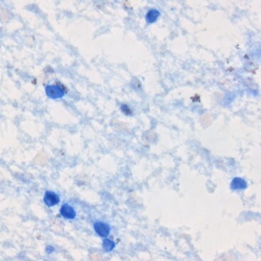
Application: ImmunohistochemistrySample Tested: Adult brainSpecies: HumanVerified Customer | Posted 04/13/2018Published in https://www.ncbi.nlm.nih.gov/pubmed/28169287 Used at 10ug/ml. Briefly, frozen brain sections were fixed in 4% PFA (Fisher Scientific), followed by antigen retrieval using heating in acid citric buffer (Vector, Burlingame, CA, USA). Endogenous avidin-biotin was blocked for 15 min (Vector). Sections were incubated with 10% horse serum in PBS (Biosera, Boussens, France) and Fc Receptor Blocking Solution was added (Human TruStain FcX Biolegend, London, UK). Primary antibody was added overnight at 4 °C and detected with donkey anti-goat-biotin (ab6578, Abcam), followed by streptavidin-alkaline phosphatase (SA-5100, Vector) and visualised with the Vector Blue Alkaline Phosphatase Substrate Kit III (Vector).
-
Application: Block/NeutralizeSample Tested: 4T1 mouse breast cancer cell line and Breast tissueSpecies: mouse tumor-infiltrate lympocytes and MouseVerified Customer | Posted 03/23/2018
-
Verified Customer | Posted 04/21/2017Antigen retrieval pH 6.0 (20 min) Dilution-1:100, 1 h incubation. Detection with ImmPRESS HRP Anti-Goat Ig (Peroxidase) Polymer (# MP-7405-15 ) and TSA-Cy3 (#SAT704A001EA)
-
Application: ImmunohistochemistrySample Tested: Spleen tissue and Colon tissueSpecies: HumanVerified Customer | Posted 04/21/2017Staining is done in Leica Bond autostainer. Epitope retrieval at pH 6 (20 min), IL17/IL17A dilution 1:100, incubation time-1h. Detected with TSA-Cy3
-
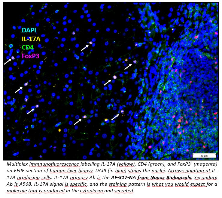
Application: ImmunocytochemistrySample Tested: FFPE and FFPE human liverSpecies: HumanVerified Customer | Posted 03/24/2017Multiplex immnunofluorescence labelling IL-17A, CD4, and FoxP3 on FFPE section of human liver biopsy. IL-17A primary Ab is the AF-317-NA from Novus Biologicals. Secondary Ab is A568. FFPE sections were dewaxed with xylene and rehydrated with ethanol solutions (decreasing %). Antigen retrieval was performed with sodium citrate pH6 in a pressure cooker. IL-17A signal is specific, and the staining pattern is what you would expect for a molecule that is produced in the cytoplasm and secreted.
-
Application: Immunohistochemistry-ParaffinSample Tested: See PMID 20426778Species: HumanVerified Customer | Posted 01/07/2015
-
Application: Immunohistochemistry-ParaffinSample Tested: See PMID 20357258Species: OtherVerified Customer | Posted 01/07/2015
-
Application: ImmunocytochemistrySample Tested: See PMID 21307293Species: HumanVerified Customer | Posted 01/07/2015
There are no reviews that match your criteria.
Protocols
Find general support by application which include: protocols, troubleshooting, illustrated assays, videos and webinars.
- 7-Amino Actinomycin D (7-AAD) Cell Viability Flow Cytometry Protocol
- Antigen Retrieval Protocol (PIER)
- Antigen Retrieval for Frozen Sections Protocol
- Appropriate Fixation of IHC/ICC Samples
- Cellular Response to Hypoxia Protocols
- Chromogenic IHC Staining of Formalin-Fixed Paraffin-Embedded (FFPE) Tissue Protocol
- Chromogenic Immunohistochemistry Staining of Frozen Tissue
- ClariTSA™ Fluorophore Kits
- Detection & Visualization of Antibody Binding
- Extracellular Membrane Flow Cytometry Protocol
- Flow Cytometry Protocol for Cell Surface Markers
- Flow Cytometry Protocol for Staining Membrane Associated Proteins
- Flow Cytometry Staining Protocols
- Flow Cytometry Troubleshooting Guide
- Fluorescent IHC Staining of Frozen Tissue Protocol
- Graphic Protocol for Heat-induced Epitope Retrieval
- Graphic Protocol for the Preparation and Fluorescent IHC Staining of Frozen Tissue Sections
- Graphic Protocol for the Preparation and Fluorescent IHC Staining of Paraffin-embedded Tissue Sections
- Graphic Protocol for the Preparation of Gelatin-coated Slides for Histological Tissue Sections
- ICC Cell Smear Protocol for Suspension Cells
- ICC Immunocytochemistry Protocol Videos
- ICC for Adherent Cells
- IHC Sample Preparation (Frozen sections vs Paraffin)
- ISH-IHC Protocol for Chromogenic Detection on Formalin Fixed Paraffin Embedded (FFPE) Tissue
- Immunocytochemistry (ICC) Protocol
- Immunocytochemistry Troubleshooting
- Immunofluorescence of Organoids Embedded in Cultrex Basement Membrane Extract
- Immunofluorescent IHC Staining of Formalin-Fixed Paraffin-Embedded (FFPE) Tissue Protocol
- Immunohistochemistry (IHC) and Immunocytochemistry (ICC) Protocols
- Immunohistochemistry Frozen Troubleshooting
- Immunohistochemistry Paraffin Troubleshooting
- Intracellular Flow Cytometry Protocol Using Alcohol (Methanol)
- Intracellular Flow Cytometry Protocol Using Detergents
- Intracellular Nuclear Staining Flow Cytometry Protocol Using Detergents
- Intracellular Staining Flow Cytometry Protocol Using Alcohol Permeabilization
- Intracellular Staining Flow Cytometry Protocol Using Detergents to Permeabilize Cells
- Preparing Samples for IHC/ICC Experiments
- Preventing Non-Specific Staining (Non-Specific Binding)
- Primary Antibody Selection & Optimization
- Propidium Iodide Cell Viability Flow Cytometry Protocol
- Protocol for Heat-Induced Epitope Retrieval (HIER)
- Protocol for Liperfluo
- Protocol for Making a 4% Formaldehyde Solution in PBS
- Protocol for VisUCyte™ HRP Polymer Detection Reagent
- Protocol for the Characterization of Human Th22 Cells
- Protocol for the Characterization of Human Th9 Cells
- Protocol for the Fluorescent ICC Staining of Cell Smears - Graphic
- Protocol for the Fluorescent ICC Staining of Cultured Cells on Coverslips - Graphic
- Protocol for the Preparation & Fixation of Cells on Coverslips
- Protocol for the Preparation and Chromogenic IHC Staining of Frozen Tissue Sections
- Protocol for the Preparation and Chromogenic IHC Staining of Frozen Tissue Sections - Graphic
- Protocol for the Preparation and Chromogenic IHC Staining of Paraffin-embedded Tissue Sections
- Protocol for the Preparation and Chromogenic IHC Staining of Paraffin-embedded Tissue Sections - Graphic
- Protocol for the Preparation and Fluorescent ICC Staining of Cells on Coverslips
- Protocol for the Preparation and Fluorescent ICC Staining of Non-adherent Cells
- Protocol for the Preparation and Fluorescent ICC Staining of Stem Cells on Coverslips
- Protocol for the Preparation and Fluorescent IHC Staining of Frozen Tissue Sections
- Protocol for the Preparation and Fluorescent IHC Staining of Paraffin-embedded Tissue Sections
- Protocol for the Preparation of Gelatin-coated Slides for Histological Tissue Sections
- Protocol for the Preparation of a Cell Smear for Non-adherent Cell ICC - Graphic
- Protocol: Annexin V and PI Staining by Flow Cytometry
- Protocol: Annexin V and PI Staining for Apoptosis by Flow Cytometry
- TUNEL and Active Caspase-3 Detection by IHC/ICC Protocol
- The Importance of IHC/ICC Controls
- Troubleshooting Guide: Fluorokine Flow Cytometry Kits
- Troubleshooting Guide: Immunohistochemistry
- View all Protocols, Troubleshooting, Illustrated assays and Webinars
Loading...
Associated Pathways


