Actin Antibody (mAbGEa) - BSA Free
Novus Biologicals | Catalog # NB100-74340

Key Product Details
Species Reactivity
Validated:
Cited:
Applications
Validated:
Cited:
Label
Antibody Source
Format
Product Specifications
Immunogen
Reactivity Notes
Localization
Specificity
Clonality
Host
Isotype
Scientific Data Images for Actin Antibody (mAbGEa) - BSA Free
Detection of Human Actin by Simple Western TM.
Left: Simple Western lane view shows lysates of human liver, loaded at 1 mg/ml. A specific band was detected for Actin at approximately 47 kDa (as indicated) using both 1:10,000 and 1:2,000 of Mouse Anti-Actin Monoclonal Antibody (Catalog # NB100-74340) followed by 1:50 HRP-conjugated Goat Anti-Mouse Secondary Antibody (Catalog # 042-205). This experiment was conducted under reducing conditions and using the 12-230kDa separation system. Right: Simple Western electropherogram showing the same Mouse Anti-Actin Monoclonal Antibody (Catalog # NB100-74340) tested at 1:10,000 (blue line) and 1:2,000 (green line) in the human liver.![Western Blot: Actin Antibody (mAbGEa) [NB100-74340]](https://resources.rndsystems.com/images/products/Actin-Antibody-mAbGEa-Western-Blot-NB100-74340-img0019.jpg "Western Blot: Actin Antibody (mAbGEa) [NB100-74340]")
Western Blot: Actin Antibody (mAbGEa) [NB100-74340]
Western Blot: Actin Antibody (mAbGEa) [NB100-74340] - Total protein from HeLa, 3T3, PC12 and Bovine normal tissue was separated on a 12% gel by SDS-PAGE, transferred to PVDF membrane and blocked in 5% non-fat milk in TBST. The membrane was probed with 1.0 ug/mL anti-Actin in 5% blocking buffer and detected with an anti-mouse IgM secondary antibody using chemiluminescence.![Immunocytochemistry/ Immunofluorescence: Actin Antibody (mAbGEa) [NB100-74340]](https://resources.rndsystems.com/images/products/Actin-Antibody-mAbGEa-Immunocytochemistry-Immunofluorescence-NB100-74340-img0018.jpg "Immunocytochemistry/ Immunofluorescence: Actin Antibody (mAbGEa) [NB100-74340]")
Immunocytochemistry/ Immunofluorescence: Actin Antibody (mAbGEa) [NB100-74340]
Immunocytochemistry/Immunofluorescence: Actin Antibody (mAbGEa) [NB100-74340] - NIH-3T3 cells were fixed for 10 minutes using 10% formalin and then permeabilized for 5 minutes using 1X PBS + 0.05% Triton X-100. The cells were incubated with anti-Actin (mAbGEa) at 5 ug/mL overnight at 4C and detected with an anti-Mouse IgM DyLight 488 (Green) at a 1:500 dilution. Nuclei were counterstained with DAPI (Blue). Cells were imaged using a 40X objective.![Simple Western: Actin Antibody (mAbGEa) [NB100-74340]](https://resources.rndsystems.com/images/products/Actin-Antibody-mAbGEa-Simple-Western-NB100-74340-img0014.jpg "Simple Western: Actin Antibody (mAbGEa) [NB100-74340]")
Simple Western: Actin Antibody (mAbGEa) [NB100-74340]
Simple Western: Actin Antibody (mAbGEa) [NB100-74340] - Image shows a specific band for Actin in 0.1 mg/mL of HeLa lysate. This experiment was performed under reducing conditions using the 12-230 kDa separation system.![Immunohistochemistry-Paraffin: Actin Antibody (mAbGEa) [NB100-74340]](https://resources.rndsystems.com/images/products/Actin-Antibody-mAbGEa-Immunohistochemistry-Paraffin-NB100-74340-img0005.jpg "Immunohistochemistry-Paraffin: Actin Antibody (mAbGEa) [NB100-74340]")
Immunohistochemistry-Paraffin: Actin Antibody (mAbGEa) [NB100-74340]
Immunohistochemistry-Paraffin: Actin Antibody (mAbGEa) [NB100-74340] - Both normal and cancer biopsies of deparaffinized Human tonsil tissues.![Flow Cytometry: Actin Antibody (mAbGEa) [NB100-74340]](https://resources.rndsystems.com/images/products/Actin-Antibody-mAbGEa-Flow-Cytometry-NB100-74340-img0015.jpg "Flow Cytometry: Actin Antibody (mAbGEa) [NB100-74340]")
Flow Cytometry: Actin Antibody (mAbGEa) [NB100-74340]
Flow Cytometry: Actin Antibody (mAbGEa) [NB100-74340] - Analysis of HeLa cells using mouse Actin antibody (Orange) and Isotype control Antibody (Blue).![Western Blot: Actin Antibody (mAbGEa) [NB100-74340]](https://resources.rndsystems.com/images/products/Actin-Antibody-mAbGEa-Western-Blot-NB100-74340-img0022.jpg "Western Blot: Actin Antibody (mAbGEa) [NB100-74340]")
Western Blot: Actin Antibody (mAbGEa) [NB100-74340]
Actin-Antibody-mAbGEa-Western-Blot-NB100-74340-img0022.jpg![Immunocytochemistry/ Immunofluorescence: Actin Antibody (mAbGEa) [NB100-74340]](https://resources.rndsystems.com/images/products/Actin-Antibody-mAbGEa-Immunocytochemistry-Immunofluorescence-NB100-74340-img0021.jpg "Immunocytochemistry/ Immunofluorescence: Actin Antibody (mAbGEa) [NB100-74340]")
Immunocytochemistry/ Immunofluorescence: Actin Antibody (mAbGEa) [NB100-74340]
Immunocytochemistry/Immunofluorescence: Actin Antibody (mAbGEa) [NB100-74340] - HeLa cells were fixed for 10 minutes using 10% formalin and then permeabilized for 5 minutes using 1X PBS + 0.05% Triton-X100. The cells were incubated with anti-Actin Antibody (mAbGEa) at 2 ug/ml overnight at 4C and detected with an anti-mouse Dylight 488 (Green) at a 1:500 dilution. Actin was detected with Phalloidin 568 (Red) at a 1:200 dilution. Nuclei were counterstained with DAPI (Blue). Cells were imaged using a 40X objective.![Flow (Intracellular): Actin Antibody (mAbGEa) [NB100-74340]](https://resources.rndsystems.com/images/products/Actin-Antibody-mAbGEa-Flow-Intracellular-NB100-74340-img0020.jpg "Flow (Intracellular): Actin Antibody (mAbGEa) [NB100-74340]")
Flow (Intracellular): Actin Antibody (mAbGEa) [NB100-74340]
Flow (Intracellular): Actin Antibody (mAbGEa) [NB100-74340] - An intracellular stain was performed on A549 cells with NB100-74340 (blue) and a matched isotype control (orange). Cells were fixed with 4% PFA and then permeablized with 0.1% saponin. Cells were incubated in an antibody dilution of 1 ug/mL for 30 minutes at room temperature, followed by mouse IgM Alexa Fluor 488-conjugated secondary antibody.![Western Blot: Actin Antibody (mAbGEa) [NB100-74340]](https://resources.rndsystems.com/images/products/Actin-Antibody-mAbGEa-Western-Blot-NB100-74340-img0008.jpg "Western Blot: Actin Antibody (mAbGEa) [NB100-74340]")
Western Blot: Actin Antibody (mAbGEa) [NB100-74340]
Western Blot: Actin Antibody (mAbGEa) [NB100-74340] - Analysis of Actin expression in 2) HeLa, 3) NTERA-2, 4) A431, 5) HepG2, 6) MCF7, 7) NIH-3T3, 8) PC-12 and 9) COS-7 whole cell lysates.![Western Blot: Actin Antibody (mAbGEa) [NB100-74340]](https://resources.rndsystems.com/images/products/Actin-Antibody-mAbGEa-Western-Blot-NB100-74340-img0012.jpg "Western Blot: Actin Antibody (mAbGEa) [NB100-74340]")
Western Blot: Actin Antibody (mAbGEa) [NB100-74340]
Western Blot: Actin Antibody (mAbGEa) [NB100-74340] - Analysis of Jurkat and CHO cell lysates using actin antibody [NB100-74340] at 1:100.![Immunocytochemistry/ Immunofluorescence: Actin Antibody (mAbGEa) [NB100-74340]](https://resources.rndsystems.com/images/products/Actin-Antibody-mAbGEa-Immunocytochemistry-Immunofluorescence-NB100-74340-img0016.jpg "Immunocytochemistry/ Immunofluorescence: Actin Antibody (mAbGEa) [NB100-74340]")
Immunocytochemistry/ Immunofluorescence: Actin Antibody (mAbGEa) [NB100-74340]
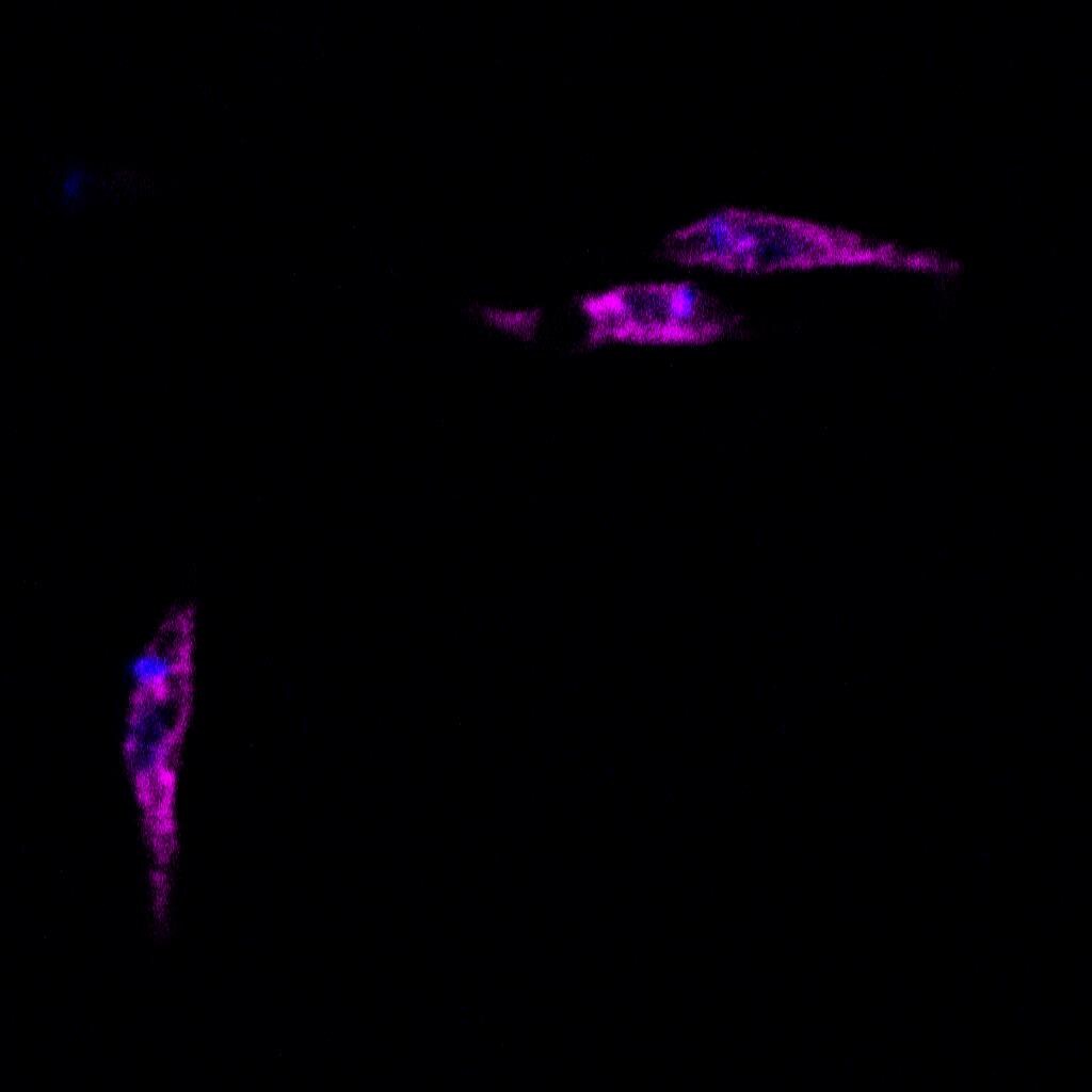
Immunocytochemistry/Immunofluorescence: Actin Antibody (mAbGEa) [NB100-74340] - Actin was detected in NIH-3T3 cells fixed with methanol using mouse anti-mouse beta-Actin monoclonal antibody (NB100-74340) at 1:1800. Cells were stained using Northern Lights 557 conjugated anti-mouse secondary antibody (NL007) and counterstained with DAPI.![Immunocytochemistry/ Immunofluorescence: Actin Antibody (mAbGEa) [NB100-74340]](https://resources.rndsystems.com/images/products/Actin-Antibody-mAbGEa-Immunocytochemistry-Immunofluorescence-NB100-74340-img0017.jpg "Immunocytochemistry/ Immunofluorescence: Actin Antibody (mAbGEa) [NB100-74340]")
Immunocytochemistry/ Immunofluorescence: Actin Antibody (mAbGEa) [NB100-74340]
Immunocytochemistry/Immunofluorescence: Actin Antibody (mAbGEa) [NB100-74340] - HeLa cells were fixed for 10 minutes using 10% formalin and then permeabilized for 5 minutes using 1X PBS + 0.05% Triton X-100. The cells were incubated with anti-Actin (mAbGEa) at 2 ug/mL overnight at 4C and detected with an anti-mouse IgM DyLight 488 (Green) at 1:500. Nuclei were counterstained with DAPI (Blue). Cells were imaged using a 40X objective.![Immunohistochemistry-Paraffin: Actin Antibody (mAbGEa) [NB100-74340]](https://resources.rndsystems.com/images/products/Actin-Antibody-mAbGEa-Immunohistochemistry-Paraffin-NB100-74340-img0010.jpg "Immunohistochemistry-Paraffin: Actin Antibody (mAbGEa) [NB100-74340]")
Immunohistochemistry-Paraffin: Actin Antibody (mAbGEa) [NB100-74340]
Immunohistochemistry-Paraffin: Actin Antibody (mAbGEa) [NB100-74340] - Analysis of Actin on human breast cancer tissue using DAB with hematoxylin counterstain.![Immunohistochemistry-Paraffin: Actin Antibody (mAbGEa) [NB100-74340]](https://resources.rndsystems.com/images/products/Actin-Antibody-mAbGEa-Immunohistochemistry-Paraffin-NB100-74340-img0003.jpg "Immunohistochemistry-Paraffin: Actin Antibody (mAbGEa) [NB100-74340]")
Immunohistochemistry-Paraffin: Actin Antibody (mAbGEa) [NB100-74340]
Immunohistochemistry-Paraffin: Actin Antibody (mAbGEa) [NB100-74340] - Both normal and cancer biopsies of deparaffinized Human colon carcinoma tissues.![Immunohistochemistry-Paraffin: Actin Antibody (mAbGEa) [NB100-74340]](https://resources.rndsystems.com/images/products/Actin-Antibody-mAbGEa-Immunohistochemistry-Paraffin-NB100-74340-img0004.jpg "Immunohistochemistry-Paraffin: Actin Antibody (mAbGEa) [NB100-74340]")
Immunohistochemistry-Paraffin: Actin Antibody (mAbGEa) [NB100-74340]
Immunohistochemistry-Paraffin: Actin Antibody (mAbGEa) [NB100-74340] - Both normal and cancer biopsies of deparaffinized Human skeletal muscle tissues. [NB100-74340] -")
Western Blot: Actin Antibody (mAbGEa) [NB100-74340] -
Western Blot: Actin Antibody (mAbGEa) [NB100-74340] - Lp(a) upregulates GPx1 & Prdx6 expression in human HepG2 cells. HepG2 cells were treated with 5 μg/mL of Lp(a) or LDL for 6 hours at 37°C. Western blots of cell lysates were performed with an anti-Gpx1 antibody (a), an anti-Prdx6 antibody (b), an anti-Sod1 antibody (c), & an anti-Park7 antibody (d) using an anti-actin antibody as a loading control. Representative blots are shown. Protein levels were normalized against actin & expressed relative to that of untreated cells. Results are expressed as mean ± SEM for pooled triplicate incubations run in quadruplicate. ∗P<0.05, relative to untreated HepG2 cells. Image collected & cropped by CiteAb from the following publication (https://pubmed.ncbi.nlm.nih.gov/30596094), licensed under a CC-BY license. Not internally tested by Novus Biologicals. [NB100-74340] -")
Western Blot: Actin Antibody (mAbGEa) [NB100-74340] -
Western Blot: Actin Antibody (mAbGEa) [NB100-74340] - DenA deneddylase activity.(A) Recombinant human DEN1 & fungal DenA deneddylate a human CUL1-Nedd8 substrate in vitro. SDS-PAGE & subsequent western analysis show cleavage of the substrate (∼60 kDa) producing the C-terminal CUL1 (∼50 kDa) as outlined in experimental procedures. (B) Deneddylation test in a heterologous yeast system. A. nidulans DenA can remove Rub1 from CulD in heterologous expression experiments in S. cerevisiae. DenA expressed as native protein or C-terminally fused w/ a V5/His6 epitope tag. Both variants driven by the inducible GAL1 promoter. CulD, N-terminal-fused w/ the LexA activation domain, expressed under control of the constitutive ADH promoter. A. nidulans proteins expressed in S. cerevisiae wild type & delta csn5 background. Western analysis w/ antibodies against Rub1 ( alpha -Rub1), the LexA epitope ( alpha -LexA) & the V5 epitope ( alpha -V5) performed. Detection w/ alpha -Rub1 generated two additional signals upon culD expression, representing LexA-CulD & a second CulD pool where LexA unspecifically cleaved off. Both signals disappeared upon co-expression of DenA indicating deneddylation activity (red arrows). The slower migrating signal of alpha -LexA western experiments corresponded to Rub1 modified LexA-CulD. This signal absent when DenA co-expressed. Detection of the V5 tag applied to monitor DenA expression. The neddylated yeast cullin migrating at around 100 kDa not affected by DenA activity. (C) Deneddylation of fungal CulA by CSN & DenA. Whole cell lysates of A. nidulans wild type, delta csnE & delta denA probed w/ alpha -CulA. The ratio of neddylated CulA (CulA*N8; ∼106 kDa*) to non-neddylated CulA (∼96 kDa**) calculated from three independent experiments. Membranes reprobed w/ alpha -Actin ( alpha -ACT) for normalization. Image collected & cropped by CiteAb from the following publication (https://dx.plos.org/10.1371/journal.pgen.1003275), licensed under a CC-BY license. Not internally tested by Novus Biologicals. [NB100-74340] -")
Western Blot: Actin Antibody (mAbGEa) [NB100-74340] -
Western Blot: Actin Antibody (mAbGEa) [NB100-74340] - Fibrillar adhesions & FN fibrils are dynamic structures governed by Rab21 & PPFIA1.(a) Living siCTL & siRAB21 ECs incubated w/ IST9 mAb for 30 min, fixed, acid washed & stained. RAB21 silencing impairs ED-A FN endocytosis as revealed by decrease of IST9 punctae co-localizing w/ endosome marker EEA1. Right panels are magnifications of boxed areas in left panels. Data are mean value±s.e.m., n=70 cells per condition pooled from two independent experiments. ***P<0.001. (b) Left panel, WB analysis of PPFIA1, RAB21 & actin on total lysates of siCTL, siPPFIA1 & siPPFIA1+siRAB21 ECs. Right panel, WB analysis of insoluble matrix fraction of ECs extracted w/ DOC buffer. PPFIA1 silencing dramatically reduces amount of DOC-insoluble fraction of endogenous ED-A FN. Of note, simultaneous silencing of Rab21 GTPase (siPPFIA1+siRAB21), which drives integrin endocytosis, rescues defective incorporation of endogenous ED-A FN in DOC-insoluble fraction of siPPFIA1 ECs. (c) Confocal microscopy analysis of patterning of endogenous cellular ED-A FN (green) in fixed confluent ECs. Before fixation, living ECs incubated for 20 min w/ exogenous SNAKA51 (red). ED-A FN polymerizes into a fibrillar network in siCTL, but not in siPPFIA1 ECs. Simultaneous silencing of Rab21 GTPase (siPPFIA1+siRAB21) fully restores ED-A FN polymerization in siPPFIA1 ECs. SNAKA51+ active alpha 5 beta 1 integrin localizes in fibrillar adhesion in siCTL, but not in siPPFIA1 ECs. Notably, simultaneous Rab21 (siPPFIA1+siRAB21) silencing promotes localization of SNAKA51 in fibrillar adhesion of siPPFIA1 ECs. Data are mean±s.e.m., n=20 cells per condition pooled from two independent experiments. Scale bar, 20 μm (a,c, right), 50μm (c, left). ***P<0.001; Student's t-test. Image collected & cropped by CiteAb from following publication (https://www.nature.com/articles/ncomms13546), licensed under a CC-BY license. Not internally tested by Novus Biologicals. [NB100-74340] -")
Western Blot: Actin Antibody (mAbGEa) [NB100-74340] -
Western Blot: Actin Antibody (mAbGEa) [NB100-74340] - Effect of the four most common herbal formulas & single herbs on the phosphorylation of myosin light chain (MLC) protein.Briefly, A10 cells were treated with herbal formulas (A) or single herbs (B). Y27632 (Y10; 10 μM) & calyculin A (A50; 50 μg/ml) were used as negative & positive controls. Western blot analysis & staining with anti-phospho-MLC, anti-total-MLC, & anti-beta actin antibodies was then performed. Phospho-MLC, total-MLC, & beta actin were all obtained with their appropriate protein size bands. The relative Phospho-MLC intensity (%) was expressed as [(Phospho-MLC/total-MLC)drug treated/ (Phospho-MLC/total-MLC)cell only x 100%]. The Mean±SEM values for at least three independent experiments along with the representative western blot were performed. Image collected & cropped by CiteAb from the following publication (https://dx.plos.org/10.1371/journal.pone.0145109), licensed under a CC-BY license. Not internally tested by Novus Biologicals. [NB100-74340] -")
Western Blot: Actin Antibody (mAbGEa) [NB100-74340] -
Western Blot: Actin Antibody (mAbGEa) [NB100-74340] - CSN targets DEN1/DenA for degradation in fungal & human cells.(A) Quantitative analysis of repeated western blot experiments displayed the differences in DenA abundance in fungal wild type & delta csnG cells. DenA levels of the different asexual developmental time points are shown relative to the vegetative (veg.) control for each strain. Anti-Actin was applied as loading control (statistics: 2-way ANOVA; n = 3; *p<0,05, **p<0,01). (B) Xpress-DEN1 was overexpressed in siGFP & siCSN1 human cells & steady state Xpress-DEN1 levels were estimated by western analysis with the alpha -Xpress antibody. Xpress-CSN1 was overexpressed in siCSN1 cells & DEN1 & CSN8 were probed with appropriate antibodies. (C) Xpress-DEN1 was overexpressed in siGFP human cells & the proteasome inhibitor MG132 was added 6 h before cell lysis at a final concentration of 10 µM. Cyclohexamide (CHX) was added in a final concentration of 10 µg/ml (D) Xpress-DEN1 was co-expressed in HeLa cells together with Xpress-CSN1wt, Xpress-CSN1(1–221) or Xpress-CSN1(222–527) in the absence or in the presence of MG132 (right panel), which was added 6 h before cell lysis. Cells were lyzed 24 h after co-transfection & lysates were analyzed by western blot using the alpha -DEN1 antibody (0 = only Xpress-DEN1). Image collected & cropped by CiteAb from the following publication (https://dx.plos.org/10.1371/journal.pgen.1003275), licensed under a CC-BY license. Not internally tested by Novus Biologicals. [NB100-74340] -")
Western Blot: Actin Antibody (mAbGEa) [NB100-74340] -
Western Blot: Actin Antibody (mAbGEa) [NB100-74340] - Analysis of Ia production by engineered S. cerevisiae.(A) Analysis of the synthesis of Ia in S. cerevisiae strains, producing actin-R177K, E270D & E270Q. Yeast strains, producing actin-R177K, E270D or E270Q & transformed with the Ia-containing plasmid (Ia) or the vector alone (Vector), were cultivated in SGal for 20 h at 30°C. Cells were broken by glass beads treatment & analyzed by 32P-ADP-ribosylation in the presence of additionally added purified wild type yeast actin (1 μg). Labeled bands represent modified yeast actin & confirm intracellular production of functionally active Ia by the S. cerevisiae strains. (B) Production of Ia by the wild type S. cerevisiae strain. Wild type yeast strains harboring the Ia-containing plasmid (Ia) or the control vector (vector) were cultivated in glucose-containing liquid medium until OD595 = 0.5. Afterwards, glucose was replaced by galactose & cultivation continued for 9 h at 30°C. Cells were lysed & the resulting extract preparations were ADP-ribosylated in the presence of Ia (+ Ia), TccC3 toxin of P. luminescens [42] (+ TccC3), purified muscle actin (+ alpha -actin) or tested in Western blotting with the anti-actin serum to show equal actin concentrations in the samples. (C, D) Mass spectrometry of actin variants. MALDI-TOF MS of wild type (C) & actin-R177K (D) protein variants isolated from S. cerevisiae. Spectra demonstrate disappearance of R177- & appearance of K177-containing peptide in mass analysis (substituted amino acid residue within identified peptides is shown in red). Image collected & cropped by CiteAb from the following publication (https://dx.plos.org/10.1371/journal.pone.0145708), licensed under a CC-BY license. Not internally tested by Novus Biologicals. [NB100-74340] -")
Western Blot: Actin Antibody (mAbGEa) [NB100-74340] -
Western Blot: Actin Antibody (mAbGEa) [NB100-74340] - alpha 5 beta 1 regulates ED-A FN secretion & polymerization.(a) Western blot analysis of lysates of ECs control (siCTL) & alpha 5 integrin subunit (siITGA5) silenced ECs. Cells were lysed 24 hours after the second siRNA oligofection & proteins were separated by SDS–PAGE & probed for alpha 5 integrin subunit or actin (for control purposes). (b) Confocal microscopy analysis of IST9 mAb-labelled endogenous ED-A FN (green) in confluent ECs. ED-A FN polymerizes into a fibrillar network in siCTL, but not in siITGA5 ECs in which it accumulates in the TGN46+ (red) TGN cisternae. The relative amount of fibrillar ED-A FN area was calculated in siCTL & siITGA5 ECs. Data are mean±s.e.m., n=20 cells per condition pooled from two independent experiments. ***P<0.001; Student's t-test. (c) Western blot analysis of soluble ED-A FN released by confluent ECs seeded on Transwell inserts. An equal percentage of apical & basolateral volumes of medium were collected after 72 h of culture, from different wells of siCTL or siITGA5 ECs. Equal amounts of exogenous rabbit IgG were added to samples (spike normalization) for loading control purposes. Quantification of the ratio between apical or basolateral amount of ED-A FN released by siCTL over siITGA5 ECs. alpha 5 integrin subunit silencing impairs basolateral, but not apical ED-A FN secretion. Data are mean±s.e.m., n=8 wells per condition pooled from four independent experiments. **P<0.01; Student's t-test. Scale bar, 50 μm (b). Image collected & cropped by CiteAb from the following publication (https://www.nature.com/articles/ncomms13546), licensed under a CC-BY license. Not internally tested by Novus Biologicals. [NB100-74340] -")
Western Blot: Actin Antibody (mAbGEa) [NB100-74340] -
Western Blot: Actin Antibody (mAbGEa) [NB100-74340] - Effect of the four most common herbal formulas & single herbs on the phosphorylation of myosin light chain (MLC) protein.Briefly, A10 cells were treated with herbal formulas (A) or single herbs (B). Y27632 (Y10; 10 μM) & calyculin A (A50; 50 μg/ml) were used as negative & positive controls. Western blot analysis & staining with anti-phospho-MLC, anti-total-MLC, & anti-beta actin antibodies was then performed. Phospho-MLC, total-MLC, & beta actin were all obtained with their appropriate protein size bands. The relative Phospho-MLC intensity (%) was expressed as [(Phospho-MLC/total-MLC)drug treated/ (Phospho-MLC/total-MLC)cell only x 100%]. The Mean±SEM values for at least three independent experiments along with the representative western blot were performed. Image collected & cropped by CiteAb from the following publication (https://dx.plos.org/10.1371/journal.pone.0145109), licensed under a CC-BY license. Not internally tested by Novus Biologicals.Applications for Actin Antibody (mAbGEa) - BSA Free
ELISA
Flow (Intracellular)
Flow Cytometry
Immunocytochemistry/ Immunofluorescence
Immunohistochemistry
Immunohistochemistry-Paraffin
Simple Western
Western Blot
See Simple Western Antibody Database for Simple Western validation: Tested in HeLa lysate 0.1 mg/mL, separated by Size, antibody dilution of 1:25, apparent MW was 50 kDa. Separated by Size-Wes, Sally Sue/Peggy Sue.
Reviewed Applications
Read 8 reviews rated 4.1 using NB100-74340 in the following applications:
Flow Cytometry Panel Builder
Bio-Techne Knows Flow Cytometry
Save time and reduce costly mistakes by quickly finding compatible reagents using the Panel Builder Tool.
Advanced Features
- Spectra Viewer - Custom analysis of spectra from multiple fluorochromes
- Spillover Popups - Visualize the spectra of individual fluorochromes
- Antigen Density Selector - Match fluorochrome brightness with antigen density

Formulation, Preparation, and Storage
Purification
Formulation
Format
Preservative
Concentration
Shipping
Stability & Storage
Background: Actin
Alternate Names
Gene Symbol
Additional Actin Products
Product Documents for Actin Antibody (mAbGEa) - BSA Free
Certificate of Analysis
To download a Certificate of Analysis, please enter a lot or batch number in the search box below.
Product Specific Notices for Actin Antibody (mAbGEa) - BSA Free
This product is for research use only and is not approved for use in humans or in clinical diagnosis. Primary Antibodies are guaranteed for 1 year from date of receipt.
Related Research Areas
Citations for Actin Antibody (mAbGEa) - BSA Free
 Powered by Bioz
Customer Reviews for Actin Antibody (mAbGEa) - BSA Free (8)
Have you used Actin Antibody (mAbGEa) - BSA Free?
Submit a review and receive an Amazon gift card!
$25/€18/£15/$25CAN/¥2500 Yen for a review with an image
$10/€7/£6/$10CAN/¥1110 Yen for a review without an image
Submit a review
Customer Images


-(01-ml)_NB100-74340_8181.bmp)
-(01-ml)_NB100-74340_8176.jpg)
-
Application: Simple WesternSample Tested: brain and spinal cordSpecies: MouseVerified Customer | Posted 03/27/2018Baseline ran higher on the samples, however the peaks were consistent across samples.
-
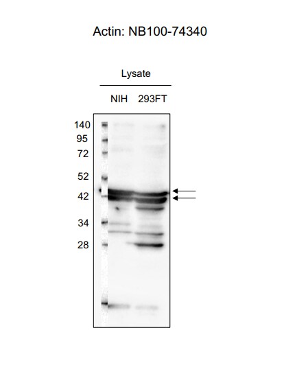
Application: Western BlotSample Tested: 293ft lysate and NIH 3T3 cell lysateSpecies: Mouse and HumanVerified Customer | Posted 06/29/2017

-
Application: Western BlotSample Tested: Whole embryos lysateSpecies: DrosophilaVerified Customer | Posted 03/21/2017Lane 1-4 and 9-12 : Embryos stage 11-13 ; +/- lambda PPase ; +/- PP2A inhibitor Lane 5-8 and 13-16 : Embryos stage 15-17 ; +/- lambda PPase ; +/- PP2A inhibitorThe antibody was diluted 1/10000 and it worked well.
-
Application: Western BlotSample Tested: SW480 (colon)Species: HumanVerified Customer | Posted 12/24/2016Marker, SW480 (colon), HT29 (colon), Colo320 (Colon) (25ug protein per well)Primary mouse anti Actin antibody diluted 1:1000 in TBS. Membrane incubation for 2 hours at RT. Secondary IRDye 800CW goat anti mouse antibody diluted 1:10000 in TBS. Membrane incubation for 1 hour at RT. Detection by Li-Cor Odyssey
-
Application: ImmunocytochemistrySample Tested:Species: OtherVerified Customer | Posted 06/11/2014Confocal immunofluorescent analysis of CHO cells using Actin antibody (NB100-74340, 1:5).
-
Application: Western BlotSample Tested:Species: HumanVerified Customer | Posted 06/11/2014Western blot analysis of extracts from Jurkat and CHO cells using Actin antibody (NB100-74340, 1:100).
-
Application: ImmunocytochemistrySample Tested: Leishmania promastigotesSpecies: OtherVerified Customer | Posted 10/17/2013ICC/IF analysis of Leishmania promastigotes in protozoa
-
Application: Western BlotSample Tested: Baking Yeast - Saccharomyces Cerevisiae, Sample Amount: Control Lane - #7 is 1ugSpecies: OtherVerified Customer | Posted 11/18/2009
There are no reviews that match your criteria.
Protocols
View specific protocols for Actin Antibody (mAbGEa) - BSA Free (NB100-74340):
Sample Preparation.
1. Grow cells to 60-85% confluency. Flow cytometry requires between 2 x 10⁵ and 1 x 10⁶ cells for optimal performance.
2. If cells are adherent, harvest gently by washing once with staining buffer and then scraping. Avoid using trypsin as this can disrupt certain epitopes of interest. If enzymatic harvest is required, use Accutase, Collagenase, or TrypLE Express for a less damaging option.
3. Reserve 100 uL for counting, then transfer cell volume into a 50 mL conical tube and centrifuge for 8 minutes at 400 RCF.
a. Count cells using a hemocytometer and a 1:1 trypan blue exclusion stain to determine cell viability before starting the flow protocol. If cells appear blue, do not proceed.
4. Re-suspend cells to a concentration of 1 x 10⁶ cells/mL in staining buffer (NBP2-26247).
5. Aliquot out 100 uL samples in accordance with your experimental samples.
Tip: When cell surface and intracellular staining are required in the same sample, it is advisable that the cell surface staining be performed first since the fixation and permeablization steps might reduce the availability of surface antigens.
Intracellular Staining.
Tip: When performing intracellular staining, it is important to use appropriate fixation and permeabilization reagents based upon the target and its subcellular location. Generally, our Intracellular Flow Assay Kit (NBP2-29450) is a good place to start as it contains an optimized combination of reagents for intracellular staining as well as an inhibitor of intracellular protein transport (necessary if staining secreted proteins). Certain targets may require more gentle or transient permeabilization protocols such as the commonly employed methanol or saponin-based methods.
Protocol for Cytoplasmic Targets:
1. Fix the cells by adding 100 uL fixation solution (such as 4% PFA) to each sample for 10-15 minutes.
2. Permeabilize cells by adding 100 uL of a permeabization buffer to every 1 x 10⁶ cells present in the sample. Mix well and incubate at room temperature for 15 minutes.
a. For cytoplasmic targets, use a gentle permeabilization solution such as 1X PBS + 0.5% Saponin or 1X PBS + 0.5% Tween-20.
b. To maintain the permeabilized state throughout your experiment, use staining buffer + 0.1% of the permeabilization reagent (i.e. 0.1% Tween-20 or 0.1% Saponin).
3. Following the 15 minute incubation, add 2 mL of the staining buffer + 0.1% permeabilizer to each sample.
4. Centrifuge for 1 minute at 400 RCF.
5. Discard supernatant and re-suspend in 100 uL of staining buffer + 0.1% permeabilizer.
6. Add appropriate amount of each antibody (eg. 1 test or 1 ug per sample, as experimentally determined).
7. Mix well and incubate at room temperature for 30 minutes- 1 hour. Gently mix samples every 10-15 minutes.
8. Following the primary/conjugate incubation, add 1-2 mL/sample of staining buffer +0.1% permeabilizer and centrifuge for 1 minute at 400 RCF.
9. Wash twice by re-suspending cells in staining buffer (2 mL for tubes or 200 uL for wells) and centrifuging at 400 RCF for 5 minutes. Discard supernatant.
10. Add appropriate amount of secondary antibody (as experimentally determined) to each sample.
11. Incubate at room temperature in dark for 20 minutes.
12. Add 1-2 mL of staining buffer and centrifuge at 400 RCF for 1 minute and discard supernatant.
13. Wash twice by re-suspending cells in staining buffer (2 mL for tubes or 200 uL for wells) and centrifuging at 400 RCF for 5 minutes. Discard supernatant.
14. Resuspend in an appropriate volume of staining buffer (usually 500 uL per sample) and proceed with analysis on your flow cytometer.
Immunocytochemistry Protocol
Culture cells to appropriate density in 35 mm culture dishes or 6-well plates.
1. Remove culture medium and add 10% formalin to the dish. Fix at room temperature for 30 minutes.
2. Remove the formalin and add ice cold methanol. Incubate for 5-10 minutes.
3. Remove methanol and add washing solution (i.e. PBS). Be sure to not let the specimen dry out. Wash three times for 10 minutes.
4. To block nonspecific antibody binding incubate in 10% normal goat serum from 1 hour to overnight at room temperature.
5. Add primary antibody at appropriate dilution and incubate at room temperature from 2 hours to overnight at room temperature.
6. Remove primary antibody and replace with washing solution. Wash three times for 10 minutes.
7. Add secondary antibody at appropriate dilution. Incubate for 1 hour at room temperature.
8. Remove antibody and replace with wash solution, then wash for 10 minutes. Add Hoechst 33258 to wash solution at 1:25,0000 and incubate for 10 minutes. Wash a third time for 10 minutes.
9. Cells can be viewed directly after washing. The plates can also be stored in PBS containing Azide covered in Parafilm (TM). Cells can also be cover-slipped using Fluoromount, with appropriate sealing.
*The above information is only intended as a guide. The researcher should determine what protocol best meets their needs. Please follow safe laboratory procedures.
Immunohistochemistry-Paraffin Embedded Sections
Antigen Unmasking:
Bring slides to a boil in 10 mM sodium citrate buffer (pH 6.0) then maintain at a sub-boiling temperature for 10 minutes. Cool slides on bench-top for 30 minutes.
Staining:
1. Wash sections in deionized water three times for 5 minutes each.
2. Wash sections in wash buffer for 5 minutes.
3. Block each section with 100-400 ul blocking solution for 1 hour at room temperature.
4. Remove blocking solution and add 100-400 ul diluted primary antibody. Incubate overnight at 4 C.
5. Remove antibody solution and wash sections in wash buffer three times for 5 minutes each.
6. Add 100-400 ul biotinylated diluted secondary antibody. Incubate 30 minutes at room temperature.
7. Remove secondary antibody solution and wash sections three times with wash buffer for 5 minutes each.
8. Add 100-400 ul Streptavidin-HRP reagent to each section and incubate for 30 minutes at room temperature.
9. Wash sections three times in wash buffer for 5 minutes each.
10. Add 100-400 ul DAB substrate to each section and monitor staining closely.
11. As soon as the sections develop, immerse slides in deionized water.
12. Counterstain sections in hematoxylin.
13. Wash sections in deionized water two times for 5 minutes each.
14. Dehydrate sections.
15. Mount coverslips.
*The above information is only intended as a guide. The researcher should determine what protocol best meets their needs. Please follow safe laboratory procedures.
Western Blot Protocol
1. Perform SDS-PAGE on samples to be analyzed, loading 40 ug of total protein per lane.
2. Transfer proteins to membrane according to the instructions provided by the manufacturer of the membrane and transfer apparatus.
3. Stain according to standard Ponceau S procedure (or similar product) to assess transfer success, and mark molecular weight standards where appropriate.
4. Rinse the blot.
5. Block the membrane using standard blocking buffer for at least 1 hour.
6. Wash the membrane in wash buffer three times for 10 minutes each.
7. Dilute primary antibody in blocking buffer and incubate 1 hour at room temperature.
8. Wash the membrane in wash buffer three times for 10 minutes each.
9. Apply the diluted HRP conjugated secondary antibody in blocking buffer (as per manufacturers instructions) and incubate 1 hour at room temperature.
10. Wash the blot in wash buffer three times for 10 minutes each (this step can be repeated as required to reduce background).
11. Apply the detection reagent of choice in accordance with the manufacturers instructions.
Note: Tween-20 can be added to the blocking or antibody dilution buffer at a final concentration of 0.05-0.2%.
*The above information is only intended as a guide. The researcher should determine what protocol best meets their needs. Please follow safe laboratory procedures.
Find general support by application which include: protocols, troubleshooting, illustrated assays, videos and webinars.
- 7-Amino Actinomycin D (7-AAD) Cell Viability Flow Cytometry Protocol
- Antigen Retrieval Protocol (PIER)
- Antigen Retrieval for Frozen Sections Protocol
- Appropriate Fixation of IHC/ICC Samples
- Cellular Response to Hypoxia Protocols
- Chromogenic IHC Staining of Formalin-Fixed Paraffin-Embedded (FFPE) Tissue Protocol
- Chromogenic Immunohistochemistry Staining of Frozen Tissue
- ClariTSA™ Fluorophore Kits
- Detection & Visualization of Antibody Binding
- ELISA Sample Preparation & Collection Guide
- ELISA Troubleshooting Guide
- Extracellular Membrane Flow Cytometry Protocol
- Flow Cytometry Protocol for Cell Surface Markers
- Flow Cytometry Protocol for Staining Membrane Associated Proteins
- Flow Cytometry Staining Protocols
- Flow Cytometry Troubleshooting Guide
- Fluorescent IHC Staining of Frozen Tissue Protocol
- Graphic Protocol for Heat-induced Epitope Retrieval
- Graphic Protocol for the Preparation and Fluorescent IHC Staining of Frozen Tissue Sections
- Graphic Protocol for the Preparation and Fluorescent IHC Staining of Paraffin-embedded Tissue Sections
- Graphic Protocol for the Preparation of Gelatin-coated Slides for Histological Tissue Sections
- How to Run an R&D Systems DuoSet ELISA
- How to Run an R&D Systems Quantikine ELISA
- How to Run an R&D Systems Quantikine™ QuicKit™ ELISA
- ICC Cell Smear Protocol for Suspension Cells
- ICC Immunocytochemistry Protocol Videos
- ICC for Adherent Cells
- IHC Sample Preparation (Frozen sections vs Paraffin)
- Immunocytochemistry (ICC) Protocol
- Immunocytochemistry Troubleshooting
- Immunofluorescence of Organoids Embedded in Cultrex Basement Membrane Extract
- Immunofluorescent IHC Staining of Formalin-Fixed Paraffin-Embedded (FFPE) Tissue Protocol
- Immunohistochemistry (IHC) and Immunocytochemistry (ICC) Protocols
- Immunohistochemistry Frozen Troubleshooting
- Immunohistochemistry Paraffin Troubleshooting
- Intracellular Flow Cytometry Protocol Using Alcohol (Methanol)
- Intracellular Flow Cytometry Protocol Using Detergents
- Intracellular Nuclear Staining Flow Cytometry Protocol Using Detergents
- Intracellular Staining Flow Cytometry Protocol Using Alcohol Permeabilization
- Intracellular Staining Flow Cytometry Protocol Using Detergents to Permeabilize Cells
- Preparing Samples for IHC/ICC Experiments
- Preventing Non-Specific Staining (Non-Specific Binding)
- Primary Antibody Selection & Optimization
- Propidium Iodide Cell Viability Flow Cytometry Protocol
- Protocol for Heat-Induced Epitope Retrieval (HIER)
- Protocol for Liperfluo
- Protocol for Making a 4% Formaldehyde Solution in PBS
- Protocol for VisUCyte™ HRP Polymer Detection Reagent
- Protocol for the Characterization of Human Th22 Cells
- Protocol for the Characterization of Human Th9 Cells
- Protocol for the Fluorescent ICC Staining of Cell Smears - Graphic
- Protocol for the Fluorescent ICC Staining of Cultured Cells on Coverslips - Graphic
- Protocol for the Preparation & Fixation of Cells on Coverslips
- Protocol for the Preparation and Chromogenic IHC Staining of Frozen Tissue Sections
- Protocol for the Preparation and Chromogenic IHC Staining of Frozen Tissue Sections - Graphic
- Protocol for the Preparation and Chromogenic IHC Staining of Paraffin-embedded Tissue Sections
- Protocol for the Preparation and Chromogenic IHC Staining of Paraffin-embedded Tissue Sections - Graphic
- Protocol for the Preparation and Fluorescent ICC Staining of Cells on Coverslips
- Protocol for the Preparation and Fluorescent ICC Staining of Non-adherent Cells
- Protocol for the Preparation and Fluorescent ICC Staining of Stem Cells on Coverslips
- Protocol for the Preparation and Fluorescent IHC Staining of Frozen Tissue Sections
- Protocol for the Preparation and Fluorescent IHC Staining of Paraffin-embedded Tissue Sections
- Protocol for the Preparation of Gelatin-coated Slides for Histological Tissue Sections
- Protocol for the Preparation of a Cell Smear for Non-adherent Cell ICC - Graphic
- Protocol: Annexin V and PI Staining by Flow Cytometry
- Protocol: Annexin V and PI Staining for Apoptosis by Flow Cytometry
- Quantikine HS ELISA Kit Assay Principle, Alkaline Phosphatase
- Quantikine HS ELISA Kit Principle, Streptavidin-HRP Polymer
- R&D Systems Quality Control Western Blot Protocol
- Sandwich ELISA (Colorimetric) – Biotin/Streptavidin Detection Protocol
- Sandwich ELISA (Colorimetric) – Direct Detection Protocol
- TUNEL and Active Caspase-3 Detection by IHC/ICC Protocol
- The Importance of IHC/ICC Controls
- Troubleshooting Guide: ELISA
- Troubleshooting Guide: Fluorokine Flow Cytometry Kits
- Troubleshooting Guide: Immunohistochemistry
- Troubleshooting Guide: Western Blot Figures
- Western Blot Conditions
- Western Blot Protocol
- Western Blot Protocol for Cell Lysates
- Western Blot Troubleshooting
- Western Blot Troubleshooting Guide
- View all Protocols, Troubleshooting, Illustrated assays and Webinars
FAQs for Actin Antibody (mAbGEa) - BSA Free
-
Q: Do you know whether this antibody reacts with Leishmania actin?
A:
NB100-74340, was raised against the entire Arabidopsis actin protein and I did an alignment between Arabidopsis Actin and Leishmania Actin, these two sequences are 71.47 percent identical so I can't guarantee this antibody works for Leishmania but since the homology is high, we can predict cross reactivity. I suggest you look into our Innovator's reward program if you are planning to test this antibody in Leishmania. Learn more about our Innovator's Reward program.
-
Q: I am interested in buying an Actin antibody to use in Western blot as a loading control. We would like to use it on rat heart samples. Can you make a recommendation?
A:
I would recommend the Actin Antibody (mAbGEa) catalog # NB100-74340. This antibody works great as a loading control in Western blot and has been validated for use with rat samples.
-
Q: I would like to know the approximate concentration.
A: The concentration is 1mg/ml.
-
Q: One of our customers is interested in one anti-Actin-2, that recognize plant (Arabidopsis and other plants), valid for WB and IP. I have found that Ab at your catalog: Actin Antibody (NB100-74340) - clone: mAbGEa. Please, confirm me if you have available this Ab, and if you have any other that could be valid for this customer.
A: NB100-74340 is an excellent choice for Western blot, but we have not tested it in IP. It is the only antibody that is tested to work in plants, and therefore the only one that I would recommend to your customer.
-
Q: Do you know whether this antibody reacts with Leishmania actin?
A:
NB100-74340, was raised against the entire Arabidopsis actin protein and I did an alignment between Arabidopsis Actin and Leishmania Actin, these two sequences are 71.47 percent identical so I can't guarantee this antibody works for Leishmania but since the homology is high, we can predict cross reactivity. I suggest you look into our Innovator's reward program if you are planning to test this antibody in Leishmania. Learn more about our Innovator's Reward program.
-
Q: I am interested in buying an Actin antibody to use in Western blot as a loading control. We would like to use it on rat heart samples. Can you make a recommendation?
A:
I would recommend the Actin Antibody (mAbGEa) catalog # NB100-74340. This antibody works great as a loading control in Western blot and has been validated for use with rat samples.
-
Q: I would like to know the approximate concentration.
A: The concentration is 1mg/ml.
-
Q: One of our customers is interested in one anti-Actin-2, that recognize plant (Arabidopsis and other plants), valid for WB and IP. I have found that Ab at your catalog: Actin Antibody (NB100-74340) - clone: mAbGEa. Please, confirm me if you have available this Ab, and if you have any other that could be valid for this customer.
A: NB100-74340 is an excellent choice for Western blot, but we have not tested it in IP. It is the only antibody that is tested to work in plants, and therefore the only one that I would recommend to your customer.