Adiponectin/Acrp30 Antibody (19F1)
Novus Biologicals | Catalog # NBP2-22450
![Western Blot: Adiponectin/Acrp30 Antibody (19F1) [NBP2-22450]](https://resources.rndsystems.com/images/products/Adiponectin-Acrp30-Antibody-19F1-Western-Blot-NBP2-22450-img0007.jpg "Western Blot: Adiponectin/Acrp30 Antibody (19F1) [NBP2-22450]")
Loading...
Key Product Details
Validated by
Orthogonal Validation
Species Reactivity
Validated:
Human, Mouse, Rat, Rabbit
Cited:
Mouse, Rat
Applications
Validated:
Immunohistochemistry, Immunohistochemistry-Paraffin, Western Blot, ELISA, Immunocytochemistry/ Immunofluorescence
Cited:
Western Blot
Label
Unconjugated
Antibody Source
Monoclonal Mouse IgG Clone # 19F1
Loading...
Product Specifications
Immunogen
Full length, recombinant, human adiponectin.
Reactivity Notes
Rat reactivity reported in scientific literature (PMID: 30357746).Please note that this antibody is reactive to Mouse and derived from the same host, Mouse. Additional Mouse on Mouse blocking steps may be required for IHC and ICC experiments. Please contact Technical Support for more information.
Clonality
Monoclonal
Host
Mouse
Isotype
IgG
Scientific Data Images for Adiponectin/Acrp30 Antibody (19F1)
Western Blot: Adiponectin/Acrp30 Antibody (19F1) [NBP2-22450]
Western Blot: Adiponectin/Acrp30 Antibody (19F1) [NBP2-22450] - Analysis of 8.4ug of rabbit subcutaneous white adipose tissue (scWAT), gonadal white adipose tissue (gWAT), perirenal white adipose tissue (pWAT), tibia marrow adipose tissue (tibMAT), tibia red marrow (tibRM), and 3T3-L1 positive control adipocyte lysates per well. Data courtesy of the Innovators Program.![Western Blot: Adiponectin/Acrp30 Antibody (19F1) [NBP2-22450]](https://resources.rndsystems.com/images/products/Adiponectin-Acrp30-Antibody-19F1-Western-Blot-NBP2-22450-img0012.jpg "Western Blot: Adiponectin/Acrp30 Antibody (19F1) [NBP2-22450]")
![Immunocytochemistry/ Immunofluorescence: Adiponectin/Acrp30 Antibody (19F1) [NBP2-22450]](https://resources.rndsystems.com/images/products/Adiponectin-Acrp30-Antibody-19F1-Immunocytochemistry-Immunofluorescence-NBP2-22450-img0011.jpg "Immunocytochemistry/ Immunofluorescence: Adiponectin/Acrp30 Antibody (19F1) [NBP2-22450]")
Immunocytochemistry/ Immunofluorescence: Adiponectin/Acrp30 Antibody (19F1) [NBP2-22450]
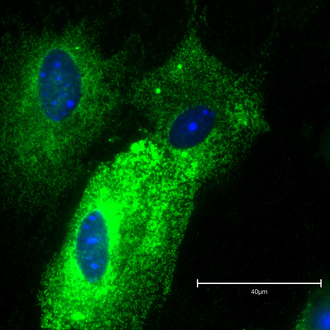
Immunocytochemistry/Immunofluorescence: Adiponectin/Acrp30 Antibody (19F1) [NBP2-22450] - Analysis of Adiponectin was performed using 10% confluent 3T3-L1 cells differentiated with Adipogenesis supplement for 5 days.![Immunohistochemistry-Paraffin: Adiponectin/Acrp30 Antibody (19F1) [NBP2-22450]](https://resources.rndsystems.com/images/products/Adiponectin-Acrp30-Antibody-19F1-Immunohistochemistry-Paraffin-NBP2-22450-img0010.jpg "Immunohistochemistry-Paraffin: Adiponectin/Acrp30 Antibody (19F1) [NBP2-22450]")
Immunohistochemistry-Paraffin: Adiponectin/Acrp30 Antibody (19F1) [NBP2-22450]
Immunohistochemistry-Paraffin: Adiponectin/Acrp30 Antibody (19F1) [NBP2-22450] - Analysis was performed on biopsies of deparaffinized Human skin tissue.![Western Blot: Adiponectin/Acrp30 Antibody (19F1) [NBP2-22450]](https://resources.rndsystems.com/images/products/Adiponectin-Acrp30-Antibody-19F1-Western-Blot-NBP2-22450-img0009.jpg "Western Blot: Adiponectin/Acrp30 Antibody (19F1) [NBP2-22450]")
Western Blot: Adiponectin/Acrp30 Antibody (19F1) [NBP2-22450]
Western Blot: Adiponectin/Acrp30 Antibody (19F1) [NBP2-22450] - Analysis was performed using 10 ul of conditioned media of Hep G2 cell line (lane 1).![Immunohistochemistry: Adiponectin/Acrp30 Antibody (19F1) [NBP2-22450]](https://resources.rndsystems.com/images/products/Adiponectin-Acrp30-Antibody-19F1-Immunohistochemistry-NBP2-22450-img0002.jpg "Immunohistochemistry: Adiponectin/Acrp30 Antibody (19F1) [NBP2-22450]")
Immunohistochemistry: Adiponectin/Acrp30 Antibody (19F1) [NBP2-22450]
Immunohistochemistry: Adiponectin/Acrp30 Antibody (19F1) [NBP2-22450] - Biopsies of deparaffinized Human skin tissue. [NBP2-22450]")
Immunocytochemistry/Immunofluorescence: Mouse Monoclonal Adiponectin/Acrp30 Antibody (19F1) [NBP2-22450]
Immunocytochemistry/Immunofluorescence: Mouse Monoclonal Adiponectin/Acrp30 Antibody (19F1) [NBP2-22450] - Adipocytes isolated from the pelvic area of the male mouse. Image from a verified customer review.Applications for Adiponectin/Acrp30 Antibody (19F1)
Application
Recommended Usage
ELISA
1:100 - 1:2000
Immunocytochemistry/ Immunofluorescence
2 - 4 ug/ml
Immunohistochemistry
1:20 - 100
Immunohistochemistry-Paraffin
1:10 - 1:500
Western Blot
1 ug/ml
Reviewed Applications
Read 1 review rated 5 using NBP2-22450 in the following applications:
Formulation, Preparation, and Storage
Purification
Protein G purified
Formulation
PBS with 1 mg/ml BSA
Preservative
0.05% Sodium Azide
Concentration
1 mg/ml
Shipping
The product is shipped with polar packs. Upon receipt, store it immediately at the temperature recommended below.
Stability & Storage
Store at -20C. Avoid freeze-thaw cycles.
Background: Adiponectin/Acrp30
Additional Adiponectin/Acrp30 Products
Product Documents for Adiponectin/Acrp30 Antibody (19F1)
Certificate of Analysis
To download a Certificate of Analysis, please enter a lot or batch number in the search box below.
Product Specific Notices for Adiponectin/Acrp30 Antibody (19F1)
This product is for research use only and is not approved for use in humans or in clinical diagnosis. Primary Antibodies are guaranteed for 1 year from date of receipt.
Related Research Areas
Citations for Adiponectin/Acrp30 Antibody (19F1)
 Powered by Bioz
Customer Reviews for Adiponectin/Acrp30 Antibody (19F1) (1)
5 out of 5
1 Customer Rating
Have you used Adiponectin/Acrp30 Antibody (19F1)?
Submit a review and receive an Amazon gift card!
$25/€18/£15/$25CAN/¥2500 Yen for a review with an image
$10/€7/£6/$10CAN/¥1110 Yen for a review without an image
Submit a review
Customer Images
Showing
1
-
1 of
1 review
Showing All
Filter By:
-
Application: ImmunofluorescenceSample Tested: AdipocytesSpecies: MouseVerified Customer | Posted 11/06/2024Adipocytes isolated from the pelvic area of the male mouse.

There are no reviews that match your criteria.
Protocols
Find general support by application which include: protocols, troubleshooting, illustrated assays, videos and webinars.
- Antigen Retrieval Protocol (PIER)
- Antigen Retrieval for Frozen Sections Protocol
- Appropriate Fixation of IHC/ICC Samples
- Cellular Response to Hypoxia Protocols
- Chromogenic IHC Staining of Formalin-Fixed Paraffin-Embedded (FFPE) Tissue Protocol
- Chromogenic Immunohistochemistry Staining of Frozen Tissue
- ClariTSA™ Fluorophore Kits
- Detection & Visualization of Antibody Binding
- ELISA Sample Preparation & Collection Guide
- ELISA Troubleshooting Guide
- Fluorescent IHC Staining of Frozen Tissue Protocol
- Graphic Protocol for Heat-induced Epitope Retrieval
- Graphic Protocol for the Preparation and Fluorescent IHC Staining of Frozen Tissue Sections
- Graphic Protocol for the Preparation and Fluorescent IHC Staining of Paraffin-embedded Tissue Sections
- Graphic Protocol for the Preparation of Gelatin-coated Slides for Histological Tissue Sections
- How to Run an R&D Systems DuoSet ELISA
- How to Run an R&D Systems Quantikine ELISA
- How to Run an R&D Systems Quantikine™ QuicKit™ ELISA
- ICC Cell Smear Protocol for Suspension Cells
- ICC Immunocytochemistry Protocol Videos
- ICC for Adherent Cells
- IHC Sample Preparation (Frozen sections vs Paraffin)
- Immunocytochemistry (ICC) Protocol
- Immunocytochemistry Troubleshooting
- Immunofluorescence of Organoids Embedded in Cultrex Basement Membrane Extract
- Immunofluorescent IHC Staining of Formalin-Fixed Paraffin-Embedded (FFPE) Tissue Protocol
- Immunohistochemistry (IHC) and Immunocytochemistry (ICC) Protocols
- Immunohistochemistry Frozen Troubleshooting
- Immunohistochemistry Paraffin Troubleshooting
- Preparing Samples for IHC/ICC Experiments
- Preventing Non-Specific Staining (Non-Specific Binding)
- Primary Antibody Selection & Optimization
- Protocol for Heat-Induced Epitope Retrieval (HIER)
- Protocol for Making a 4% Formaldehyde Solution in PBS
- Protocol for VisUCyte™ HRP Polymer Detection Reagent
- Protocol for the Fluorescent ICC Staining of Cell Smears - Graphic
- Protocol for the Fluorescent ICC Staining of Cultured Cells on Coverslips - Graphic
- Protocol for the Preparation & Fixation of Cells on Coverslips
- Protocol for the Preparation and Chromogenic IHC Staining of Frozen Tissue Sections
- Protocol for the Preparation and Chromogenic IHC Staining of Frozen Tissue Sections - Graphic
- Protocol for the Preparation and Chromogenic IHC Staining of Paraffin-embedded Tissue Sections
- Protocol for the Preparation and Chromogenic IHC Staining of Paraffin-embedded Tissue Sections - Graphic
- Protocol for the Preparation and Fluorescent ICC Staining of Cells on Coverslips
- Protocol for the Preparation and Fluorescent ICC Staining of Non-adherent Cells
- Protocol for the Preparation and Fluorescent ICC Staining of Stem Cells on Coverslips
- Protocol for the Preparation and Fluorescent IHC Staining of Frozen Tissue Sections
- Protocol for the Preparation and Fluorescent IHC Staining of Paraffin-embedded Tissue Sections
- Protocol for the Preparation of Gelatin-coated Slides for Histological Tissue Sections
- Protocol for the Preparation of a Cell Smear for Non-adherent Cell ICC - Graphic
- Quantikine HS ELISA Kit Assay Principle, Alkaline Phosphatase
- Quantikine HS ELISA Kit Principle, Streptavidin-HRP Polymer
- R&D Systems Quality Control Western Blot Protocol
- Sandwich ELISA (Colorimetric) – Biotin/Streptavidin Detection Protocol
- Sandwich ELISA (Colorimetric) – Direct Detection Protocol
- TUNEL and Active Caspase-3 Detection by IHC/ICC Protocol
- The Importance of IHC/ICC Controls
- Troubleshooting Guide: ELISA
- Troubleshooting Guide: Immunohistochemistry
- Troubleshooting Guide: Western Blot Figures
- Western Blot Conditions
- Western Blot Protocol
- Western Blot Protocol for Cell Lysates
- Western Blot Troubleshooting
- Western Blot Troubleshooting Guide
- View all Protocols, Troubleshooting, Illustrated assays and Webinars
Loading...
Associated Pathways

