AKT1 [p Ser473] Antibody (104A282) - BSA Free
Novus Biologicals | Catalog # NB100-56749
Key Product Details
Validated by
Independent Antibodies, Biological Validation
Species Reactivity
Validated:
Human, Mouse, Rat, Rabbit
Cited:
Human, Mouse, Rat, Rabbit
Predicted:
Amphibian (100%), Chicken (100%), Zebrafish (100%). Backed by our 100% Guarantee.
Applications
Validated:
Immunohistochemistry, Immunohistochemistry-Paraffin, Immunohistochemistry-Frozen, Western Blot
Cited:
Immunohistochemistry-Paraffin, Western Blot
Label
Unconjugated
Antibody Source
Monoclonal Mouse IgG1 kappa Clone # 104A282
Format
BSA Free
Loading...
Product Specifications
Immunogen
This AKT1 phospho Ser473 antibody was raised against a synthetic peptide containing phosphorylated serines at amino acid residues 473 of human AKT1.
Reactivity Notes
Rat reactivity reported in scientific literature (PMID: 31092832). Rabbit reactivity reported in scientific literature (PMID: 32936958)
Modification
p Ser473
Specificity
Clone 104A282 detects specifically the Ser473 phosphorylated form of AKT1.
Clonality
Monoclonal
Host
Mouse
Isotype
IgG1 kappa
Theoretical MW
55.7 kDa.
Disclaimer note: The observed molecular weight of the protein may vary from the listed predicted molecular weight due to post translational modifications, post translation cleavages, relative charges, and other experimental factors.
Disclaimer note: The observed molecular weight of the protein may vary from the listed predicted molecular weight due to post translational modifications, post translation cleavages, relative charges, and other experimental factors.
Scientific Data Images for AKT1 [p Ser473] Antibody (104A282) - BSA Free
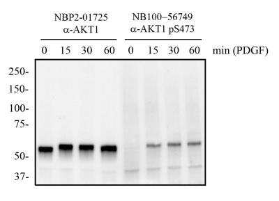
Western Blot: AKT1 [p Ser473] Antibody (104A282) [NB100-56749] - Total protein from mouse 3T3 cells treated with and without PDGF (50 ng/mL) for the indicated times was separated on a 7.5% gel by SDS-PAGE, transferred to PVDF membrane and blocked in 5% non-fat milk in TBST. The membrane was probed with 2.0 ug/mL anti-AKT1 (NBP2-01725) and 2 ug/mL pS473 AKT1 in 1% BSA in TBST and detected with an anti-mouse HRP secondary antibody using chemiluminescence. Note the detection of phosphorylated AKT1 in response to PDGF treatment compared to total AKT1 protein.
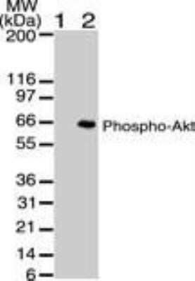
Western Blot: AKT1 [p Ser473] Antibody (104A282) [NB100-56749] - WB of phospho AKT using phospho AKT antibody at 2 ug/mL against untreated (lane 1) and PDGF treated (lane 2) NIH-3T3 lysate. HRP conjugated secondary antibody and ECL substrate solution were used for this test. Image using the Azide and BSA Free form of this antibody.
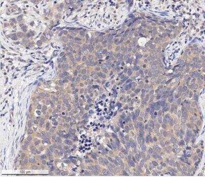
Immunohistochemistry-Paraffin: AKT1 [p Ser473] Antibody (104A282) [NB100-56749] - IHC analysis of an FFPE human breast carcinoma tissue section using 1:250 dilution of phospho Ser473 AKT1 antibody (clone 104A282) on a Bond Rx autostainer (Leica Biosystems). The assay involved 20 minutes of heat induced antigen retrieval (HIER) with 10 mM sodium citrate buffer (pH 6.0) and endogenous peroxidase quenching using peroxide block. The sections were incubated with primary antibody for 30 minutes. Bond Polymer Refine Detection (Leica Biosystems) and DAB were used for signal detection which followed counterstaining with hematoxylin. Whole slide scanning and capturing of representative images (20X) were performed using Aperio AT2 (Leica Biosystems). This antibody generated a diffused cytoplasmic staining of phosphor-AKT (Ser-473) in the cancer cells as well as the stromal cells. Staining was performed by Histowiz.
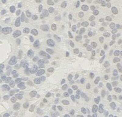
Immunohistochemistry-Frozen: AKT1 [p Ser473] Antibody (104A282) [NB100-56749] - Rat mammary carcinoma tissue section stained with AKT1 [p Ser473] Antibody (104A282). IHC-Fr image submitted by a verified customer review.
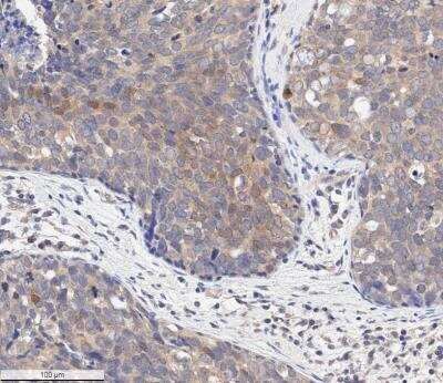
Immunohistochemistry-Paraffin: AKT1 [p Ser473] Antibody (104A282) [NB100-56749] - IHC analysis of an FFPE human breast carcinoma tissue section using 1:250 dilution of pSer473 AKT1 antibody (clone 104A282) on a Bond Rx autostainer (Leica Biosystems). The assay involved 20 minutes of heat induced antigen retrieval (HIER) with 10 mM sodium citrate buffer (pH 6.0) and endogenous peroxidase quenching using peroxide block. The sections were incubated with primary antibody for 30 minutes. Bond Polymer Refine Detection (Leica Biosystems) and DAB were used for signal detection which followed counterstaining with hematoxylin. Whole slide scanning and capturing of representative images (20X) were performed using Aperio AT2 (Leica Biosystems). This antibody generated a diffused cytoplasmic staining of phosphor-AKT (Ser-473) in the cancer cells as well as the stromal cells. Some cancer cells depicted nuclear stianing also. Staining was performed by Histowiz.
![AKT1 [p Ser473] Antibody (104A282) - BSA Free](https://resources.rndsystems.com/images/products/nb100-56749_mouse-monoclonal-akt1-p-ser473-antibody-104a282-western-blot-132202617551020.jpg "Western Blot: AKT1 [p Ser473] Antibody (104A282) - BSA Free [NB100-56749] -")
Western Blot: AKT1 [p Ser473] Antibody (104A282) - BSA Free [NB100-56749] -
SP1/PTEN mediates lung infection in mice and the activity of the Akt signaling pathway. A, survival days of the mice after si-SP1 and si-PTEN administration (p < 0.01, the Kaplan-Meier analysis); B, bacterial load in murine lung tissues examined by CFU analysis (*p < 0.05, the one-way ANOVA); C, infiltration of inflammatory cells in murine lung tissues examined by HE staining (*p < 0.05, the one-way ANOVA); D, epithelial cell apoptosis in the murine lung tissues after Mtb infection examined by the TUNEL assay (*p < 0.05, the one-way ANOVA); E, fibrosis in murine lung tissues determined by Masson’s trichrome staining (*p < 0.05, the one-way ANOVA); F-G, protein levels of SP1 and PTEN and the Akt phosphorylation in murine lung tissues determined by western blot analysis (*p < 0.05, the one-way ANOVA). For animal studies, n = 6 in each group. Image collected and cropped by CiteAb from the following open publication (https://pubmed.ncbi.nlm.nih.gov/35420971), licensed under a CC-BY license. Not internally tested by Novus Biologicals.![AKT1 [p Ser473] Antibody (104A282) - BSA Free](https://resources.rndsystems.com/images/products/nb100-56749_mouse-monoclonal-akt1-p-ser473-antibody-104a282-western-blot-13220261820240.jpg "Western Blot: AKT1 [p Ser473] Antibody (104A282) - BSA Free [NB100-56749] -")
Western Blot: AKT1 [p Ser473] Antibody (104A282) - BSA Free [NB100-56749] -
EZH2/HMGA2 regulates the PI3K/AKT signaling. MI rats were subjected to injection of EZH2-OE or EZH2-NC (MI + MSC-EXO + EZH2 NC/EZH2 OE) and EZH2-OE + HMGA2-OE or EZH2-OE + HMGA2-NC. A HMGA2 protein expression in myocardial tissues in response to EZH2-OE + HMGA2-OE examined using western blot (Additional file 5: Fig. S5). B Weight change in rats after EZH2-OE + HMGA2-OE treatment. C Changes in HMI in rats after EZH2-OE + HMGA2-OE treatment. D Measurement of Dd, Sd, and LVEF by echocardiography at 2 weeks after LAD in rats. E Measurement of LVMI by echocardiography in rats. F KEGG pathway analysis of gene enrichment pathways [29]. G Changes in PI3K/AKT pathway in rat myocardium examined using western blot (Additional file 6: Fig. S6). All data are expressed as mean +/- SD (n = 6/group, *p < 0.05 vs. Sham, DMSO, MI + MSC-EXO + EZH2-NC or EZH2-OE + HMGA2-NC group determined by unpaired t test or one-way ANOVA) Image collected and cropped by CiteAb from the following open publication (https://pubmed.ncbi.nlm.nih.gov/35264108), licensed under a CC-BY license. Not internally tested by Novus Biologicals.![AKT1 [p Ser473] Antibody (104A282) - BSA Free](https://resources.rndsystems.com/images/products/nb100-56749_mouse-monoclonal-akt1-p-ser473-antibody-104a282-western-blot-13220261817569.jpg "Western Blot: AKT1 [p Ser473] Antibody (104A282) - BSA Free [NB100-56749] -")
Western Blot: AKT1 [p Ser473] Antibody (104A282) - BSA Free [NB100-56749] -
SP1/PTEN mediates lung infection in mice and the activity of the Akt signaling pathway. A, survival days of the mice after si-SP1 and si-PTEN administration (p < 0.01, the Kaplan-Meier analysis); B, bacterial load in murine lung tissues examined by CFU analysis (*p < 0.05, the one-way ANOVA); C, infiltration of inflammatory cells in murine lung tissues examined by HE staining (*p < 0.05, the one-way ANOVA); D, epithelial cell apoptosis in the murine lung tissues after Mtb infection examined by the TUNEL assay (*p < 0.05, the one-way ANOVA); E, fibrosis in murine lung tissues determined by Masson’s trichrome staining (*p < 0.05, the one-way ANOVA); F-G, protein levels of SP1 and PTEN and the Akt phosphorylation in murine lung tissues determined by western blot analysis (*p < 0.05, the one-way ANOVA). For animal studies, n = 6 in each group. Image collected and cropped by CiteAb from the following open publication (https://pubmed.ncbi.nlm.nih.gov/35420971), licensed under a CC-BY license. Not internally tested by Novus Biologicals.Applications for AKT1 [p Ser473] Antibody (104A282) - BSA Free
Application
Recommended Usage
Immunohistochemistry
1:200 - 1:250
Immunohistochemistry-Frozen
reported by customer review
Immunohistochemistry-Paraffin
1:200 - 1:250
Reviewed Applications
Read 2 reviews rated 5 using NB100-56749 in the following applications:
Formulation, Preparation, and Storage
Purification
Protein G purified
Formulation
PBS
Format
BSA Free
Preservative
0.05% Sodium Azide
Concentration
1 mg/ml
Shipping
The product is shipped with polar packs. Upon receipt, store it immediately at the temperature recommended below.
Stability & Storage
Store at 4C short term. Aliquot and store at -20C long term. Avoid freeze-thaw cycles.
Background: Akt1
The main function of AKT is to control inhibition of apoptosis and promote cell proliferation. Survival factors can activate AKT Ser473 and Thr308 phosphorylation sites in a transcription-independent manner, resulting in the inactivation of apoptotic signaling transduction through the tumor suppressor PTEN, an antagonist to PI3-K (5). PTEN exerts enzymatic activity as a phosphatidylinositol-3,4,5-trisphosphate (PIP3) phosphatase, opposing PI3K activity by decreasing availability of PIP3 to proliferating cells, leading to overexpression and inappropriate activation of AKT noted in many types of cancer.
AKT1 function has been linked to overall physiological growth and function (2). AKT1 has been correlated with proteus syndrome, a rare disorder characterized by overgrowth of various tissues caused by a mosaic variant in the AKT1 gene in humans.
AKT2 is strongly correlated with Type II diabetes, including phenotypes of insulin resistance, hyperglycemia and atherosclerosis (2, 6).
The function of AKT3 is specifically associated to brain development, where disruptions to AKT3 are correlated with microcephaly, hemimegalencephaly, megalencephaly and intellectual disabilities (2).
References
1. Ersahin, T., Tuncbag, N., & Cetin-Atalay, R. (2015). The PI3K/AKT/mTOR interactive pathway. Mol Biosyst, 11(7), 1946-1954. doi:10.1039/c5mb00101c
2. Cohen, M. M., Jr. (2013). The AKT genes and their roles in various disorders. Am J Med Genet A, 161a(12), 2931-2937. doi:10.1002/ajmg.a.36101
3. Georgescu, M. M. (2010). PTEN Tumor Suppressor Network in PI3K-Akt Pathway Control. Genes Cancer, 1(12), 1170-1177. doi:10.1177/1947601911407325
4. Mishra, P., Paital, B., Jena, S., Swain, S. S., Kumar, S., Yadav, M. K.,... Samanta, L. (2019). Possible activation of NRF2 by Vitamin E/Curcumin against altered thyroid hormone induced oxidative stress via NFkB/AKT/mTOR/KEAP1 signalling in rat heart. Sci Rep, 9(1), 7408. doi:10.1038/s41598-019-43320-5
5. Wedel, S., Hudak, L., Seibel, J. M., Juengel, E., Oppermann, E., Haferkamp, A., & Blaheta, R. A. (2011). Critical analysis of simultaneous blockage of histone deacetylase and multiple receptor tyrosine kinase in the treatment of prostate cancer. Prostate, 71(7), 722-735. doi:10.1002/pros.21288
6. Rotllan, N., Chamorro-Jorganes, A., Araldi, E., Wanschel, A. C., Aryal, B., Aranda, J. F.,... Fernandez-Hernando, C. (2015). Hematopoietic Akt2 deficiency attenuates the progression of atherosclerosis. Faseb j, 29(2), 597-610. doi:10.1096/fj.14-262097
Long Name
v-Akt Murine Thymoma Viral Oncogene Homolog 1
Alternate Names
PKB alpha, PRKBA, RAC-alpha
Gene Symbol
AKT1
UniProt
Additional Akt1 Products
Product Documents for AKT1 [p Ser473] Antibody (104A282) - BSA Free
Certificate of Analysis
To download a Certificate of Analysis, please enter a lot or batch number in the search box below.
Product Specific Notices for AKT1 [p Ser473] Antibody (104A282) - BSA Free
This product is for research use only and is not approved for use in humans or in clinical diagnosis. Primary Antibodies are guaranteed for 1 year from date of receipt.
Citations for AKT1 [p Ser473] Antibody (104A282) - BSA Free
 Powered by Bioz
Customer Reviews for AKT1 [p Ser473] Antibody (104A282) - BSA Free (2)
5 out of 5
2 Customer Ratings
Have you used AKT1 [p Ser473] Antibody (104A282) - BSA Free?
Submit a review and receive an Amazon gift card!
$25/€18/£15/$25CAN/¥2500 Yen for a review with an image
$10/€7/£6/$10CAN/¥1110 Yen for a review without an image
Submit a review
Customer Images
![AKT1 [p Ser473] Antibody (104A282) - BSA Free NB100-56749](https://resources.rndsystems.com/images/reviews/review_nb100-56749_56711_0_0_0_0_0.jpg)
![AKT1 [p Ser473] Antibody (104A282) - BSA Free NB100-56749](https://resources.rndsystems.com/images/reviews/Western-Blot_AKT1-Antibody-(NB100-56749)-(01-mg)_NB100-56749_9386.jpg)
Showing
1
-
2 of
2 reviews
Showing All
Filter By:
-
Application: Immunohistochemistry-FrozenSample Tested: Mammary carcinomaSpecies: RatVerified Customer | Posted 08/10/2021Mammary carcinoma
![AKT1 [p Ser473] Antibody (104A282) - BSA Free NB100-56749](data:image/png;base64,R0lGODlhAQABAAD/ACwAAAAAAQABAAACADs=) Bio-Techne ResponseThis review was submitted through the legacy Novus Innovators Program, reflecting a new species or application tested on a primary antibody.
Bio-Techne ResponseThis review was submitted through the legacy Novus Innovators Program, reflecting a new species or application tested on a primary antibody. -
Application: Western BlotSample Tested:Species: HumanVerified Customer | Posted 08/16/2014A review for p-S473-AKT antibody (NB100-56749)
There are no reviews that match your criteria.
Protocols
Find general support by application which include: protocols, troubleshooting, illustrated assays, videos and webinars.
- Antigen Retrieval Protocol (PIER)
- Antigen Retrieval for Frozen Sections Protocol
- Appropriate Fixation of IHC/ICC Samples
- Cellular Response to Hypoxia Protocols
- Chromogenic IHC Staining of Formalin-Fixed Paraffin-Embedded (FFPE) Tissue Protocol
- Chromogenic Immunohistochemistry Staining of Frozen Tissue
- ClariTSA™ Fluorophore Kits
- Detection & Visualization of Antibody Binding
- Fluorescent IHC Staining of Frozen Tissue Protocol
- Graphic Protocol for Heat-induced Epitope Retrieval
- Graphic Protocol for the Preparation and Fluorescent IHC Staining of Frozen Tissue Sections
- Graphic Protocol for the Preparation and Fluorescent IHC Staining of Paraffin-embedded Tissue Sections
- Graphic Protocol for the Preparation of Gelatin-coated Slides for Histological Tissue Sections
- IHC Sample Preparation (Frozen sections vs Paraffin)
- Immunofluorescent IHC Staining of Formalin-Fixed Paraffin-Embedded (FFPE) Tissue Protocol
- Immunohistochemistry (IHC) and Immunocytochemistry (ICC) Protocols
- Immunohistochemistry Frozen Troubleshooting
- Immunohistochemistry Paraffin Troubleshooting
- Preparing Samples for IHC/ICC Experiments
- Preventing Non-Specific Staining (Non-Specific Binding)
- Primary Antibody Selection & Optimization
- Protocol for Heat-Induced Epitope Retrieval (HIER)
- Protocol for Making a 4% Formaldehyde Solution in PBS
- Protocol for VisUCyte™ HRP Polymer Detection Reagent
- Protocol for the Preparation & Fixation of Cells on Coverslips
- Protocol for the Preparation and Chromogenic IHC Staining of Frozen Tissue Sections
- Protocol for the Preparation and Chromogenic IHC Staining of Frozen Tissue Sections - Graphic
- Protocol for the Preparation and Chromogenic IHC Staining of Paraffin-embedded Tissue Sections
- Protocol for the Preparation and Chromogenic IHC Staining of Paraffin-embedded Tissue Sections - Graphic
- Protocol for the Preparation and Fluorescent IHC Staining of Frozen Tissue Sections
- Protocol for the Preparation and Fluorescent IHC Staining of Paraffin-embedded Tissue Sections
- Protocol for the Preparation of Gelatin-coated Slides for Histological Tissue Sections
- R&D Systems Quality Control Western Blot Protocol
- TUNEL and Active Caspase-3 Detection by IHC/ICC Protocol
- The Importance of IHC/ICC Controls
- Troubleshooting Guide: Immunohistochemistry
- Troubleshooting Guide: Western Blot Figures
- Western Blot Conditions
- Western Blot Protocol
- Western Blot Protocol for Cell Lysates
- Western Blot Troubleshooting
- Western Blot Troubleshooting Guide
- View all Protocols, Troubleshooting, Illustrated assays and Webinars
FAQs for AKT1 [p Ser473] Antibody (104A282) - BSA Free
Showing
1
-
5 of
5 FAQs
Showing All
-
Q: Do your HRP-conjugated antibodies contain sodium azide?
A: No. None of our HRP-conjugated antibodies contain sodium azide as this agent inhibits the activity of HRP.
-
Q: How do I choose secondary antibodies to label the same cells when I have two primary antibodies from the same host?
A: Use isotype-specific secondary antibodies if the primary antibodies are of different isotypes. You can also make direct conjugates of the primary antibodies by use of antibody labeling kits, dyes, or custom conjugations (please contact Technical Support for custom orders).
-
Q: I am looking for a antibody that recognizes human Akt1 but NOT Akt2 or 3, for Western blot analyses. I also want that antibody to recognize Akt1 regardless of its phosphorylated form.
A: At the moment we do not have an AKT1 antibody that definitively does not react with either AKT2 or AKT3.
-
Q: What is the molecular weight of your antibodies?
A: All IgG antibodies are approximately 150 kDa (each heavy chain is about 50 kDa and each light chain is about 25 kDa).
-
Q: Why are many of your antibodies formulated with sodium azide and BSA?
A: Sodium azide is a preservative which is added to prevent bacterial growth. BSA is added as a protein stabilizer.
-
Q: Do your HRP-conjugated antibodies contain sodium azide?
A: No. None of our HRP-conjugated antibodies contain sodium azide as this agent inhibits the activity of HRP.
-
Q: How do I choose secondary antibodies to label the same cells when I have two primary antibodies from the same host?
A: Use isotype-specific secondary antibodies if the primary antibodies are of different isotypes. You can also make direct conjugates of the primary antibodies by use of antibody labeling kits, dyes, or custom conjugations (please contact Technical Support for custom orders).
-
Q: I am looking for a antibody that recognizes human Akt1 but NOT Akt2 or 3, for Western blot analyses. I also want that antibody to recognize Akt1 regardless of its phosphorylated form.
A: At the moment we do not have an AKT1 antibody that definitively does not react with either AKT2 or AKT3.
-
Q: What is the molecular weight of your antibodies?
A: All IgG antibodies are approximately 150 kDa (each heavy chain is about 50 kDa and each light chain is about 25 kDa).
-
Q: Why are many of your antibodies formulated with sodium azide and BSA?
A: Sodium azide is a preservative which is added to prevent bacterial growth. BSA is added as a protein stabilizer.
Loading...
Associated Pathways


IL-2 Signaling Pathways
 IL-4 Signaling Pathways
 IL-7 Signaling Pathways
 IL-9 Signaling Pathways
 IL-15 Signaling Pathways
 IL-21 Signaling Pathways
 mTOR Signaling Pathway
 Notch Signaling Pathways
 TGF-beta Signaling Pathways
 VEGF - VEGF R2 Signaling Pathways
