AKT1 [p Ser473] Antibody - BSA Free
Novus Biologicals | Catalog # NB600-590
Loading...
Key Product Details
Validated by
Biological Validation
Species Reactivity
Validated:
Human, Mouse, Rat
Cited:
Human
Applications
Validated:
Immunohistochemistry, Immunohistochemistry-Paraffin, Western Blot, ELISA, Immunocytochemistry/ Immunofluorescence, Dot Blot
Cited:
Western Blot, IF/IHC
Label
Unconjugated
Antibody Source
Polyclonal Rabbit IgG
Format
BSA Free
Loading...
Product Specifications
Immunogen
AKT1 [p Ser473] Antibody was prepared by repeated immunizations in rabbits with a synthetic peptide corresponding to a C-terminus region near phospho Serine 473 of the human, mouse, rat and chicken AKT1 proteins conjugated to KLH. (Uniprot: P31749)
Reactivity Notes
This antibody is specific for phosphorylated human AKT1 pS473. Minimal reactivity occurs against non-phosphorylated AKT. Reactivity against AKT1 from other species may occur but has not yet been tested.
Modification
p Ser473
Localization
Cytoplasmic and Nuclear
Specificity
This antibody is specific for phosphorylated human AKTpS473. Minimal reactivity occurs against non-phosphorylated AKT. Reactivity against AKT from other species may occur but has not yet been tested.
Clonality
Polyclonal
Host
Rabbit
Isotype
IgG
Description
This product was prepared from monospecific antiserum by immunoaffinity chromatography using a phospho peptide coupled to agarose beads followed by solid phase adsorption(s) against a non-phospho peptide and non-specific peptide to remove any unwanted reactivities. Assay by immunoelectrophoresis resulted in a single precipitin arc against anti-Rabbit Serum
Store vial at -20C prior to opening. Aliquot contents and freeze at -20C or below for extended storage. Avoid cycles of freezing and thawing. Centrifuge product if not completely clear after standing at room temperature. This product is stable for several weeks at 4C as an undiluted liquid. Dilute only prior to immediate use.
Store vial at -20C prior to opening. Aliquot contents and freeze at -20C or below for extended storage. Avoid cycles of freezing and thawing. Centrifuge product if not completely clear after standing at room temperature. This product is stable for several weeks at 4C as an undiluted liquid. Dilute only prior to immediate use.
Scientific Data Images for AKT1 [p Ser473] Antibody - BSA Free
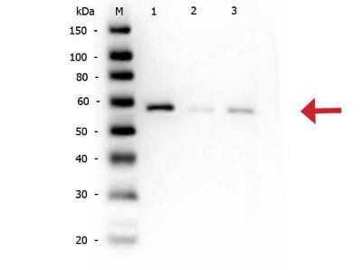
Western Blot: AKT1 [p Ser473] Antibody [NB600-590] - Western Blot of AKT11 [p Ser473] antibody. Lane 1: AKT11 Recombinant Protein. Lane 2: AKT11 Mutant Human Recombinant Protein.Lane 3: AKT11 (phosphatase treated) Human Recombinant Protein. Load: 50 ng per lane.Primary antibody: AKT1 pS473 antibody at 1:1,000 for overnight at 4C.Secondary antibody: Peroxidase rabbit secondary antibody at 1:40,000 for 30 min at RT.Block: Blocking Buffer for Fluorescent Western Blotting for 30 min at RT.Predicted/Observed size: ~56 kDa for AKT1pS473.
![Immunocytochemistry/ Immunofluorescence: AKT1 [p Ser473] Antibody [NB600-590]](https://resources.rndsystems.com/images/products/AKT1-[p-Ser473]-Antibody-Immunocytochemistry-Immunofluorescence-NB600-590-img0019.jpg "Immunocytochemistry/ Immunofluorescence: AKT1 [p Ser473] Antibody [NB600-590]")
Immunocytochemistry/ Immunofluorescence: AKT1 [p Ser473] Antibody [NB600-590]
Immunocytochemistry/Immunofluorescence: AKT1 [p Ser473] Antibody [NB600-590] - Cardiomyocytes infected with adenovirus expressing wild-type AKT. Fixation: 0.5% PFA. Antigen retrieval: not required. Primary antibody: AKT pS473 antibody at 1:40 for 1 h at RT. Secondary antibody: texas-red conjugated rabbit secondary antibody at 1:10,000 for 45 min at RT. Localization: AKT pS473 is nuclear. Staining: AKT pS473 as green fluorescent signal with texas-red conjugated phalloidin (red) to label filamentous actin.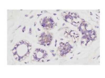
Immunohistochemistry: AKT1 [p Ser473] Antibody [NB600-590] - Immunohistochemistry of AKT11 [p Ser473] antibody. Tissue: human breast carcinoma. Fixation: formalin fixed paraffin embedded. Antigen retrieval: not required.Primary antibody: AKT1 pS473 antibody at 1:100 for 1 h at RT.Secondary antibody: Dako's Techmate streptavidin-biotin reagents at 1:10,000 for 45 min at RT.Localization: AKT1 pS473 is nuclear and occasionally cytoplasmic.
![Western Blot: AKT1 [p Ser473] Antibody [NB600-590]](https://resources.rndsystems.com/images/products/AKT1-[p-Ser473]-Antibody-Western-Blot-NB600-590-img0010.jpg "Western Blot: AKT1 [p Ser473] Antibody [NB600-590]")
Western Blot: AKT1 [p Ser473] Antibody [NB600-590]
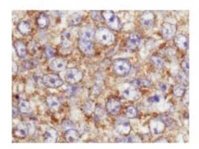
Western Blot: AKT1 [p Ser473] Antibody [NB600-590] - Lane 1: nuclear extract from cells infected with adenovirus expressing nuclear-targeted AKT kinase. Load: 35 ug per lane. Primary antibody: AKT pS473 antibody at 1:200 dilution for overnight at 4C. Secondary antibody: IRDye800 rabbit secondary antibody at 1:10,000 for 45 min at RT. Block: 5% BLOTTO overnight at 4C. Predicted/Observed size: 56 kDa for AKT pS473. Other band(s): unspecific.![Western Blot: AKT1 [p Ser473] Antibody [NB600-590]](https://resources.rndsystems.com/images/products/AKT1-[p-Ser473]-Antibody-Western-Blot-NB600-590-img0012.jpg "Western Blot: AKT1 [p Ser473] Antibody [NB600-590]")
Immunohistochemistry: AKT1 [p Ser473] Antibody [NB600-590] - Immunohistochemistry of AKT1 [p Ser473] antibody. Tissue: human breast carcinoma. Fixation: formalin fixed paraffin embedded. Antigen retrieval: not required.Primary antibody: AKT1 pS473 antibody at 100 dilution for 1 h at RT.Secondary antibody: Dako's Techmate streptavidin-biotin reagents at 1:10,000 for 45 min at RT.Localization: AKT1 pS473 is nuclear and occasionally cytoplasmic.
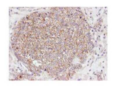
Immunohistochemistry: AKT1 [p Ser473] Antibody [NB600-590] - Immunohistochemistry at higher magnification of AKT1 [p Ser473] antibody. Tissue: human breast carcinoma. Fixation: formalin fixed paraffin embedded. Antigen retrieval: not required.Primary antibody: AKT1 pS473 antibody at 100 dilution for 1 h at RT.Secondary antibody: Dako's Techmate streptavidin-biotin reagents at 1:10,000 for 45 min at RT.Localization: AKT1 pS473 is nuclear and occasionally cytoplasmic.
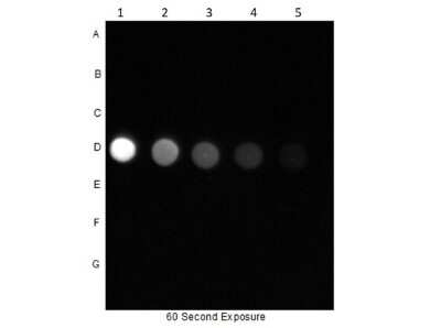
Dot Blot: AKT1 [p Ser473] Antibody [NB600-590]
![AKT1 [p Ser473] Antibody](https://resources.rndsystems.com/images/products/nb600-590_rabbit-polyclonal-akt1-p-ser473-antibody-235202317492110.jpg "AKT1 [p Ser473] Antibody")
AKT1 [p Ser473] Antibody
Dot Blot of Rabbit Anti-AKT pS473 Antibody. Dilutions in Columns: (1) 100ng, (2) 33.33ng, (3) 11.11ng, (4) 3.7ng, (5) 1.23ng. Tested BSA Peptide Reactivity in Rows: (A) AKT1-BSA, (B) AKT1 pT308-BSA, (C) AKT1 S473-BSA, (D) AKT1 pS473-BSA, (E) CDC27 T244-BSA, (F) CDC27 pT244-BSA, (G) BSA control. Primary Antibody: Anti-AKTpS473 at 1ug/mL overnight at 2-8C. Secondary Antibody: Goat anti-Rabbit IgG HRP![AKT1 [p Ser473] Antibody](https://resources.rndsystems.com/images/products/nb600-590_rabbit-polyclonal-akt1-p-ser473-antibody-235202318143128.jpg "AKT1 [p Ser473] Antibody")
AKT1 [p Ser473] Antibody
Immunohistochemistry of Rabbit Anti-Akt pS473 antibody. Tissue: human breast carcinoma. Fixation: formalin fixed paraffin embedded. Antigen retrieval: not required. Primary antibody: Akt pS473 antibody at 100 dilution for 1 h at RT. Secondary antibody: Dako's Techmate streptavidin-biotin reagents at 1:10,000 for 45 min at RT. Localization: Akt pS473 is nuclear and occasionally cytoplasmic.![AKT1 [p Ser473] Antibody](https://resources.rndsystems.com/images/products/nb600-590_rabbit-polyclonal-akt1-p-ser473-antibody-235202318143133.jpg "AKT1 [p Ser473] Antibody")
AKT1 [p Ser473] Antibody
Immunohistochemistry at higher magnification of Rabbit Anti-Akt pS473 antibody. Tissue: human breast carcinoma. Fixation: formalin fixed paraffin embedded. Antigen retrieval: not required. Primary antibody: Akt pS473 antibody at 100 dilution for 1 h at RT. Secondary antibody: Dako's Techmate streptavidin-biotin reagents at 1:10,000 for 45 min at RT. Localization: Akt pS473 is nuclear and occasionally cytoplasmic.![AKT1 [p Ser473] Antibody](https://resources.rndsystems.com/images/products/nb600-590_rabbit-polyclonal-akt1-p-ser473-antibody-2552023131331.jpg "AKT1 [p Ser473] Antibody")
AKT1 [p Ser473] Antibody
Western Blot of Rabbit anti-AKT pS473 antibody. Lane 1: AKT1 Recombinant ProteinApplications for AKT1 [p Ser473] Antibody - BSA Free
Application
Recommended Usage
ELISA
1:15000-1:60000
Immunocytochemistry/ Immunofluorescence
1:40
Immunohistochemistry
1:100-1:500
Immunohistochemistry-Paraffin
1:10-1:500
Western Blot
1:200-1:1000
Application Notes
This This product is phospho specific for pS473 and is tested in ELISA, western blotting, immunohistochemistry (formalin-fixed paraffin-embedded sections), and immunofluorescence. By immunoblot a single band of the expected apparent molecular weight ~56kDa is observed. For immunohistochemistry no pre-treatment of sample is required.
Formulation, Preparation, and Storage
Purification
Immunogen affinity purified
Formulation
0.02 M Potassium Phosphate, 0.15 M Sodium Chloride, pH 7.2
Format
BSA Free
Preservative
0.01% Sodium Azide
Concentration
Please see the vial label for concentration. If unlisted please contact technical services.
Shipping
The product is shipped with polar packs. Upon receipt, store it immediately at the temperature recommended below.
Stability & Storage
Store at -20C short term. Aliquot and store at -80C long term. Avoid freeze-thaw cycles.
Background: Akt1
The main function of AKT is to control inhibition of apoptosis and promote cell proliferation. Survival factors can activate AKT Ser473 and Thr308 phosphorylation sites in a transcription-independent manner, resulting in the inactivation of apoptotic signaling transduction through the tumor suppressor PTEN, an antagonist to PI3-K (5). PTEN exerts enzymatic activity as a phosphatidylinositol-3,4,5-trisphosphate (PIP3) phosphatase, opposing PI3K activity by decreasing availability of PIP3 to proliferating cells, leading to overexpression and inappropriate activation of AKT noted in many types of cancer.
AKT1 function has been linked to overall physiological growth and function (2). AKT1 has been correlated with proteus syndrome, a rare disorder characterized by overgrowth of various tissues caused by a mosaic variant in the AKT1 gene in humans.
AKT2 is strongly correlated with Type II diabetes, including phenotypes of insulin resistance, hyperglycemia and atherosclerosis (2, 6).
The function of AKT3 is specifically associated to brain development, where disruptions to AKT3 are correlated with microcephaly, hemimegalencephaly, megalencephaly and intellectual disabilities (2).
References
1. Ersahin, T., Tuncbag, N., & Cetin-Atalay, R. (2015). The PI3K/AKT/mTOR interactive pathway. Mol Biosyst, 11(7), 1946-1954. doi:10.1039/c5mb00101c
2. Cohen, M. M., Jr. (2013). The AKT genes and their roles in various disorders. Am J Med Genet A, 161a(12), 2931-2937. doi:10.1002/ajmg.a.36101
3. Georgescu, M. M. (2010). PTEN Tumor Suppressor Network in PI3K-Akt Pathway Control. Genes Cancer, 1(12), 1170-1177. doi:10.1177/1947601911407325
4. Mishra, P., Paital, B., Jena, S., Swain, S. S., Kumar, S., Yadav, M. K.,... Samanta, L. (2019). Possible activation of NRF2 by Vitamin E/Curcumin against altered thyroid hormone induced oxidative stress via NFkB/AKT/mTOR/KEAP1 signalling in rat heart. Sci Rep, 9(1), 7408. doi:10.1038/s41598-019-43320-5
5. Wedel, S., Hudak, L., Seibel, J. M., Juengel, E., Oppermann, E., Haferkamp, A., & Blaheta, R. A. (2011). Critical analysis of simultaneous blockage of histone deacetylase and multiple receptor tyrosine kinase in the treatment of prostate cancer. Prostate, 71(7), 722-735. doi:10.1002/pros.21288
6. Rotllan, N., Chamorro-Jorganes, A., Araldi, E., Wanschel, A. C., Aryal, B., Aranda, J. F.,... Fernandez-Hernando, C. (2015). Hematopoietic Akt2 deficiency attenuates the progression of atherosclerosis. Faseb j, 29(2), 597-610. doi:10.1096/fj.14-262097
Long Name
v-Akt Murine Thymoma Viral Oncogene Homolog 1
Alternate Names
PKB alpha, PRKBA, RAC-alpha
Entrez Gene IDs
207 (Human)
Gene Symbol
AKT1
UniProt
Additional Akt1 Products
Product Documents for AKT1 [p Ser473] Antibody - BSA Free
Certificate of Analysis
To download a Certificate of Analysis, please enter a lot or batch number in the search box below.
Product Specific Notices for AKT1 [p Ser473] Antibody - BSA Free
This product is for research use only and is not approved for use in humans or in clinical diagnosis. Primary Antibodies are guaranteed for 1 year from date of receipt.
Citations for AKT1 [p Ser473] Antibody - BSA Free
 Powered by Bioz
Customer Reviews for AKT1 [p Ser473] Antibody - BSA Free
There are currently no reviews for this product. Be the first to review AKT1 [p Ser473] Antibody - BSA Free and earn rewards!
Have you used AKT1 [p Ser473] Antibody - BSA Free?
Submit a review and receive an Amazon gift card!
$25/€18/£15/$25CAN/¥2500 Yen for a review with an image
$10/€7/£6/$10CAN/¥1110 Yen for a review without an image
Submit a review
Protocols
Find general support by application which include: protocols, troubleshooting, illustrated assays, videos and webinars.
- Antigen Retrieval Protocol (PIER)
- Antigen Retrieval for Frozen Sections Protocol
- Appropriate Fixation of IHC/ICC Samples
- Cellular Response to Hypoxia Protocols
- Chromogenic IHC Staining of Formalin-Fixed Paraffin-Embedded (FFPE) Tissue Protocol
- Chromogenic Immunohistochemistry Staining of Frozen Tissue
- ClariTSA™ Fluorophore Kits
- Detection & Visualization of Antibody Binding
- ELISA Sample Preparation & Collection Guide
- ELISA Troubleshooting Guide
- Fluorescent IHC Staining of Frozen Tissue Protocol
- Graphic Protocol for Heat-induced Epitope Retrieval
- Graphic Protocol for the Preparation and Fluorescent IHC Staining of Frozen Tissue Sections
- Graphic Protocol for the Preparation and Fluorescent IHC Staining of Paraffin-embedded Tissue Sections
- Graphic Protocol for the Preparation of Gelatin-coated Slides for Histological Tissue Sections
- How to Run an R&D Systems DuoSet ELISA
- How to Run an R&D Systems Quantikine ELISA
- How to Run an R&D Systems Quantikine™ QuicKit™ ELISA
- ICC Cell Smear Protocol for Suspension Cells
- ICC Immunocytochemistry Protocol Videos
- ICC for Adherent Cells
- IHC Sample Preparation (Frozen sections vs Paraffin)
- Immunocytochemistry (ICC) Protocol
- Immunocytochemistry Troubleshooting
- Immunofluorescence of Organoids Embedded in Cultrex Basement Membrane Extract
- Immunofluorescent IHC Staining of Formalin-Fixed Paraffin-Embedded (FFPE) Tissue Protocol
- Immunohistochemistry (IHC) and Immunocytochemistry (ICC) Protocols
- Immunohistochemistry Frozen Troubleshooting
- Immunohistochemistry Paraffin Troubleshooting
- Preparing Samples for IHC/ICC Experiments
- Preventing Non-Specific Staining (Non-Specific Binding)
- Primary Antibody Selection & Optimization
- Protocol for Heat-Induced Epitope Retrieval (HIER)
- Protocol for Making a 4% Formaldehyde Solution in PBS
- Protocol for VisUCyte™ HRP Polymer Detection Reagent
- Protocol for the Fluorescent ICC Staining of Cell Smears - Graphic
- Protocol for the Fluorescent ICC Staining of Cultured Cells on Coverslips - Graphic
- Protocol for the Preparation & Fixation of Cells on Coverslips
- Protocol for the Preparation and Chromogenic IHC Staining of Frozen Tissue Sections
- Protocol for the Preparation and Chromogenic IHC Staining of Frozen Tissue Sections - Graphic
- Protocol for the Preparation and Chromogenic IHC Staining of Paraffin-embedded Tissue Sections
- Protocol for the Preparation and Chromogenic IHC Staining of Paraffin-embedded Tissue Sections - Graphic
- Protocol for the Preparation and Fluorescent ICC Staining of Cells on Coverslips
- Protocol for the Preparation and Fluorescent ICC Staining of Non-adherent Cells
- Protocol for the Preparation and Fluorescent ICC Staining of Stem Cells on Coverslips
- Protocol for the Preparation and Fluorescent IHC Staining of Frozen Tissue Sections
- Protocol for the Preparation and Fluorescent IHC Staining of Paraffin-embedded Tissue Sections
- Protocol for the Preparation of Gelatin-coated Slides for Histological Tissue Sections
- Protocol for the Preparation of a Cell Smear for Non-adherent Cell ICC - Graphic
- Quantikine HS ELISA Kit Assay Principle, Alkaline Phosphatase
- Quantikine HS ELISA Kit Principle, Streptavidin-HRP Polymer
- R&D Systems Quality Control Western Blot Protocol
- Sandwich ELISA (Colorimetric) – Biotin/Streptavidin Detection Protocol
- Sandwich ELISA (Colorimetric) – Direct Detection Protocol
- TUNEL and Active Caspase-3 Detection by IHC/ICC Protocol
- The Importance of IHC/ICC Controls
- Troubleshooting Guide: ELISA
- Troubleshooting Guide: Immunohistochemistry
- Troubleshooting Guide: Western Blot Figures
- Western Blot Conditions
- Western Blot Protocol
- Western Blot Protocol for Cell Lysates
- Western Blot Troubleshooting
- Western Blot Troubleshooting Guide
- View all Protocols, Troubleshooting, Illustrated assays and Webinars
FAQs for AKT1 [p Ser473] Antibody - BSA Free
Showing
1
-
5 of
5 FAQs
Showing All
-
Q: Do your HRP-conjugated antibodies contain sodium azide?
A: No. None of our HRP-conjugated antibodies contain sodium azide as this agent inhibits the activity of HRP.
-
Q: How do I choose secondary antibodies to label the same cells when I have two primary antibodies from the same host?
A: Use isotype-specific secondary antibodies if the primary antibodies are of different isotypes. You can also make direct conjugates of the primary antibodies by use of antibody labeling kits, dyes, or custom conjugations (please contact Technical Support for custom orders).
-
Q: I am looking for a antibody that recognizes human Akt1 but NOT Akt2 or 3, for Western blot analyses. I also want that antibody to recognize Akt1 regardless of its phosphorylated form.
A: At the moment we do not have an AKT1 antibody that definitively does not react with either AKT2 or AKT3.
-
Q: What is the molecular weight of your antibodies?
A: All IgG antibodies are approximately 150 kDa (each heavy chain is about 50 kDa and each light chain is about 25 kDa).
-
Q: Why are many of your antibodies formulated with sodium azide and BSA?
A: Sodium azide is a preservative which is added to prevent bacterial growth. BSA is added as a protein stabilizer.
-
Q: Do your HRP-conjugated antibodies contain sodium azide?
A: No. None of our HRP-conjugated antibodies contain sodium azide as this agent inhibits the activity of HRP.
-
Q: How do I choose secondary antibodies to label the same cells when I have two primary antibodies from the same host?
A: Use isotype-specific secondary antibodies if the primary antibodies are of different isotypes. You can also make direct conjugates of the primary antibodies by use of antibody labeling kits, dyes, or custom conjugations (please contact Technical Support for custom orders).
-
Q: I am looking for a antibody that recognizes human Akt1 but NOT Akt2 or 3, for Western blot analyses. I also want that antibody to recognize Akt1 regardless of its phosphorylated form.
A: At the moment we do not have an AKT1 antibody that definitively does not react with either AKT2 or AKT3.
-
Q: What is the molecular weight of your antibodies?
A: All IgG antibodies are approximately 150 kDa (each heavy chain is about 50 kDa and each light chain is about 25 kDa).
-
Q: Why are many of your antibodies formulated with sodium azide and BSA?
A: Sodium azide is a preservative which is added to prevent bacterial growth. BSA is added as a protein stabilizer.
Loading...
Associated Pathways


IL-2 Signaling Pathways
 IL-4 Signaling Pathways
 IL-7 Signaling Pathways
 IL-9 Signaling Pathways
 IL-15 Signaling Pathways
 IL-21 Signaling Pathways
 mTOR Signaling Pathway
 Notch Signaling Pathways
 TGF-beta Signaling Pathways
 VEGF - VEGF R2 Signaling Pathways
