CD14 Antibody (4B4F12) - BSA Free
Novus Biologicals | Catalog # NBP2-37291
Loading...
Key Product Details
Validated by
Orthogonal Validation, Biological Validation
Species Reactivity
Human, Mouse
Applications
Immunohistochemistry, Immunohistochemistry-Paraffin, Western Blot, ELISA, Flow Cytometry, Dual RNAscope ISH-IHC, Immunocytochemistry/ Immunofluorescence, CyTOF-ready
Label
Unconjugated
Antibody Source
Monoclonal Mouse IgG1 Clone # 4B4F12
Format
BSA Free
Loading...
Product Specifications
Immunogen
Purified recombinant fragment of human CD14 (AA: 20-214) expressed in E. Coli.
Reactivity Notes
Please note that this antibody is reactive to Mouse and derived from the same host, Mouse. Mouse-On-Mouse blocking reagent may be needed for IHC and ICC experiments to reduce high background signal. You can find these reagents under catalog numbers PK-2200-NB and MP-2400-NB. Please contact Technical Support if you have any questions.
Clonality
Monoclonal
Host
Mouse
Isotype
IgG1
Theoretical MW
40 kDa.
Disclaimer note: The observed molecular weight of the protein may vary from the listed predicted molecular weight due to post translational modifications, post translation cleavages, relative charges, and other experimental factors.
Disclaimer note: The observed molecular weight of the protein may vary from the listed predicted molecular weight due to post translational modifications, post translation cleavages, relative charges, and other experimental factors.
Scientific Data Images for CD14 Antibody (4B4F12) - BSA Free
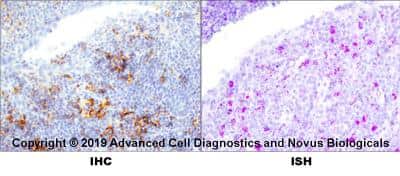
Dual RNAscope ISH-IHC: CD14 Antibody (4B4F12) - BSA Free [NBP2-37291] - CD14 Antibody (4B4F12) [NBP2-37291] - Tissue sections of human tonsil were probed for CD14 mRNA (ACD RNAScope probe, catalog # 418801; Fast Red chromogen, ACD catalog # 322500). Adjacent tissue section was processes for immunohistochemistry using mouse monoclonal (Novus catalog # NBP2-37291) at 1:100 dilution for 1 hour at room temperature followed by incubation with the anti-mouse IgG VisUCyte HRP Polymer Antibody (Catalog # VC001) and DAB chromogen (yellow-brown). Tissue was counterstained with hematoxylin (blue).
![Immunocytochemistry/ Immunofluorescence: CD14 Antibody (4B4F12) - BSA Free [NBP2-37291]](https://resources.rndsystems.com/images/products/CD14-Antibody-4B4F12-Immunocytochemistry-NBP2-37291-img0007.jpg "Immunocytochemistry/ Immunofluorescence: CD14 Antibody (4B4F12) - BSA Free [NBP2-37291]")
Immunocytochemistry/ Immunofluorescence: CD14 Antibody (4B4F12) - BSA Free [NBP2-37291]
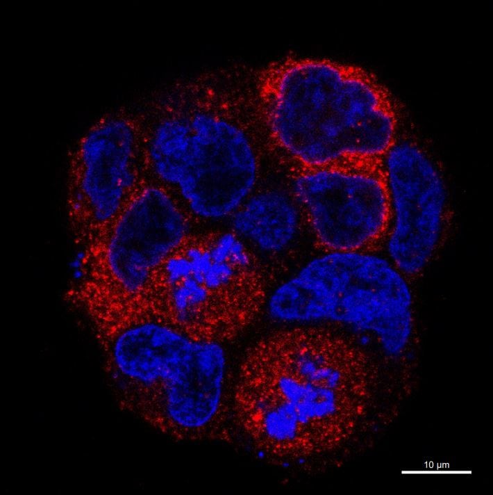
Immunocytochemistry/Immunofluorescence: CD14 Antibody (4B4F12) - BSA Free [NBP2-37291] - CD14 Antibody (4B4F12) [NBP2-37291] - Confocal image of hCD14-transfected HEKT cells, 4% formalin fixed, indirectly labeled with anti-CD14 1:200, and AF568-conjugated Ab 1:1000. Note that cells have different immunoreactivity due to various expression level of CD14 in single cells. Image from verified customer review.![Flow Cytometry: CD14 Antibody (4B4F12) - BSA Free [NBP2-37291]](https://resources.rndsystems.com/images/products/CD14-Antibody-4B4F12-Flow-Cytometry-NBP2-37291-img0008.jpg "Flow Cytometry: CD14 Antibody (4B4F12) - BSA Free [NBP2-37291]")
Flow Cytometry: CD14 Antibody (4B4F12) - BSA Free [NBP2-37291]
Flow Cytometry: CD14 Antibody (4B4F12) - BSA Free [NBP2-37291] - CD14 Antibody (4B4F12) [NBP2-37291] - An intracellular stain was performed on Jurkat cells with NBP2-37291AF647 and a matched isotype control. Cells were fixed with 4% PFA and then permeablized with 0.1% saponin. Cells were incubated in an antibody dilution of 5 ug/mL for 30 minutes at room temperature. Both antibodies were conjugated to Alexa Fluor 647.![Western Blot: CD14 Antibody (4B4F12)BSA Free [NBP2-37291]](https://resources.rndsystems.com/images/products/CD14-Antibody-4B4F12-Western-Blot-NBP2-37291-img0002.jpg "Western Blot: CD14 Antibody (4B4F12)BSA Free [NBP2-37291]")
Western Blot: CD14 Antibody (4B4F12)BSA Free [NBP2-37291]
Western Blot: CD14 Antibody (4B4F12) - BSA Free [NBP2-37291] - CD14 Antibody (4B4F12) [NBP2-37291] - Analysis using CD14 mAb against human CD14 (AA: 20-214) recombinant protein. (Expected MW is 46.8 kDa)![Western Blot: CD14 Antibody (4B4F12)BSA Free [NBP2-37291]](https://resources.rndsystems.com/images/products/CD14-Antibody-4B4F12-Western-Blot-NBP2-37291-img0004.jpg "Western Blot: CD14 Antibody (4B4F12)BSA Free [NBP2-37291]")
Western Blot: CD14 Antibody (4B4F12)BSA Free [NBP2-37291]
Western Blot: CD14 Antibody (4B4F12) - BSA Free [NBP2-37291] - CD14 Antibody (4B4F12) [NBP2-37291] - Analysis using CD14 mouse mAb against HepG2 (1), A549 (2), HL60 (3), RAW264.7 (4), Hela (5), HEK293 (6) and NIH/3T3 (7) cell lysate.![Western Blot: CD14 Antibody (4B4F12)BSA Free [NBP2-37291]](https://resources.rndsystems.com/images/products/CD14-Antibody-4B4F12-Western-Blot-NBP2-37291-img0010.jpg "Western Blot: CD14 Antibody (4B4F12)BSA Free [NBP2-37291]")
![Western Blot: CD14 Antibody (4B4F12)BSA Free [NBP2-37291]](https://resources.rndsystems.com/images/products/CD14-Antibody-4B4F12-Western-Blot-NBP2-37291-img0003.jpg "Western Blot: CD14 Antibody (4B4F12)BSA Free [NBP2-37291]")
Western Blot: CD14 Antibody (4B4F12)BSA Free [NBP2-37291]
Western Blot: CD14 Antibody (4B4F12) - BSA Free [NBP2-37291] - CD14 Antibody (4B4F12) [NBP2-37291] - Analysis using CD14 mAb against HEK293 (1) and CD14 (AA: 20-214)-hIgGFc transfected HEK293 (2) cell lysate.![ELISA: CD14 Antibody (4B4F12) - BSA Free [NBP2-37291]](https://resources.rndsystems.com/images/products/CD14-Antibody-4B4F12-ELISA-NBP2-37291-img0001.jpg "ELISA: CD14 Antibody (4B4F12) - BSA Free [NBP2-37291]")
ELISA: CD14 Antibody (4B4F12) - BSA Free [NBP2-37291]
ELISA: CD14 Antibody (4B4F12) - BSA Free [NBP2-37291] - CD14 Antibody (4B4F12) [NBP2-37291] - Red: Control Antigen (100ng); Purple: Antigen (10ng); Green: Antigen (50ng); Blue: Antigen (100ng);Applications for CD14 Antibody (4B4F12) - BSA Free
Application
Recommended Usage
ELISA
1:10000
Flow Cytometry
1:200 - 1:400
Immunocytochemistry/ Immunofluorescence
1:200
Immunohistochemistry
1:200 - 1:1000
Immunohistochemistry-Paraffin
1:200 - 1:1000
Western Blot
1:500 - 1:2000
Application Notes
This antibody is Cytof ready. CD14 antibody validated for ICC/IF from a verified customer review.
Reviewed Applications
Read 1 review rated 5 using NBP2-37291 in the following applications:
Flow Cytometry Panel Builder
Bio-Techne Knows Flow Cytometry
Save time and reduce costly mistakes by quickly finding compatible reagents using the Panel Builder Tool.
Advanced Features
- Spectra Viewer - Custom analysis of spectra from multiple fluorochromes
- Spillover Popups - Visualize the spectra of individual fluorochromes
- Antigen Density Selector - Match fluorochrome brightness with antigen density

Formulation, Preparation, and Storage
Purification
Protein G purified
Formulation
PBS
Format
BSA Free
Preservative
0.05% Sodium Azide
Concentration
1 mg/ml
Shipping
The product is shipped with polar packs. Upon receipt, store it immediately at the temperature recommended below.
Stability & Storage
Store at 4C short term. Aliquot and store at -20C long term. Avoid freeze-thaw cycles.
Background: CD14
Alternate Names
CD14
Gene Symbol
CD14
Additional CD14 Products
Product Documents for CD14 Antibody (4B4F12) - BSA Free
Certificate of Analysis
To download a Certificate of Analysis, please enter a lot or batch number in the search box below.
Product Specific Notices for CD14 Antibody (4B4F12) - BSA Free
This product is for research use only and is not approved for use in humans or in clinical diagnosis. Primary Antibodies are guaranteed for 1 year from date of receipt.
Citations for CD14 Antibody (4B4F12) - BSA Free
 Powered by Bioz
Customer Reviews for CD14 Antibody (4B4F12) - BSA Free (1)
5 out of 5
1 Customer Rating
Have you used CD14 Antibody (4B4F12) - BSA Free?
Submit a review and receive an Amazon gift card!
$25/€18/£15/$25CAN/¥2500 Yen for a review with an image
$10/€7/£6/$10CAN/¥1110 Yen for a review without an image
Submit a review
Customer Images
Showing
1
-
1 of
1 review
Showing All
Filter By:
-
Application: ImmunocytochemistrySample Tested: Hek 293TSpecies: HumanVerified Customer | Posted 04/18/2018Confocal image of hCD14-transfected HEKT cells,4% formalin fixed, indirectly labeled with anti-CD14 1:200, and AF568-conjugated Ab 1:1000. Note that cells have different immunoreactivity due to various expression level of CD14 in single cells.

There are no reviews that match your criteria.
Protocols
Find general support by application which include: protocols, troubleshooting, illustrated assays, videos and webinars.
- 7-Amino Actinomycin D (7-AAD) Cell Viability Flow Cytometry Protocol
- Antigen Retrieval Protocol (PIER)
- Antigen Retrieval for Frozen Sections Protocol
- Appropriate Fixation of IHC/ICC Samples
- Cellular Response to Hypoxia Protocols
- Chromogenic IHC Staining of Formalin-Fixed Paraffin-Embedded (FFPE) Tissue Protocol
- Chromogenic Immunohistochemistry Staining of Frozen Tissue
- ClariTSA™ Fluorophore Kits
- Detection & Visualization of Antibody Binding
- ELISA Sample Preparation & Collection Guide
- ELISA Troubleshooting Guide
- Extracellular Membrane Flow Cytometry Protocol
- Flow Cytometry Protocol for Cell Surface Markers
- Flow Cytometry Protocol for Staining Membrane Associated Proteins
- Flow Cytometry Staining Protocols
- Flow Cytometry Troubleshooting Guide
- Fluorescent IHC Staining of Frozen Tissue Protocol
- Graphic Protocol for Heat-induced Epitope Retrieval
- Graphic Protocol for the Preparation and Fluorescent IHC Staining of Frozen Tissue Sections
- Graphic Protocol for the Preparation and Fluorescent IHC Staining of Paraffin-embedded Tissue Sections
- Graphic Protocol for the Preparation of Gelatin-coated Slides for Histological Tissue Sections
- How to Run an R&D Systems DuoSet ELISA
- How to Run an R&D Systems Quantikine ELISA
- How to Run an R&D Systems Quantikine™ QuicKit™ ELISA
- ICC Cell Smear Protocol for Suspension Cells
- ICC Immunocytochemistry Protocol Videos
- ICC for Adherent Cells
- IHC Sample Preparation (Frozen sections vs Paraffin)
- ISH-IHC Protocol for Chromogenic Detection on Formalin Fixed Paraffin Embedded (FFPE) Tissue
- Immunocytochemistry (ICC) Protocol
- Immunocytochemistry Troubleshooting
- Immunofluorescence of Organoids Embedded in Cultrex Basement Membrane Extract
- Immunofluorescent IHC Staining of Formalin-Fixed Paraffin-Embedded (FFPE) Tissue Protocol
- Immunohistochemistry (IHC) and Immunocytochemistry (ICC) Protocols
- Immunohistochemistry Frozen Troubleshooting
- Immunohistochemistry Paraffin Troubleshooting
- Intracellular Flow Cytometry Protocol Using Alcohol (Methanol)
- Intracellular Flow Cytometry Protocol Using Detergents
- Intracellular Nuclear Staining Flow Cytometry Protocol Using Detergents
- Intracellular Staining Flow Cytometry Protocol Using Alcohol Permeabilization
- Intracellular Staining Flow Cytometry Protocol Using Detergents to Permeabilize Cells
- Preparing Samples for IHC/ICC Experiments
- Preventing Non-Specific Staining (Non-Specific Binding)
- Primary Antibody Selection & Optimization
- Propidium Iodide Cell Viability Flow Cytometry Protocol
- Protocol for Heat-Induced Epitope Retrieval (HIER)
- Protocol for Liperfluo
- Protocol for Making a 4% Formaldehyde Solution in PBS
- Protocol for VisUCyte™ HRP Polymer Detection Reagent
- Protocol for the Characterization of Human Th22 Cells
- Protocol for the Characterization of Human Th9 Cells
- Protocol for the Fluorescent ICC Staining of Cell Smears - Graphic
- Protocol for the Fluorescent ICC Staining of Cultured Cells on Coverslips - Graphic
- Protocol for the Preparation & Fixation of Cells on Coverslips
- Protocol for the Preparation and Chromogenic IHC Staining of Frozen Tissue Sections
- Protocol for the Preparation and Chromogenic IHC Staining of Frozen Tissue Sections - Graphic
- Protocol for the Preparation and Chromogenic IHC Staining of Paraffin-embedded Tissue Sections
- Protocol for the Preparation and Chromogenic IHC Staining of Paraffin-embedded Tissue Sections - Graphic
- Protocol for the Preparation and Fluorescent ICC Staining of Cells on Coverslips
- Protocol for the Preparation and Fluorescent ICC Staining of Non-adherent Cells
- Protocol for the Preparation and Fluorescent ICC Staining of Stem Cells on Coverslips
- Protocol for the Preparation and Fluorescent IHC Staining of Frozen Tissue Sections
- Protocol for the Preparation and Fluorescent IHC Staining of Paraffin-embedded Tissue Sections
- Protocol for the Preparation of Gelatin-coated Slides for Histological Tissue Sections
- Protocol for the Preparation of a Cell Smear for Non-adherent Cell ICC - Graphic
- Protocol: Annexin V and PI Staining by Flow Cytometry
- Protocol: Annexin V and PI Staining for Apoptosis by Flow Cytometry
- Quantikine HS ELISA Kit Assay Principle, Alkaline Phosphatase
- Quantikine HS ELISA Kit Principle, Streptavidin-HRP Polymer
- R&D Systems Quality Control Western Blot Protocol
- Sandwich ELISA (Colorimetric) – Biotin/Streptavidin Detection Protocol
- Sandwich ELISA (Colorimetric) – Direct Detection Protocol
- TUNEL and Active Caspase-3 Detection by IHC/ICC Protocol
- The Importance of IHC/ICC Controls
- Troubleshooting Guide: ELISA
- Troubleshooting Guide: Fluorokine Flow Cytometry Kits
- Troubleshooting Guide: Immunohistochemistry
- Troubleshooting Guide: Western Blot Figures
- Western Blot Conditions
- Western Blot Protocol
- Western Blot Protocol for Cell Lysates
- Western Blot Troubleshooting
- Western Blot Troubleshooting Guide
- View all Protocols, Troubleshooting, Illustrated assays and Webinars
Loading...
Associated Pathways


