CD44 Antibody (8E2F3) - BSA Free
Novus Biologicals | Catalog # NBP1-47386
Key Product Details
Validated by
Knockout/Knockdown
Species Reactivity
Validated:
Human, Mouse, Rabbit (Negative)
Cited:
Human, Mouse, Rat, Canine
Predicted:
Baboon (94%), Canine (93%), Equine (93%). Backed by our 100% Guarantee.
Applications
Validated:
Knockout Validated, Immunohistochemistry, Immunohistochemistry-Paraffin, Western Blot, ELISA, Flow Cytometry, Flow (Intracellular), Immunocytochemistry/ Immunofluorescence, Immunoprecipitation
Cited:
Immunohistochemistry-Paraffin, Western Blot, Flow Cytometry, Immunocytochemistry/ Immunofluorescence, IF/IHC, SDS-Page
Label
Unconjugated
Antibody Source
Monoclonal Mouse IgG1 Clone # 8E2F3
Format
BSA Free
Loading...
Product Specifications
Immunogen
Purified recombinant fragment of human CD44 (628-699) expressed in E. coli. [Uniprot: P16070]
Reactivity Notes
Not reactive to rabbit per customer review.
Localization
Membrane; Single-pass type I membrane protein.
Marker
Cell Membrane Marker
Clonality
Monoclonal
Host
Mouse
Isotype
IgG1
Theoretical MW
82 kDa.
Disclaimer note: The observed molecular weight of the protein may vary from the listed predicted molecular weight due to post translational modifications, post translation cleavages, relative charges, and other experimental factors.
Disclaimer note: The observed molecular weight of the protein may vary from the listed predicted molecular weight due to post translational modifications, post translation cleavages, relative charges, and other experimental factors.
Scientific Data Images for CD44 Antibody (8E2F3) - BSA Free
![Immunocytochemistry/ Immunofluorescence: CD44 Antibody (8E2F3) - BSA Free [NBP1-47386]](https://resources.rndsystems.com/images/products/CD44-Antibody-8E2F3-Immunocytochemistry-Immunofluorescence-NBP1-47386-img0011.jpg "Immunocytochemistry/ Immunofluorescence: CD44 Antibody (8E2F3) - BSA Free [NBP1-47386]")
Immunocytochemistry/ Immunofluorescence: CD44 Antibody (8E2F3) - BSA Free [NBP1-47386]
Immunocytochemistry/Immunofluorescence: CD44 Antibody (8E2F3) [NBP1-47386] - Analysis of paraffin-embedded human lung cancer tissues using anti-CD44 mAb (green), showing membrane localization. DRAQ5 fluorescent DNA dye (blue).![Immunohistochemistry-Paraffin: CD44 Antibody (8E2F3) - BSA Free [NBP1-47386]](https://resources.rndsystems.com/images/products/CD44-Antibody-8E2F3-Immunohistochemistry-Paraffin-NBP1-47386-img0014.jpg "Immunohistochemistry-Paraffin: CD44 Antibody (8E2F3) - BSA Free [NBP1-47386]")
Immunohistochemistry-Paraffin: CD44 Antibody (8E2F3) - BSA Free [NBP1-47386]
Immunohistochemistry-Paraffin: CD44 Antibody (8E2F3) [NBP1-47386] - Analysis of FFPE human breast carcinoma tissues using CD44 antibody (8E2F3). The signal was developed using DAB based detection and the sections were processed for counterstaining with hematoxylin. The antibody generated mainly a membrane staining representative of CD44 protein.![Flow Cytometry: CD44 Antibody (8E2F3) - BSA Free [NBP1-47386]](https://resources.rndsystems.com/images/products/CD44-Antibody-8E2F3-Flow-Cytometry-NBP1-47386-img0008.jpg "Flow Cytometry: CD44 Antibody (8E2F3) - BSA Free [NBP1-47386]")
Flow Cytometry: CD44 Antibody (8E2F3) - BSA Free [NBP1-47386]
Flow Cytometry: CD44 Antibody (8E2F3) [NBP1-47386] - Analysis of HeLa cells using anti-CD44 mAb (right) and negative control (left).![Immunocytochemistry/ Immunofluorescence: CD44 Antibody (8E2F3) - BSA Free [NBP1-47386]](https://resources.rndsystems.com/images/products/CD44-Antibody-8E2F3-Immunocytochemistry-Immunofluorescence-NBP1-47386-img0018.jpg "Immunocytochemistry/ Immunofluorescence: CD44 Antibody (8E2F3) - BSA Free [NBP1-47386]")
Immunocytochemistry/ Immunofluorescence: CD44 Antibody (8E2F3) - BSA Free [NBP1-47386]
Immunocytochemistry/Immunofluorescence: CD44 Antibody (8E2F3) [NBP1-47386] - HeLa cells were fixed in 4% paraformaldehyde for 10 minutes and permeabilized in 0.05% Triton X-100 in PBS for 5 minutes. The cells were incubated with anti-CD44 Antibody [8E2F3] conjugated to DyLight 550 (NBP1-47386R) at 5 ug/ml for 1 hour at room temperature. Nuclei were counterstained with DAPI (Blue). Cells were imaged using a 100X objective and digitally deconvolved.![Western Blot: CD44 Antibody (8E2F3)BSA Free [NBP1-47386]](https://resources.rndsystems.com/images/products/CD44-Antibody-8E2F3-BSA-Free-Western-Blot-NBP1-47386-img0021.jpg "Western Blot: CD44 Antibody (8E2F3)BSA Free [NBP1-47386]")
Western Blot: CD44 Antibody (8E2F3)BSA Free [NBP1-47386]
CD44-Antibody-8E2F3-BSA-Free-Western-Blot-NBP1-47386-img0021.jpg![Immunocytochemistry/ Immunofluorescence: CD44 Antibody (8E2F3) - BSA Free [NBP1-47386]](https://resources.rndsystems.com/images/products/CD44-Antibody-8E2F3-Immunocytochemistry-Immunofluorescence-NBP1-47386-img0019.jpg "Immunocytochemistry/ Immunofluorescence: CD44 Antibody (8E2F3) - BSA Free [NBP1-47386]")
Immunocytochemistry/ Immunofluorescence: CD44 Antibody (8E2F3) - BSA Free [NBP1-47386]
Immunocytochemistry/Immunofluorescence: CD44 Antibody (8E2F3) [NBP1-47386] - HeLa cells were fixed in 4% paraformaldehyde for 10 minutes and permeabilized in 0.05% Triton X-100 in PBS for 5 minutes. The cells were incubated with CD44 Antibody [8E2F3] conjugated to Alexa Fluor 647 (NBP1-47386AF647) at 5 ug/ml for 1 hour at room temperature. Nuclei were counterstained with DAPI (Blue). Cells were imaged using a 100X objective and digitally deconvolved.![Flow Cytometry: CD44 Antibody (8E2F3) - BSA Free [NBP1-47386]](https://resources.rndsystems.com/images/products/CD44-Antibody-8E2F3-Flow-Cytometry-NBP1-47386-img0017.jpg "Flow Cytometry: CD44 Antibody (8E2F3) - BSA Free [NBP1-47386]")
Flow Cytometry: CD44 Antibody (8E2F3) - BSA Free [NBP1-47386]
Flow Cytometry: CD44 Antibody (8E2F3) [NBP1-47386] - An intracellular stain was performed on HeLa cells with NBP1-47386AF488 (blue) and a matched isotype control (orange). Cells were fixed with 4% PFA and then permeablized with 0.1% saponin. Cells were incubated in an antibody dilution of 5 ug/mL for 30 minutes at room temperature. Both antibodies were conjugated to Alexa Fluor 488.![Immunocytochemistry/ Immunofluorescence: CD44 Antibody (8E2F3) - BSA Free [NBP1-47386]](https://resources.rndsystems.com/images/products/CD44-Antibody-8E2F3-Immunocytochemistry-Immunofluorescence-NBP1-47386-img0009.jpg "Immunocytochemistry/ Immunofluorescence: CD44 Antibody (8E2F3) - BSA Free [NBP1-47386]")
Immunocytochemistry/ Immunofluorescence: CD44 Antibody (8E2F3) - BSA Free [NBP1-47386]
Immunocytochemistry/Immunofluorescence: CD44 Antibody (8E2F3) [NBP1-47386] - Analysis of methanol-fixed A431 (A), HeLa (B), PANC-1 (C) and EC (D) cells using anti-CD44 mAb (green), showing membrane localization. DRAQ5 fluorescent DNA dye (blue).![Immunocytochemistry/ Immunofluorescence: CD44 Antibody (8E2F3) - BSA Free [NBP1-47386]](https://resources.rndsystems.com/images/products/CD44-Antibody-8E2F3-Immunocytochemistry-Immunofluorescence-NBP1-47386-img0010.jpg "Immunocytochemistry/ Immunofluorescence: CD44 Antibody (8E2F3) - BSA Free [NBP1-47386]")
Immunocytochemistry/ Immunofluorescence: CD44 Antibody (8E2F3) - BSA Free [NBP1-47386]
Immunocytochemistry/Immunofluorescence: CD44 Antibody (8E2F3) [NBP1-47386] - Analysis of PANC-1 cells using anti-CD44 mAb (green). Actin filaments have been labeled with DY-554 phalloidin (red). DRAQ5 fluorescent DNA dye (blue).![Flow (Intracellular): CD44 Antibody (8E2F3) - BSA Free [NBP1-47386]](https://resources.rndsystems.com/images/products/CD44-Antibody-8E2F3-Flow-Intracellular-NBP1-47386-img0016.jpg "Flow (Intracellular): CD44 Antibody (8E2F3) - BSA Free [NBP1-47386]")
Flow (Intracellular): CD44 Antibody (8E2F3) - BSA Free [NBP1-47386]
Flow (Intracellular): CD44 Antibody (8E2F3) [NBP1-47386] - An intracellular stain was performed on HeLa cells with NBP1-47386AF647 (blue) and a matched isotype control (orange). Cells were fixed with 4% PFA and then permeablized with 0.1% saponin. Cells were incubated in an antibody dilution of 2.5 ug/mL for 30 minutes at room temperature. Both antibodies were conjugated to Alexa Fluor 647.![Immunoprecipitation: CD44 Antibody (8E2F3) - BSA Free [NBP1-47386]](https://resources.rndsystems.com/images/products/CD44-Antibody-8E2F3-BSA-Free-Immunoprecipitation-NBP1-47386-img0020.jpg "Immunoprecipitation: CD44 Antibody (8E2F3) - BSA Free [NBP1-47386]")
Immunoprecipitation: CD44 Antibody (8E2F3) - BSA Free [NBP1-47386]
Immunoprecipitation: CD44 Antibody (8E2F3) - BSA Free [NBP1-47386] - HAP1 lysates were prepared and immunoprecipitation was performed using 1.0 ug of the CD44 Antibody (NBP1-47386) pre-coupled to either protein G or protein A Sepharose beads. Ability of the antibodies to capture CD44 antigen was first assessed by comparing the level of CD44 antigen from the starting material (SM) to its level remaining in the unbound fractions (UB). Anti-CD44 antigen at 1/2000 was used for each immunoblot. Immunoprecipitate for CD44 Antibody (NBP1-47386) that showed depleted CD44 antigen in the UB can be seen. Image, protocol and testing courtesy of YCharOS Inc. (ycharos.com). in A431 Human Cell Line -")
CD44 (8E2F3) in A431 Human Cell Line -
CD44 (8E2F3) was detected in immersion fixed A431 human skin carcinoma cell line using Mouse anti- CD44 (8E2F3) Protein-G purified Monoclonal Antibody conjugated to Biotin (Catalog # NBP1-47386B) at 2 µg/mL overnight at 4C. Cells were stained using Streptavidin conjugated to DyLight 550 (red) and counterstained with DAPI (blue). Cells were imaged using a 100X objective and digitally deconvolved. in A431 Human Cell Line -")
CD44 (8E2F3) in A431 Human Cell Line -
CD44 (8E2F3) was detected in immersion fixed A431 human skin carcinoma cell line using Mouse anti-CD44 (8E2F3) Protein-G purified Monoclonal Antibody conjugated to DyLight 550 (Catalog # NBP1-47386R) (red) at 5 µg/mL overnight at 4C. Cells were counterstained with DAPI (blue). Cells were imaged using a 100X objective and digitally deconvolved. in NIH-3T3 Mouse Cell Line.")
CD44 (8E2F3) in NIH-3T3 Mouse Cell Line.
CD44 (8E2F3) was detected in immersion fixed NIH3T3 Mouse fibroblast cell line using Mouse anti- CD44 (8E2F3) Protein-G purified Monoclonal Antibody conjugated to DyLight 550 (Catalog # NBP1-47386R) (red) at 2 µg/mL overnight at 4C. Cells were counterstained with DAPI (blue). Cells were imaged using a 100X objective and digitally deconvolved. in U-251 MG Human Cell Line by Flow Cytometry.")
Detection of CD44 (8E2F3) in U-251 MG Human Cell Line by Flow Cytometry.
U-251 MG human glioblastoma cell line was stained with Mouse anti-CD44 (8E2F3) Protein-G purified Monoclonal Antibody conjugated to Alexa Fluor® 647 (Catalog # NBP1-47386AF647, blue histogram) or matched control antibody (orange histogram). in A431 Human Cell Line by Flow Cytometry.")
Detection of CD44 (8E2F3) in A431 Human Cell Line by Flow Cytometry.
An intracellular stain was performed on A431 human skin carcinoma cell line using Mouse anti-CD44 (8E2F3) Protein-G purified Monoclonal Antibody conjugated to DyLight 550 (Catalog # NBP1-47386R, blue histogram) or matched control antibody (orange histogram) at 2.5 µg/mL for 30 minutes at RT. in U-2 OS Human Cell Line.")
CD44 (8E2F3) in U-2 OS Human Cell Line.
CD44 (8E2F3) was detected in immersion fixed U-2 OS human osteosarcoma cell line using Mouse anti-CD44 (8E2F3) Protein-G purified Monoclonal Antibody conjugated to Biotin (Catalog # NBP1-47386B) at 5 µg/mL overnight at 4C. Cells were stained using Streptavidin conjugated to DyLight 550 (red) and counterstained with DAPI (blue). Cells were imaged using a 100X objective and digitally deconvolved.
Detection of CD44 by Immunoprecipitation.
MDA‑MB‑231 human breast cancer cell line lysates were prepared and immunoprecipitation was performed using 2.0 μg of CD44 Antibody (8E2F3) - BSA Free (Catalog # NBP1-47386) pre-coupled to Dynabeads Protein G. Immunoprecipitated CD44 was detected in Western Blot with a rabbit CD44 antibody used at 1/3000. The Ponceau stained transfer of the blot is shown. SM=4% starting material; UB=4% unbound fraction; IP=immunoprecipitate; HC=antibody heavy chain. Image, protocol and testing courtesy of YCharOS Inc. (ycharos.com).
CD44 Specificity is Shown by Immunocytochemistry in Knockout Cell Line.
MDA‑MB‑231 human breast cancer parental cell line WT and CD44 MDA-MB-231 KO cells were labelled with a green or a far-red fluorescent dye, respectively. Cells were stained with CD44 Antibody (8E2F3) - BSA Free (Catalog # NBP1-47386) followed by incubation with an Alexa-fluor 555 conjugated secondary antibody (upper panel). DAPI-only counterstained cells shown on a lower panel. Acquisition of the blue (nucleus-DAPI), green (identification of WT cells), red (antibody staining) and far-red (identification of KO cells) channels was performed. Representative images of the blue and red (grayscale) channels are shown. WT and KO cells are outlined with green and magenta dashed line, respectively. Primary antibody dilution used: 1/1000. Image, protocol and testing courtesy of YCharOS Inc. (ycharos.com).![CD44 Antibody (8E2F3) - BSA Free Immunohistochemistry-Paraffin: CD44 Antibody (8E2F3) - BSA Free [NBP1-47386]](https://resources.rndsystems.com/images/products/antibody/nbp1-47386_mouse-monoclonal-cd44-antibody-8e2f3-immunohistochemistry-paraffin-2322026134732.png "Immunohistochemistry-Paraffin: CD44 Antibody (8E2F3) - BSA Free [NBP1-47386]")
Immunohistochemistry-Paraffin: CD44 Antibody (8E2F3) - BSA Free [NBP1-47386]
Analysis of a FFPE tissue section of human skin using 1:200 dilution of CD44 (8E2F3) antibody. The staining was developed using HRP labeled anti-mouse secondary antibody and DAB reagent, and nuclei of cells were counter-stained with hematoxylin.Applications for CD44 Antibody (8E2F3) - BSA Free
Application
Recommended Usage
ELISA
1:10000
Flow Cytometry
1:200-1:400
Immunocytochemistry/ Immunofluorescence
1:200-1:1000
Immunohistochemistry
1:200-1:1000
Immunohistochemistry-Paraffin
1:200-1:1000
Knockout Validated
Knockout validated from YCharOS Inc. (YCharOS.com)
Reviewed Applications
Read 1 review rated 5 using NBP1-47386 in the following applications:
Flow Cytometry Panel Builder
Bio-Techne Knows Flow Cytometry
Save time and reduce costly mistakes by quickly finding compatible reagents using the Panel Builder Tool.
Advanced Features
- Spectra Viewer - Custom analysis of spectra from multiple fluorochromes
- Spillover Popups - Visualize the spectra of individual fluorochromes
- Antigen Density Selector - Match fluorochrome brightness with antigen density

Formulation, Preparation, and Storage
Purification
Ammonium sulfate precipitation
Formulation
PBS
Format
BSA Free
Preservative
0.02% Sodium Azide
Concentration
1.0 mg/ml
Shipping
The product is shipped with polar packs. Upon receipt, store it immediately at the temperature recommended below.
Stability & Storage
Store at 4C short term. Aliquot and store at -20C long term. Avoid freeze-thaw cycles.
Background: CD44
Alternate Names
CD44, ECMR-III, HCAM, HCELL, LHR, MDU2, MDU3, MIC4, MUTCH-I, Pgp1
Entrez Gene IDs
960 (Human)
Gene Symbol
CD44
UniProt
Additional CD44 Products
Product Documents for CD44 Antibody (8E2F3) - BSA Free
Certificate of Analysis
To download a Certificate of Analysis, please enter a lot or batch number in the search box below.
Product Specific Notices for CD44 Antibody (8E2F3) - BSA Free
This product is for research use only and is not approved for use in humans or in clinical diagnosis. Primary Antibodies are guaranteed for 1 year from date of receipt.
Related Research Areas
Citations for CD44 Antibody (8E2F3) - BSA Free
 Powered by Bioz
Customer Reviews for CD44 Antibody (8E2F3) - BSA Free (1)
5 out of 5
1 Customer Rating
Have you used CD44 Antibody (8E2F3) - BSA Free?
Submit a review and receive an Amazon gift card!
$25/€18/£15/$25CAN/¥2500 Yen for a review with an image
$10/€7/£6/$10CAN/¥1110 Yen for a review without an image
Submit a review
Customer Images
Showing
1
-
1 of
1 review
Showing All
Filter By:
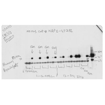
-
Application: Western BlotSample Tested: Human Brain TissueSpecies: HumanVerified Customer | Posted 07/12/2013Western blot analysis of CD44 in brain tissue

There are no reviews that match your criteria.
Protocols
View specific protocols for CD44 Antibody (8E2F3) - BSA Free (NBP1-47386):
Immunohistochemistry-Paraffin Embedded Sections
Antigen Unmasking:
Bring slides to a boil in 10 mM sodium citrate buffer (pH 6.0) then maintain at a sub-boiling temperature for 10 minutes. Cool slides on bench-top for 30 minutes (keep slides in the sodium citrate buffer at all times).
Staining:
1. Wash sections in deionized water three times for 5 minutes each.
2. Wash sections in PBS for 5 minutes.
3. Block each section with 100-400 ul blocking solution (1% BSA in PBS) for 1 hour at room temperature.
4. Remove blocking solution and add 100-400 ul diluted primary antibody. Incubate overnight at 4 C.
5. Remove antibody solution and wash sections in wash buffer three times for 5 minutes each.
6. Add 100-400 ul HRP polymer conjugated secondary antibody. Incubate 30 minutes at room temperature.
7. Wash sections three times in wash buffer for 5 minutes each.
8. Add 100-400 ul DAB substrate to each section and monitor staining closely.
9. As soon as the sections develop, immerse slides in deionized water.
10. Counterstain sections in hematoxylin.
11. Wash sections in deionized water two times for 5 minutes each.
12. Dehydrate sections.
13. Mount coverslips.
Western Blot Protocol
1. Perform SDS-PAGE on samples to be analyzed, loading 10-25 ug of total protein per lane.
2. Transfer proteins to PVDF membrane according to the instructions provided by the manufacturer of the membrane and transfer apparatus.
3. Stain the membrane with Ponceau S (or similar product) to assess transfer success, and mark molecular weight standards where appropriate.
4. Rinse the blot TBS -0.05% Tween 20 (TBST).
5. Block the membrane in 5% Non-fat milk in TBST (blocking buffer) for at least 1 hour.
6. Wash the membrane in TBST three times for 10 minutes each.
7. Dilute primary antibody in blocking buffer and incubate overnight at 4C with gentle rocking.
8. Wash the membrane in TBST three times for 10 minutes each.
9. Incubate the membrane in diluted HRP conjugated secondary antibody in blocking buffer (as per manufacturer's instructions) for 1 hour at room temperature.
10. Wash the blot in TBST three times for 10 minutes each (this step can be repeated as required to reduce background).
11. Apply the detection reagent of choice in accordance with the manufacturer's instructions.
Find general support by application which include: protocols, troubleshooting, illustrated assays, videos and webinars.
- 7-Amino Actinomycin D (7-AAD) Cell Viability Flow Cytometry Protocol
- Antigen Retrieval Protocol (PIER)
- Antigen Retrieval for Frozen Sections Protocol
- Appropriate Fixation of IHC/ICC Samples
- Cellular Response to Hypoxia Protocols
- Chromogenic IHC Staining of Formalin-Fixed Paraffin-Embedded (FFPE) Tissue Protocol
- Chromogenic Immunohistochemistry Staining of Frozen Tissue
- ClariTSA™ Fluorophore Kits
- Detection & Visualization of Antibody Binding
- ELISA Sample Preparation & Collection Guide
- ELISA Troubleshooting Guide
- Extracellular Membrane Flow Cytometry Protocol
- Flow Cytometry Protocol for Cell Surface Markers
- Flow Cytometry Protocol for Staining Membrane Associated Proteins
- Flow Cytometry Staining Protocols
- Flow Cytometry Troubleshooting Guide
- Fluorescent IHC Staining of Frozen Tissue Protocol
- Graphic Protocol for Heat-induced Epitope Retrieval
- Graphic Protocol for the Preparation and Fluorescent IHC Staining of Frozen Tissue Sections
- Graphic Protocol for the Preparation and Fluorescent IHC Staining of Paraffin-embedded Tissue Sections
- Graphic Protocol for the Preparation of Gelatin-coated Slides for Histological Tissue Sections
- How to Run an R&D Systems DuoSet ELISA
- How to Run an R&D Systems Quantikine ELISA
- How to Run an R&D Systems Quantikine™ QuicKit™ ELISA
- ICC Cell Smear Protocol for Suspension Cells
- ICC Immunocytochemistry Protocol Videos
- ICC for Adherent Cells
- IHC Sample Preparation (Frozen sections vs Paraffin)
- Immunocytochemistry (ICC) Protocol
- Immunocytochemistry Troubleshooting
- Immunofluorescence of Organoids Embedded in Cultrex Basement Membrane Extract
- Immunofluorescent IHC Staining of Formalin-Fixed Paraffin-Embedded (FFPE) Tissue Protocol
- Immunohistochemistry (IHC) and Immunocytochemistry (ICC) Protocols
- Immunohistochemistry Frozen Troubleshooting
- Immunohistochemistry Paraffin Troubleshooting
- Immunoprecipitation Protocol
- Intracellular Flow Cytometry Protocol Using Alcohol (Methanol)
- Intracellular Flow Cytometry Protocol Using Detergents
- Intracellular Nuclear Staining Flow Cytometry Protocol Using Detergents
- Intracellular Staining Flow Cytometry Protocol Using Alcohol Permeabilization
- Intracellular Staining Flow Cytometry Protocol Using Detergents to Permeabilize Cells
- Preparing Samples for IHC/ICC Experiments
- Preventing Non-Specific Staining (Non-Specific Binding)
- Primary Antibody Selection & Optimization
- Propidium Iodide Cell Viability Flow Cytometry Protocol
- Protocol for Heat-Induced Epitope Retrieval (HIER)
- Protocol for Liperfluo
- Protocol for Making a 4% Formaldehyde Solution in PBS
- Protocol for VisUCyte™ HRP Polymer Detection Reagent
- Protocol for the Characterization of Human Th22 Cells
- Protocol for the Characterization of Human Th9 Cells
- Protocol for the Fluorescent ICC Staining of Cell Smears - Graphic
- Protocol for the Fluorescent ICC Staining of Cultured Cells on Coverslips - Graphic
- Protocol for the Preparation & Fixation of Cells on Coverslips
- Protocol for the Preparation and Chromogenic IHC Staining of Frozen Tissue Sections
- Protocol for the Preparation and Chromogenic IHC Staining of Frozen Tissue Sections - Graphic
- Protocol for the Preparation and Chromogenic IHC Staining of Paraffin-embedded Tissue Sections
- Protocol for the Preparation and Chromogenic IHC Staining of Paraffin-embedded Tissue Sections - Graphic
- Protocol for the Preparation and Fluorescent ICC Staining of Cells on Coverslips
- Protocol for the Preparation and Fluorescent ICC Staining of Non-adherent Cells
- Protocol for the Preparation and Fluorescent ICC Staining of Stem Cells on Coverslips
- Protocol for the Preparation and Fluorescent IHC Staining of Frozen Tissue Sections
- Protocol for the Preparation and Fluorescent IHC Staining of Paraffin-embedded Tissue Sections
- Protocol for the Preparation of Gelatin-coated Slides for Histological Tissue Sections
- Protocol for the Preparation of a Cell Smear for Non-adherent Cell ICC - Graphic
- Protocol: Annexin V and PI Staining by Flow Cytometry
- Protocol: Annexin V and PI Staining for Apoptosis by Flow Cytometry
- Quantikine HS ELISA Kit Assay Principle, Alkaline Phosphatase
- Quantikine HS ELISA Kit Principle, Streptavidin-HRP Polymer
- R&D Systems Quality Control Western Blot Protocol
- Sandwich ELISA (Colorimetric) – Biotin/Streptavidin Detection Protocol
- Sandwich ELISA (Colorimetric) – Direct Detection Protocol
- TUNEL and Active Caspase-3 Detection by IHC/ICC Protocol
- The Importance of IHC/ICC Controls
- Troubleshooting Guide: ELISA
- Troubleshooting Guide: Fluorokine Flow Cytometry Kits
- Troubleshooting Guide: Immunohistochemistry
- Troubleshooting Guide: Western Blot Figures
- Western Blot Conditions
- Western Blot Protocol
- Western Blot Protocol for Cell Lysates
- Western Blot Troubleshooting
- Western Blot Troubleshooting Guide
- View all Protocols, Troubleshooting, Illustrated assays and Webinars
Loading...
Associated Pathways


