FOXO3 Antibody - BSA Free
Novus Biologicals | Catalog # NB100-614
![Western Blot: FOXO3 Antibody [NB100-614]](https://resources.rndsystems.com/images/products/FOXO3-Antibody-Western-Blot-NB100-614-img0014.jpg "Western Blot: FOXO3 Antibody [NB100-614]")
Key Product Details
Validated by
Species Reactivity
Validated:
Cited:
Applications
Validated:
Cited:
Label
Antibody Source
Format
Product Specifications
Immunogen
Reactivity Notes
Specificity
Clonality
Host
Isotype
Theoretical MW
Disclaimer note: The observed molecular weight of the protein may vary from the listed predicted molecular weight due to post translational modifications, post translation cleavages, relative charges, and other experimental factors.
Scientific Data Images for FOXO3 Antibody - BSA Free
Western Blot: FOXO3 Antibody [NB100-614]
FOXO3-Antibody-Western-Blot-NB100-614-img0014.jpg![Immunocytochemistry/ Immunofluorescence: FOXO3 Antibody [NB100-614]](https://resources.rndsystems.com/images/products/FOXO3-Antibody-Immunocytochemistry-Immunofluorescence-NB100-614-img0007.jpg "Immunocytochemistry/ Immunofluorescence: FOXO3 Antibody [NB100-614]")
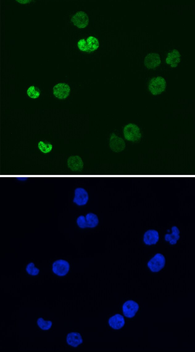
Immunocytochemistry/ Immunofluorescence: FOXO3 Antibody [NB100-614]
Immunocytochemistry/Immunofluorescence: FOXO3 Antibody [NB100-614] - Analysis of FOXO3 in human B cell lymphoma using anti-FOXO3 antibody.Image submitted by a verified customer review.![Immunohistochemistry-Paraffin: FOXO3 Antibody [NB100-614]](https://resources.rndsystems.com/images/products/FOXO3-Antibody-Immunohistochemistry-NB100-614-img0009.jpg "Immunohistochemistry-Paraffin: FOXO3 Antibody [NB100-614]")
Immunohistochemistry-Paraffin: FOXO3 Antibody [NB100-614]
Immunohistochemistry-Paraffin: FOXO3 Antibody [NB100-614] - Sample: FFPE section of human ovarian carcinoma (left) and mouse renal cell carcinoma (right). Antibody: Affinity purified rabbit anti-FOXO3a used at a dilution of 1:1,000 (1ug/ml). Detection: DAB![Western Blot: FOXO3 Antibody [NB100-614]](https://resources.rndsystems.com/images/products/FOXO3-Antibody-Western-Blot-NB100-614-img0011.jpg "Western Blot: FOXO3 Antibody [NB100-614]")
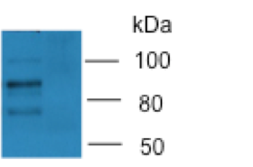
Western Blot: FOXO3 Antibody [NB100-614]
Western Blot: FOXO3 Antibody [NB100-614] - Detection of Human and Mouse FOXO3a by Western Blot. Samples: Whole cell lysate (50 ug) from HeLa, 293T, Jurkat, mouse TCMK-1, and mouse NIH3T3 cells. Antibodies: Affinity purified rabbit anti-FOXO3a antibody NB100-614 used for WB at 0.1 ug/ml. Detection: Chemiluminescence with an exposure time of 3 minutes.![Western Blot: FOXO3 Antibody [NB100-614]](https://resources.rndsystems.com/images/products/FOXO3-Antibody-Western-Blot-NB100-614-img0012.jpg "Western Blot: FOXO3 Antibody [NB100-614]")
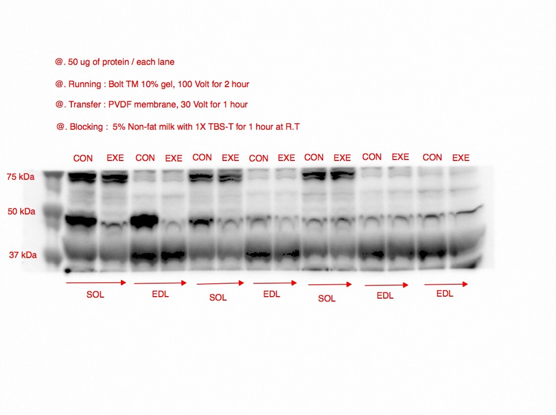
Western Blot: FOXO3 Antibody [NB100-614]
Western Blot: FOXO3 Antibody [NB100-614] - FOXO3 Protein expression levels both control group and exercise group for skeletal muscles. Image submitted by a verified customer review *{Mouse WB}*![Immunoprecipitation: FOXO3 Antibody [NB100-614]](https://resources.rndsystems.com/images/products/FOXO3-Antibody-Immunoprecipitation-NB100-614-img0013.jpg "Immunoprecipitation: FOXO3 Antibody [NB100-614]")
Immunoprecipitation: FOXO3 Antibody [NB100-614]
Immunoprecipitation: FOXO3 Antibody [NB100-614] - Detection of human FOXO3a by western blot of immunoprecipitates. Samples: Whole cell lysate (1 mg for IP; 20% of IP loaded) from MCF-7 cells. Antibodies: Affinity purified rabbit anti-FOXO3a antibody NB100-614 used for IP at 6 ug/mg lysate. FOXO3a was also immunoprecipitated by rabbit anti-FOXO3a antibody NB100-613. For blotting immunoprecipitated FOXO3a, NB100-614 was used at 1 ug/ml. Detection: Chemiluminescence with an exposure time of 3 minutes.![Knockout Validated: FOXO3 Antibody [NB100-614]](https://resources.rndsystems.com/images/products/FOXO3-Antibody-Knockout-Validated-NB100-614-img0015.jpg "Western Blot: FOXO3 Antibody [NB100-614]")
Western Blot: FOXO3 Antibody [NB100-614]
Western Blot: FOXO3 Antibody [NB100-614] - Expression of FOXO increases in aging brain. Representative Western blotting results. Foxo1- or Foxo1/3-specific knockouts (1KO or 1/3KO) selectively lost targeted isoforms. CBM-cerebellum, STR-striatum, SCD-spinal cord, CTX-cortex, BST-brain stem, MDB-midbrain, HPC-hippocampus.
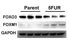
Western Blot: Rabbit Polyclonal FOXO3 Antibody [NB100-614] -
Western Blot: Rabbit Polyclonal FOXO3 Antibody [NB100-614] - Analysis of FOXO3 and FOXM1in parental HCT-116 and HCT-116-FUR cells. Image from a verified customer review.
Western Blot: FOXO3 Antibody [NB100-614] -
Western Blot: FOXO3 Antibody [NB100-614] - Increased proteotoxic stress & defective autophagy in Foxo1/3/4 knockout mice. (a) Tissue lysates from 12‐month‐old WT & KO brain cortex were analyzed by WB of indicated proteins. (c) p62 IF & (d) p62 & Ub IHC on cortical neurons from 12‐month‐old WT & KO brain cortex. Red arrows point to labeling positive inclusions. Scale bar = 200 μm. (e) Quantitation of representative results for (d). (f) Indicated genotype of neural progenitor cultures was differentiated into neurons for 5 days. Some cultures were treated with rapamycin (100 ng/ml) for 24 hr on day 4 of differentiation. (h) The autophagy activity was analyzed by flow cytometry. The bar graph shows autophagy indices as % of control of (f). (i) Cultures as in (f) were treated with chloroquine (100 ng/ml) for 4 hr & analyzed for the expression levels of LC3 by WB. Representative result from three to five independent experiments is shown. (b, g, j) Quantitation of WB band densities. Only chloroquine‐treated cultures were statistically compared in (j). Error bars, mean ± SEM. *p < .05. **p < .01, ***p < .005; ****p < .001. Statistical significance was determined by one‐way ANOVA Image collected & cropped by CiteAb from the following publication (https://pubmed.ncbi.nlm.nih.gov/29178390), licensed under a CC-BY license. Not internally tested by Novus Biologicals.
Western Blot: FOXO3 Antibody [NB100-614] -
Western Blot: FOXO3 Antibody [NB100-614] - Expression of FOXO increases in aging brain. (a) XY plots of FOXO1 or FOXO3 mRNA expression within the noted brain regions vs. age of the subjects at time of death. (b) Pearson correlation coefficients (r) & p‐values for the correlation of FOXO1 or FOXO3 mRNA expression in various regions of the human brain with the age. (c) The mRNA expressions of FOXO1 & FOXO3 were measured in human cerebellums (n = 33). Blue & green dots indicate samples used for WB in (d). The mRNA (e) & protein (f) expression of Foxo1, Foxo3, & phospho‐T24/32 Foxo1/3 in young (<3‐month, n = 6), adult (3‐18‐month, n = 6), & old (18–20‐month, n = 6) FVB/B6 mixed‐strain mouse cerebellums is shown. Each dot represents individual animal. Error bars, mean ± SEM. *p < .05; **p < .01; ***p < .005. Statistical significance was determined by unpaired t‐test. (g) FOXO1 IHC analysis of brain sections of WT & Foxo 1/3/4 KO mice. Residual FOXO1 immunoreactivity in KO mice is visible in endothelial cells (inset). Scale bar = 200 μm. (h) RT‐qPCR results for Foxo1 & Foxo3 mRNA. Empty bars represent WT, & colored bars represent KO tissues (n = 4). (i) Representative Western blotting results. Foxo1‐ or Foxo1/3‐specific knockouts (1KO or 1/3KO) selectively lost targeted isoforms. CBM—cerebellum, STR—striatum, SCD—spinal cord, CTX—cortex, BST—brain stem, MDB—midbrain, HPC—hippocampus Image collected & cropped by CiteAb from the following publication (https://pubmed.ncbi.nlm.nih.gov/29178390), licensed under a CC-BY license. Not internally tested by Novus Biologicals.
Western Blot: FOXO3 Antibody [NB100-614] -
Western Blot: FOXO3 Antibody [NB100-614] - Increased proteotoxic stress & defective autophagy in Foxo1/3/4 knockout mice. (a) Tissue lysates from 12‐month‐old WT & KO brain cortex were analyzed by WB of indicated proteins. (c) p62 IF & (d) p62 & Ub IHC on cortical neurons from 12‐month‐old WT & KO brain cortex. Red arrows point to labeling positive inclusions. Scale bar = 200 μm. (e) Quantitation of representative results for (d). (f) Indicated genotype of neural progenitor cultures was differentiated into neurons for 5 days. Some cultures were treated with rapamycin (100 ng/ml) for 24 hr on day 4 of differentiation. (h) The autophagy activity was analyzed by flow cytometry. The bar graph shows autophagy indices as % of control of (f). (i) Cultures as in (f) were treated with chloroquine (100 ng/ml) for 4 hr & analyzed for the expression levels of LC3 by WB. Representative result from three to five independent experiments is shown. (b, g, j) Quantitation of WB band densities. Only chloroquine‐treated cultures were statistically compared in (j). Error bars, mean ± SEM. *p < .05. **p < .01, ***p < .005; ****p < .001. Statistical significance was determined by one‐way ANOVA Image collected & cropped by CiteAb from the following publication (https://pubmed.ncbi.nlm.nih.gov/29178390), licensed under a CC-BY license. Not internally tested by Novus Biologicals.
Western Blot: FOXO3 Antibody [NB100-614] -
Western Blot: FOXO3 Antibody [NB100-614] - Expression of FOXO increases in aging brain. (a) XY plots of FOXO1 or FOXO3 mRNA expression within the noted brain regions vs. age of the subjects at time of death. (b) Pearson correlation coefficients (r) & p‐values for the correlation of FOXO1 or FOXO3 mRNA expression in various regions of the human brain with the age. (c) The mRNA expressions of FOXO1 & FOXO3 were measured in human cerebellums (n = 33). Blue & green dots indicate samples used for WB in (d). The mRNA (e) & protein (f) expression of Foxo1, Foxo3, & phospho‐T24/32 Foxo1/3 in young (<3‐month, n = 6), adult (3‐18‐month, n = 6), & old (18–20‐month, n = 6) FVB/B6 mixed‐strain mouse cerebellums is shown. Each dot represents individual animal. Error bars, mean ± SEM. *p < .05; **p < .01; ***p < .005. Statistical significance was determined by unpaired t‐test. (g) FOXO1 IHC analysis of brain sections of WT & Foxo 1/3/4 KO mice. Residual FOXO1 immunoreactivity in KO mice is visible in endothelial cells (inset). Scale bar = 200 μm. (h) RT‐qPCR results for Foxo1 & Foxo3 mRNA. Empty bars represent WT, & colored bars represent KO tissues (n = 4). (i) Representative Western blotting results. Foxo1‐ or Foxo1/3‐specific knockouts (1KO or 1/3KO) selectively lost targeted isoforms. CBM—cerebellum, STR—striatum, SCD—spinal cord, CTX—cortex, BST—brain stem, MDB—midbrain, HPC—hippocampus Image collected & cropped by CiteAb from the following publication (https://pubmed.ncbi.nlm.nih.gov/29178390), licensed under a CC-BY license. Not internally tested by Novus Biologicals.Applications for FOXO3 Antibody - BSA Free
Immunocytochemistry/ Immunofluorescence
Immunohistochemistry-Paraffin
Immunoprecipitation
Western Blot
Reviewed Applications
Read 4 reviews rated 4.5 using NB100-614 in the following applications:
Formulation, Preparation, and Storage
Purification
Formulation
Format
Preservative
Concentration
Shipping
Stability & Storage
Background: FoxO3
Long Name
Alternate Names
Entrez Gene IDs
Gene Symbol
UniProt
Additional FoxO3 Products
Product Documents for FOXO3 Antibody - BSA Free
Certificate of Analysis
To download a Certificate of Analysis, please enter a lot or batch number in the search box below.
Product Specific Notices for FOXO3 Antibody - BSA Free
This product is for research use only and is not approved for use in humans or in clinical diagnosis. Primary Antibodies are guaranteed for 1 year from date of receipt.
Citations for FOXO3 Antibody - BSA Free
 Powered by Bioz
Customer Reviews for FOXO3 Antibody - BSA Free (4)
Have you used FOXO3 Antibody - BSA Free?
Submit a review and receive an Amazon gift card!
$25/€18/£15/$25CAN/¥2500 Yen for a review with an image
$10/€7/£6/$10CAN/¥1110 Yen for a review without an image
Submit a review
Customer Images
-
Application: Western BlotSample Tested: Cancer Cell lysate and mouse tissue homogenatesSpecies: Mouse and HumanVerified Customer | Posted 05/10/2024Western blot analysis of FOXO3 and FOXM1in parental HCT-116 and HCT-116-FUR cells.

-
Application: Western BlotSample Tested: Mouse skeletal muscle homogenateSpecies: MouseVerified Customer | Posted 02/06/2017FOXO3 Protein expression levels both control group and exercise group for skeletal muscles.
-
Application: ImmunofluorescenceSample Tested: Human B cell lymphomaSpecies: HumanVerified Customer | Posted 06/04/2016FOXO3 expression in human B cell lymphoma
-
Application: Western BlotSample Tested: human brain lysateSpecies: HumanVerified Customer | Posted 06/04/2016FOXO3 expression in human brain lysate
There are no reviews that match your criteria.
Protocols
Find general support by application which include: protocols, troubleshooting, illustrated assays, videos and webinars.
- Antigen Retrieval Protocol (PIER)
- Antigen Retrieval for Frozen Sections Protocol
- Appropriate Fixation of IHC/ICC Samples
- Cellular Response to Hypoxia Protocols
- Chromogenic IHC Staining of Formalin-Fixed Paraffin-Embedded (FFPE) Tissue Protocol
- Chromogenic Immunohistochemistry Staining of Frozen Tissue
- ClariTSA™ Fluorophore Kits
- Detection & Visualization of Antibody Binding
- Fluorescent IHC Staining of Frozen Tissue Protocol
- Graphic Protocol for Heat-induced Epitope Retrieval
- Graphic Protocol for the Preparation and Fluorescent IHC Staining of Frozen Tissue Sections
- Graphic Protocol for the Preparation and Fluorescent IHC Staining of Paraffin-embedded Tissue Sections
- Graphic Protocol for the Preparation of Gelatin-coated Slides for Histological Tissue Sections
- ICC Cell Smear Protocol for Suspension Cells
- ICC Immunocytochemistry Protocol Videos
- ICC for Adherent Cells
- IHC Sample Preparation (Frozen sections vs Paraffin)
- Immunocytochemistry (ICC) Protocol
- Immunocytochemistry Troubleshooting
- Immunofluorescence of Organoids Embedded in Cultrex Basement Membrane Extract
- Immunofluorescent IHC Staining of Formalin-Fixed Paraffin-Embedded (FFPE) Tissue Protocol
- Immunohistochemistry (IHC) and Immunocytochemistry (ICC) Protocols
- Immunohistochemistry Frozen Troubleshooting
- Immunohistochemistry Paraffin Troubleshooting
- Immunoprecipitation Protocol
- Preparing Samples for IHC/ICC Experiments
- Preventing Non-Specific Staining (Non-Specific Binding)
- Primary Antibody Selection & Optimization
- Protocol for Heat-Induced Epitope Retrieval (HIER)
- Protocol for Making a 4% Formaldehyde Solution in PBS
- Protocol for VisUCyte™ HRP Polymer Detection Reagent
- Protocol for the Fluorescent ICC Staining of Cell Smears - Graphic
- Protocol for the Fluorescent ICC Staining of Cultured Cells on Coverslips - Graphic
- Protocol for the Preparation & Fixation of Cells on Coverslips
- Protocol for the Preparation and Chromogenic IHC Staining of Frozen Tissue Sections
- Protocol for the Preparation and Chromogenic IHC Staining of Frozen Tissue Sections - Graphic
- Protocol for the Preparation and Chromogenic IHC Staining of Paraffin-embedded Tissue Sections
- Protocol for the Preparation and Chromogenic IHC Staining of Paraffin-embedded Tissue Sections - Graphic
- Protocol for the Preparation and Fluorescent ICC Staining of Cells on Coverslips
- Protocol for the Preparation and Fluorescent ICC Staining of Non-adherent Cells
- Protocol for the Preparation and Fluorescent ICC Staining of Stem Cells on Coverslips
- Protocol for the Preparation and Fluorescent IHC Staining of Frozen Tissue Sections
- Protocol for the Preparation and Fluorescent IHC Staining of Paraffin-embedded Tissue Sections
- Protocol for the Preparation of Gelatin-coated Slides for Histological Tissue Sections
- Protocol for the Preparation of a Cell Smear for Non-adherent Cell ICC - Graphic
- R&D Systems Quality Control Western Blot Protocol
- TUNEL and Active Caspase-3 Detection by IHC/ICC Protocol
- The Importance of IHC/ICC Controls
- Troubleshooting Guide: Immunohistochemistry
- Troubleshooting Guide: Western Blot Figures
- Western Blot Conditions
- Western Blot Protocol
- Western Blot Protocol for Cell Lysates
- Western Blot Troubleshooting
- Western Blot Troubleshooting Guide
- View all Protocols, Troubleshooting, Illustrated assays and Webinars
FAQs for FOXO3 Antibody - BSA Free
-
Q: I've bought 2 antibodies from novus biologicals (NB100-614, NB100-2312) and now i am optimizing them for IP. Which beads did you use, protein a or protein g beads? in some cases only one kind of beads work, i have now tried protein a beads. If possible it would be also very helpful which protocol you used for the IP? (buffers, volumes, incubation times?)
A: Protein A beads were used for IP for these products. We don't have a specific protocol. We recommend for NB100-2312 a dilution of 1 to 10 mcg/mg lysate. For NB100-614 the dilution is 2 to 5 ug/mg lysate. Please see our general IP protocol.
Associated Pathways


