FOXO3 Antibody - BSA Free
Novus Biologicals | Catalog # NBP2-16521
![Western Blot: FOXO3 Antibody [NBP2-16521]](https://resources.rndsystems.com/images/products/FOXO3-Antibody-Western-Blot-NBP2-16521-img0021.jpg "Western Blot: FOXO3 Antibody [NBP2-16521]")
Loading...
Key Product Details
Validated by
Knockout/Knockdown, Orthogonal Validation
Species Reactivity
Validated:
Human, Mouse, Rat, Squirrel, Xenopus
Cited:
Human, Mouse
Predicted:
Bovine (100%), Porcine (100%), Sheep (100%). Backed by our 100% Guarantee.
Applications
Validated:
Immunohistochemistry, Immunohistochemistry-Paraffin, Western Blot, Immunocytochemistry/ Immunofluorescence, Immunoprecipitation, Chromatin Immunoprecipitation (ChIP), Knockdown Validated
Cited:
Western Blot, Immunoprecipitation, Chromatin Immunoprecipitation (ChIP), Chemotaxis, IF/IHC
Label
Unconjugated
Antibody Source
Polyclonal Rabbit IgG
Format
BSA Free
Loading...
Product Specifications
Immunogen
Carrier-protein conjugated synthetic peptide encompassing a sequence within the C-terminus region of human FOXO3. The exact sequence is proprietary.
Localization
Cytoplasm, Cytosol, Nucleus
Clonality
Polyclonal
Host
Rabbit
Isotype
IgG
Theoretical MW
71 kDa.
Disclaimer note: The observed molecular weight of the protein may vary from the listed predicted molecular weight due to post translational modifications, post translation cleavages, relative charges, and other experimental factors.
Disclaimer note: The observed molecular weight of the protein may vary from the listed predicted molecular weight due to post translational modifications, post translation cleavages, relative charges, and other experimental factors.
Scientific Data Images for FOXO3 Antibody - BSA Free
Western Blot: FOXO3 Antibody [NBP2-16521]
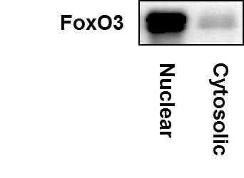
Western Blot: FOXO3 Antibody [NBP2-16521] - FoxO3 expression in nuclear and cytosolic fraction of human breast cancer cell line MDA-MB-231. Image from verified customer review.![Immunohistochemistry-Paraffin: FOXO3 Antibody [NBP2-16521]](https://resources.rndsystems.com/images/products/FOXO3-Antibody-Immunohistochemistry-Paraffin-NBP2-16521-img0028.jpg "Immunohistochemistry-Paraffin: FOXO3 Antibody [NBP2-16521]")
Immunohistochemistry-Paraffin: FOXO3 Antibody [NBP2-16521]
Immunohistochemistry-Paraffin: FOXO3 Antibody [NBP2-16521] - Mouse duodenum. FOXO3A stained by FOXO3A antibody [C3], C-term diluted at 1:2000. Antigen Retrieval: Citrate buffer, pH 6.0, 15 min.![Western Blot: FOXO3 Antibody [NBP2-16521]](https://resources.rndsystems.com/images/products/FOXO3A-Antibody-Western-Blot-NBP2-16521-img0002.jpg "Western Blot: FOXO3 Antibody [NBP2-16521]")
Western Blot: FOXO3 Antibody [NBP2-16521]
Western Blot: FOXO3 Antibody [NBP2-16521] - 50 ug of mouse brain whole cell lysate). Antibody at 1:500.![Immunohistochemistry-Paraffin: FOXO3 Antibody [NBP2-16521]](https://resources.rndsystems.com/images/products/FOXO3A-Antibody-Immunohistochemistry-Paraffin-NBP2-16521-img0003.jpg "Immunohistochemistry-Paraffin: FOXO3 Antibody [NBP2-16521]")
Immunohistochemistry-Paraffin: FOXO3 Antibody [NBP2-16521]
Immunohistochemistry-Paraffin: FOXO3 Antibody [NBP2-16521] - Huh-7 xenograft, using FOXO3A antibody at 1:500 dilution. Antigen Retrieval: Trilogy™ (EDTA based, pH 8.0) buffer, 15min.![Immunohistochemistry-Paraffin: FOXO3 Antibody [NBP2-16521]](https://resources.rndsystems.com/images/products/FOXO3-Antibody-Immunohistochemistry-Paraffin-NBP2-16521-img0024.jpg "Immunohistochemistry-Paraffin: FOXO3 Antibody [NBP2-16521]")
Immunohistochemistry-Paraffin: FOXO3 Antibody [NBP2-16521]
Immunohistochemistry-Paraffin: FOXO3 Antibody [NBP2-16521] - Rat colon. FOXO3A stained by FOXO3A antibody [C3], C-term diluted at 1:2000.Antigen Retrieval: Citrate buffer, pH 6.0, 15 min.![Immunohistochemistry-Paraffin: FOXO3 Antibody [NBP2-16521]](https://resources.rndsystems.com/images/products/FOXO3-Antibody-Immunohistochemistry-Paraffin-NBP2-16521-img0026.jpg "Immunohistochemistry-Paraffin: FOXO3 Antibody [NBP2-16521]")
Immunohistochemistry-Paraffin: FOXO3 Antibody [NBP2-16521]
Immunohistochemistry-Paraffin: FOXO3 Antibody [NBP2-16521] - Rat brain. FOXO3A stained by FOXO3A antibody [C3], C-term diluted at 1:2000.Antigen Retrieval: Citrate buffer, pH 6.0, 15 min.![Immunohistochemistry-Paraffin: FOXO3 Antibody [NBP2-16521]](https://resources.rndsystems.com/images/products/FOXO3-Antibody-Immunohistochemistry-Paraffin-Negative-NBP2-16521-img0027.jpg "Immunohistochemistry-Paraffin: FOXO3 Antibody [NBP2-16521]")
Immunohistochemistry-Paraffin: FOXO3 Antibody [NBP2-16521]
Immunohistochemistry-Paraffin: FOXO3 Antibody [NBP2-16521] - Mouse brain. FOXO3A stained by FOXO3A antibody [C3], C-term diluted at 1:2000. Antigen Retrieval: Citrate buffer, pH 6.0, 15 minutes.![Immunoprecipitation: FOXO3 Antibody [NBP2-16521]](https://resources.rndsystems.com/images/products/FOXO3-Antibody-Immunoprecipitation-NBP2-16521-img0029.jpg "Immunoprecipitation: FOXO3 Antibody [NBP2-16521]")
Immunoprecipitation: FOXO3 Antibody [NBP2-16521]
Immunoprecipitation: FOXO3 Antibody [NBP2-16521] - Jurkat whole cell lysate/extract A. Control with 2 ug of preimmune rabbit IgG B. Immunoprecipitation of FOXO3A protein by 2 ug of FOXO3A antibody 5% SDS-PAGE The immunoprecipitated FOXO3A protein was detected by FOXO3A antibody diluted at 1:1000. EasyBlot anti-rabbit IgG was used as a secondary reagent.
Western Blot: FOXO3 Antibody [NBP2-16521] -
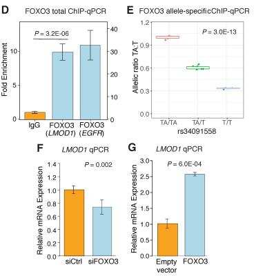
Western Blot: FOXO3 Antibody [NBP2-16521] - LMOD1 expression is mediated by PDGF-BB-FOXO3 signaling cascade.(A) UCSC Browser screenshot of the LMOD1 CAD locus at chromosome 1q32.1 highlighting the candidate causal variant, rs34091558, overlapping ATAC-seq open chromatin & RNA-seq tracks in HCASMCs treated with PDGF-BB (n = 2 biological replicates). Genomic coordinates refer to hg19 assembly. Quantitative RT-PCR (B) & Western blotting (C) data revealing PDGF-BB & phosphorylated-FOXO3 (P-FOXO3) mediated LMOD1 expression. (D) Co-expression microarray analysis performed in a cohort of carotid atherosclerotic plaques (n = 127) indicating that LMOD1 & FOXO3 are positively correlated in arterial tissues. Image collected & cropped by CiteAb from the following publication (https://pubmed.ncbi.nlm.nih.gov/30444878), licensed under a CC-BY license. Not internally tested by Novus Biologicals.
Immunoprecipitation: FOXO3 Antibody [NBP2-16521] -
Immunoprecipitation: FOXO3 Antibody [NBP2-16521] - Chemotherapy-induced A2BR expression promotes FOXO3 binding on pluripotency factor genes through decreased H3K27me3 & increased H3K27ac chromatin marks. A & B, MDA-MB-231 NTC or A2BR knockdown subclones were transfected with pLX304 (empty vector, EV) or pLX304 encoding A2BR. Cells were treated with vehicle (V) or 10 nM paclitaxel (P) for 72 h & chromatin immunoprecipitation (ChIP) was performed with antibody against FOXO3. Primers flanking the FOXO3 binding site in the NANOG, SOX2 & KLF4 gene (A) were used for qPCR (B; mean ± SEM; n = 3); **p < 0.01, ***p < 0.001 vs. NTC/EV-V; #p < 0.05, ##p < 0.01 vs. NTC/EV-P; ^^p < 0.01, ^^^p < 0.0001 vs. A2BR shRNA/EV-P; ns, not significant. C & E, MDA-MB-231 NTC or A2BR knockdown subclones were treated with V or P for 72 h & immunoblot assays were performed. D, MDA-MB-231 NTC or A2BR knockdown subclones were treated with V or P for 72 h. Cytosolic & nuclear lysates were prepared, & immunoblot assays were performed. F-H, MDA-MB-231 NTC or A2BR knockdown subclones were treated with V or 10 nM P for 72 h. ChIP was performed using antibodies against H3K27me3 (F), H3K27ac (G), or Histone H3 (H) followed by qPCR with primers flanking FOXO3 binding sites in the NANOG, SOX2 & KLF4 gene (mean ± SEM; n = 3); *p < 0.05, **p < 0.01, ***p < 0.001 vs. NTC-V; #p < 0.05, ##p < 0.01, ###p < 0.001 vs. NTC-P; ns, not significant. Image collected & cropped by CiteAb from the following publication (https://pubmed.ncbi.nlm.nih.gov/35401817), licensed under a CC-BY license. Not internally tested by Novus Biologicals.
Immunoprecipitation: FOXO3 Antibody [NBP2-16521] -
Immunoprecipitation: FOXO3 Antibody [NBP2-16521] - SMARCD3 knockdown blocks paclitaxel-induced FOXO3 binding on pluripotency factor genes & inhibits BCSC enrichment. A, MDA-MB-231 cells were transfected with vector encoding NTC or either of two shRNAs targeting SMARCD3 (#1 & #2), & immunoblot assay was performed. B-D, MDA-MB-231 NTC or SMARCD3 knockdown subclones were treated with vehicle (V) or 10 nM paclitaxel (P) for 72 h, & ALDH (B; mean ± SEM; n = 3), mammosphere (C; mean ± SEM; n = 4), & qPCR (D; mean ± SEM; n = 3) assays were performed; *p < 0.05, **p < 0.01, ***p < 0.001 vs. NTC-V; #p < 0.05, ##p < 0.01, ###p < 0.001 vs. NTC-P; ns, not significant. E-I, MDA-MB-231 NTC or SMARCD3 knockdown subclones were treated with V or 10 nM P for 72 h. ChIP was performed using antibodies against FOXO3 (E), H3K27me3 (F), H3K27ac (G), KDM6A (H), or p300 (I), followed by qPCR with primers flanking FOXO3 binding sites in the NANOG, SOX2 & KLF4 gene (mean ± SEM; n = 4); *p < 0.05, **p < 0.01, ***p < 0.001 vs. NTC-V; ##p < 0.01, ###p < 0.001 vs. NTC-P; ns, not significant. Image collected & cropped by CiteAb from the following publication (https://pubmed.ncbi.nlm.nih.gov/35401817), licensed under a CC-BY license. Not internally tested by Novus Biologicals.
Immunoprecipitation: FOXO3 Antibody [NBP2-16521] -
Immunoprecipitation: FOXO3 Antibody [NBP2-16521] - Chemotherapy-induced A2BR expression promotes FOXO3 binding on pluripotency factor genes through decreased H3K27me3 & increased H3K27ac chromatin marks. A & B, MDA-MB-231 NTC or A2BR knockdown subclones were transfected with pLX304 (empty vector, EV) or pLX304 encoding A2BR. Cells were treated with vehicle (V) or 10 nM paclitaxel (P) for 72 h & chromatin immunoprecipitation (ChIP) was performed with antibody against FOXO3. Primers flanking the FOXO3 binding site in the NANOG, SOX2 & KLF4 gene (A) were used for qPCR (B; mean ± SEM; n = 3); **p < 0.01, ***p < 0.001 vs. NTC/EV-V; #p < 0.05, ##p < 0.01 vs. NTC/EV-P; ^^p < 0.01, ^^^p < 0.0001 vs. A2BR shRNA/EV-P; ns, not significant. C & E, MDA-MB-231 NTC or A2BR knockdown subclones were treated with V or P for 72 h & immunoblot assays were performed. D, MDA-MB-231 NTC or A2BR knockdown subclones were treated with V or P for 72 h. Cytosolic & nuclear lysates were prepared, & immunoblot assays were performed. F-H, MDA-MB-231 NTC or A2BR knockdown subclones were treated with V or 10 nM P for 72 h. ChIP was performed using antibodies against H3K27me3 (F), H3K27ac (G), or Histone H3 (H) followed by qPCR with primers flanking FOXO3 binding sites in the NANOG, SOX2 & KLF4 gene (mean ± SEM; n = 3); *p < 0.05, **p < 0.01, ***p < 0.001 vs. NTC-V; #p < 0.05, ##p < 0.01, ###p < 0.001 vs. NTC-P; ns, not significant. Image collected & cropped by CiteAb from the following publication (https://pubmed.ncbi.nlm.nih.gov/35401817), licensed under a CC-BY license. Not internally tested by Novus Biologicals.
Immunoprecipitation: FOXO3 Antibody [NBP2-16521] -
Immunoprecipitation: FOXO3 Antibody [NBP2-16521] - A2BR decreases H3K27me3 & increases H3K27ac marks through recruitment of KDM6A & p300 at FOXO3 binding sites of pluripotency factor genes. A & B, MDA-MB-231 NTC or A2BR knockdown subclones were transfected with pLX304 (empty vector, EV) or pLX304 encoding A2BR. Cells were treated with vehicle (V) or 10 nM paclitaxel (P) for 72 h. ChIP was performed using antibodies against KDM6A (A) or p300 (B) followed by qPCR with primers flanking FOXO3 binding sites in the NANOG, SOX2 & KLF4 gene (mean ± SEM; n = 3); *p < 0.05, **p < 0.01, ***p < 0.001 vs. NTC-V; ##p < 0.01, ###p < 0.001 vs. NTC-P; ^^p < 0.01, ^^^p < 0.0001 vs. A2BR shRNA/EV-P; ns, not significant. C, MDA-MB-231 NTC or A2BR knockdown subclones were treated with V or P for 72 h & immunoblot assays were performed. D, MDA-MB-231 NTC or A2BR knockdown subclones were treated with V or P for 72 h & nuclear lysates were prepared. Immunoprecipitation (IP) was performed using FOXO3 antibody or control IgG followed by immunoblot assays. NL, nuclear protein lysate. Image collected & cropped by CiteAb from the following publication (https://pubmed.ncbi.nlm.nih.gov/35401817), licensed under a CC-BY license. Not internally tested by Novus Biologicals.
Western Blot: FOXO3 Antibody [NBP2-16521] -
Western Blot: FOXO3 Antibody [NBP2-16521] - Various whole cell extracts (30 ug) were separated by 7.5% SDS-PAGE, and the membrane was blotted with FOXO3A antibody [C3], C-term diluted at 1:1000. The HRP-conjugated anti-rabbit IgG antibody was used to detect the primary antibody. Corresponding RNA expression data for the same cell lines are based on Human Protein Atlas program.
Immunohistochemistry-Paraffin: FOXO3 Antibody [NBP2-16521] -
FOXO3A antibody [C3], C-term detects FOXO3A protein at cell membrane and cytoplasm by immunohistochemical analysis.Sample: Paraffin-embedded human glioblastoma.
FOXO3A stained by FOXO3A antibody [C3], C-term (NBP2-16521) diluted at 1:500.
Antigen Retrieval: Citrate buffer, pH 6.0, 15 min

Western Blot: FOXO3 Antibody [NBP2-16521] -
Non-transfected (-) and transfected (+) C2C12 whole cell extracts (30 ug) were separated by 7.5% SDS-PAGE, and the membrane was blotted with FOXO3A antibody [C3], C-term (NBP2-16521) diluted at 1:500. The HRP-conjugated anti-rabbit IgG antibody was used to detect the primary antibody.
Immunocytochemistry/ Immunofluorescence: FOXO3 Antibody [NBP2-16521] -
FOXO3A antibody [C3], C-term detects FOXO3A protein at nucleus by immunofluorescent analysis.Sample: HeLa cells were fixed in 4% paraformaldehyde at RT for 15 min.
Green: FOXO3A stained by FOXO3A antibody [C3], C-term (NBP2-16521) diluted at 1:1000.
Blue: Hoechst 33342 staining.
Scale bar= 10 um.

Western Blot: FOXO3 Antibody [NBP2-16521] -
Various whole cell extracts (30 ug) were separated by 7.5% SDS-PAGE, and the membrane was blotted with FOXO3A antibody [C3], C-term (NBP2-16521) diluted at 1:1000. The HRP-conjugated anti-rabbit IgG antibody was used to detect the primary antibody.
Immunohistochemistry-Paraffin: FOXO3 Antibody [NBP2-16521] -
FOXO3A antibody [C3], C-term detects FOXO3A protein at nucleus by immunohistochemical analysis.Sample: Paraffin-embedded mouse testis.
FOXO3A stained by FOXO3A antibody [C3], C-term (NBP2-16521) diluted at 1:500.
Antigen Retrieval: Citrate buffer, pH 6.0, 15 min
Applications for FOXO3 Antibody - BSA Free
Application
Recommended Usage
Chromatin Immunoprecipitation (ChIP)
Reported in scientific literature (PMID: 26229077)
Immunocytochemistry/ Immunofluorescence
1:100-1:1000
Immunohistochemistry
1:100-1:1000
Immunohistochemistry-Paraffin
1:100-1:1000
Immunoprecipitation
1:100-1:500
Western Blot
1:500-1:3000
Reviewed Applications
Read 4 reviews rated 4 using NBP2-16521 in the following applications:
Formulation, Preparation, and Storage
Purification
Antigen Affinity-purified
Formulation
PBS, 20% Glycerol
Format
BSA Free
Preservative
0.025% Proclin 300
Concentration
Concentrations vary lot to lot. See vial label for concentration. If unlisted please contact technical services.
Shipping
The product is shipped with polar packs. Upon receipt, store it immediately at the temperature recommended below.
Stability & Storage
Aliquot and store at -20C or -80C. Avoid freeze-thaw cycles.
Background: FoxO3
Long Name
Forkhead Box O3
Alternate Names
AF6q21, FKHRL1, FKHRL1P2, FOXO3A
Gene Symbol
FOXO3
UniProt
Additional FoxO3 Products
Product Documents for FOXO3 Antibody - BSA Free
Certificate of Analysis
To download a Certificate of Analysis, please enter a lot or batch number in the search box below.
Product Specific Notices for FOXO3 Antibody - BSA Free
This product is for research use only and is not approved for use in humans or in clinical diagnosis. Primary Antibodies are guaranteed for 1 year from date of receipt.
⚠ WARNING: This product can expose you to chemicals including mercury, which is known to the State of California to cause reproductive toxicity with developmental effects. For more information go to www.P65Warnings.ca.gov.Citations for FOXO3 Antibody - BSA Free
 Powered by Bioz
Customer Reviews for FOXO3 Antibody - BSA Free (4)
4 out of 5
4 Customer Ratings
Have you used FOXO3 Antibody - BSA Free?
Submit a review and receive an Amazon gift card!
$25/€18/£15/$25CAN/¥2500 Yen for a review with an image
$10/€7/£6/$10CAN/¥1110 Yen for a review without an image
Submit a review
Customer Images
Showing
1
-
4 of
4 reviews
Showing All
Filter By:
-
Application: Western BlotSample Tested: Nuclear fraction of human breast cancer cell linesSpecies: HumanVerified Customer | Posted 10/24/2018FoxO3 expression in nuclear and cytosolic fraction of human breast cancer cell lines MDA-MB-231

-
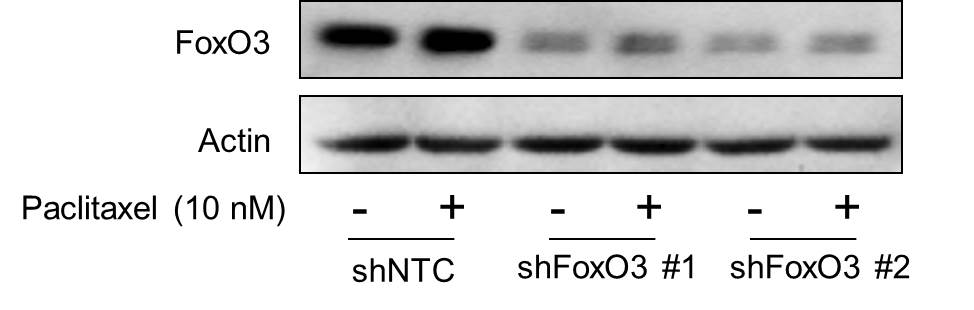
Application: Western BlotSample Tested: Human Cancer CellsSpecies: HumanVerified Customer | Posted 09/18/2018This antibody was used to detect FoxO3 expression in control (NTC) and two different FoxO3 knockdown subclones in response to paclitaxel treatment in human breast cancer cell line by western blot.
-
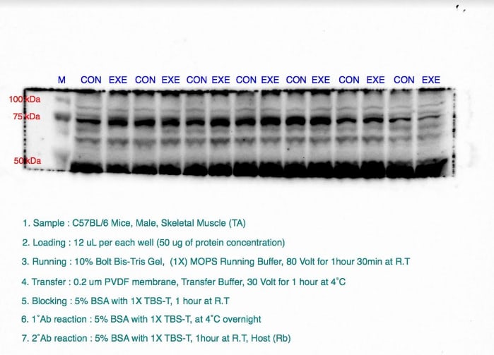
Application: Western BlotSample Tested: Mouse skeletal muscleSpecies: MouseVerified Customer | Posted 03/13/2017
-
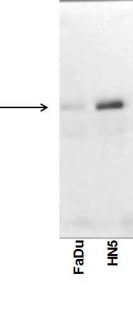
Application: Western BlotSample Tested: Human cancer cell whole cell lysateSpecies: HumanVerified Customer | Posted 07/31/2015FoxO3 expression in human HNSCC cell lines. NBP2-16521
There are no reviews that match your criteria.
Protocols
Find general support by application which include: protocols, troubleshooting, illustrated assays, videos and webinars.
- Antigen Retrieval Protocol (PIER)
- Antigen Retrieval for Frozen Sections Protocol
- Appropriate Fixation of IHC/ICC Samples
- Cellular Response to Hypoxia Protocols
- ChIP Protocol Video
- Chromatin Immunoprecipitation (ChIP) Protocol
- Chromatin Immunoprecipitation Protocol
- Chromogenic IHC Staining of Formalin-Fixed Paraffin-Embedded (FFPE) Tissue Protocol
- Chromogenic Immunohistochemistry Staining of Frozen Tissue
- ClariTSA™ Fluorophore Kits
- Detection & Visualization of Antibody Binding
- Fluorescent IHC Staining of Frozen Tissue Protocol
- Graphic Protocol for Heat-induced Epitope Retrieval
- Graphic Protocol for the Preparation and Fluorescent IHC Staining of Frozen Tissue Sections
- Graphic Protocol for the Preparation and Fluorescent IHC Staining of Paraffin-embedded Tissue Sections
- Graphic Protocol for the Preparation of Gelatin-coated Slides for Histological Tissue Sections
- ICC Cell Smear Protocol for Suspension Cells
- ICC Immunocytochemistry Protocol Videos
- ICC for Adherent Cells
- IHC Sample Preparation (Frozen sections vs Paraffin)
- Immunocytochemistry (ICC) Protocol
- Immunocytochemistry Troubleshooting
- Immunofluorescence of Organoids Embedded in Cultrex Basement Membrane Extract
- Immunofluorescent IHC Staining of Formalin-Fixed Paraffin-Embedded (FFPE) Tissue Protocol
- Immunohistochemistry (IHC) and Immunocytochemistry (ICC) Protocols
- Immunohistochemistry Frozen Troubleshooting
- Immunohistochemistry Paraffin Troubleshooting
- Immunoprecipitation Protocol
- Preparing Samples for IHC/ICC Experiments
- Preventing Non-Specific Staining (Non-Specific Binding)
- Primary Antibody Selection & Optimization
- Protocol for Heat-Induced Epitope Retrieval (HIER)
- Protocol for Making a 4% Formaldehyde Solution in PBS
- Protocol for VisUCyte™ HRP Polymer Detection Reagent
- Protocol for the Fluorescent ICC Staining of Cell Smears - Graphic
- Protocol for the Fluorescent ICC Staining of Cultured Cells on Coverslips - Graphic
- Protocol for the Preparation & Fixation of Cells on Coverslips
- Protocol for the Preparation and Chromogenic IHC Staining of Frozen Tissue Sections
- Protocol for the Preparation and Chromogenic IHC Staining of Frozen Tissue Sections - Graphic
- Protocol for the Preparation and Chromogenic IHC Staining of Paraffin-embedded Tissue Sections
- Protocol for the Preparation and Chromogenic IHC Staining of Paraffin-embedded Tissue Sections - Graphic
- Protocol for the Preparation and Fluorescent ICC Staining of Cells on Coverslips
- Protocol for the Preparation and Fluorescent ICC Staining of Non-adherent Cells
- Protocol for the Preparation and Fluorescent ICC Staining of Stem Cells on Coverslips
- Protocol for the Preparation and Fluorescent IHC Staining of Frozen Tissue Sections
- Protocol for the Preparation and Fluorescent IHC Staining of Paraffin-embedded Tissue Sections
- Protocol for the Preparation of Gelatin-coated Slides for Histological Tissue Sections
- Protocol for the Preparation of a Cell Smear for Non-adherent Cell ICC - Graphic
- R&D Systems Quality Control Western Blot Protocol
- TUNEL and Active Caspase-3 Detection by IHC/ICC Protocol
- The Importance of IHC/ICC Controls
- Troubleshooting Guide: Immunohistochemistry
- Troubleshooting Guide: Western Blot Figures
- Western Blot Conditions
- Western Blot Protocol
- Western Blot Protocol for Cell Lysates
- Western Blot Troubleshooting
- Western Blot Troubleshooting Guide
- View all Protocols, Troubleshooting, Illustrated assays and Webinars
Loading...
Associated Pathways


IL-9 Signaling Pathways
 IL-15 Signaling Pathways
 IL-21 Signaling Pathways
 MAPK Signaling: Oxidative Stress Pathway
 TGF-beta Signaling Pathways
