Glut4 Antibody - BSA Free
Novus Biologicals | Catalog # NBP1-49533
![Western Blot: Glut4 AntibodyBSA Free [NBP1-49533]](https://resources.rndsystems.com/images/products/Glut4-Antibody-Western-Blot-NBP1-49533-img0015.jpg "Western Blot: Glut4 AntibodyBSA Free [NBP1-49533]")
Key Product Details
Validated by
Species Reactivity
Validated:
Cited:
Applications
Validated:
Cited:
Label
Antibody Source
Format
Product Specifications
Immunogen
Reactivity Notes
Localization
Clonality
Host
Isotype
Scientific Data Images for Glut4 Antibody - BSA Free
Western Blot: Glut4 AntibodyBSA Free [NBP1-49533]
Glut4-Antibody-Western-Blot-NBP1-49533-img0015.jpg![Immunocytochemistry/ Immunofluorescence: Glut4 Antibody - BSA Free [NBP1-49533]](https://resources.rndsystems.com/images/products/Glut4-Antibody-Immunocytochemistry-Immunofluorescence-NBP1-49533-img0011.jpg "Immunocytochemistry/ Immunofluorescence: Glut4 Antibody - BSA Free [NBP1-49533]")
Immunocytochemistry/ Immunofluorescence: Glut4 Antibody - BSA Free [NBP1-49533]
Immunocytochemistry/Immunofluorescence: Glut4 Antibody [NBP1-49533] - HeLa cells were fixed for 10 minutes using 10% formalin and then permeabilized for 5 minutes using 1X TBS + 0.5% Triton X-100. The cells were incubated with anti-GLUT4 [NBP1-49533] at a 1:200 dilution overnight at 4C and detected with an anti-rabbit DyLight 488 (Green) at a 1:500 dilution. Alpha tubulin (DM1A) NB100-690 was used as a co-stain at a 1:1000 dilution and detected with an anti-mouse DyLight 550 (Red) at a 1:500 dilution. Nuclei were counterstained with DAPI (Blue). Cells were imaged using a 40X objective.![Immunohistochemistry: Glut4 Antibody - BSA Free [NBP1-49533]](https://resources.rndsystems.com/images/products/Glut4-Antibody-Immunohistochemistry-NBP1-49533-img0005.jpg "Immunohistochemistry: Glut4 Antibody - BSA Free [NBP1-49533]")
Immunohistochemistry: Glut4 Antibody - BSA Free [NBP1-49533]
Immunohistochemistry: Glut4 Antibody [NBP1-49533] - Analysis of GLUT4 in mouse kidney![Flow Cytometry: Glut4 Antibody - BSA Free [NBP1-49533]](https://resources.rndsystems.com/images/products/Glut4-Antibody---BSA-Free-Flow-Cytometry-NBP1-49533-img0016.jpg "Flow Cytometry: Glut4 Antibody - BSA Free [NBP1-49533]")
Flow Cytometry: Glut4 Antibody - BSA Free [NBP1-49533]
Flow Cytometry: Glut4 Antibody - BSA Free [NBP1-49533] - Analysis using the Alexa Fluor (R) 647 conjugate of NBP1-49533 (NBP1-49533AF647) on mouse Thymic Epithelial cells. Glut4 antibody in comparison to negative and IgG Control. Primary antibody dilution: 1:500. Image from verified customer review.![Western Blot: Glut4 AntibodyBSA Free [NBP1-49533]](https://resources.rndsystems.com/images/products/Glut4-Antibody-Western-Blot-NBP1-49533-img0006.jpg "Western Blot: Glut4 AntibodyBSA Free [NBP1-49533]")
Western Blot: Glut4 AntibodyBSA Free [NBP1-49533]
Western Blot: Glut4 Antibody [NBP1-49533] - Analysis of GLUT4 in A) MCF7 whole cell lysate and B) 3T3L1 whole cell lysate.![Western Blot: Glut4 AntibodyBSA Free [NBP1-49533]](https://resources.rndsystems.com/images/products/Glut4-Antibody-Western-Blot-NBP1-49533-img0010.jpg "Western Blot: Glut4 AntibodyBSA Free [NBP1-49533]")
Western Blot: Glut4 AntibodyBSA Free [NBP1-49533]
Western Blot: Glut4 Antibody [NBP1-49533] - Total protein from Human HeLa and A431, Mouse 3T3 and Rat PC12 cells was separated on a 12% gel by SDS-PAGE, transferred to PVDF membrane and blocked in 5% non-fat milk in TBST. The membrane was probed with 2.0 ug/ml anti-Glut4 in 1% non-fat milk in TBST and detected with an anti-rabbit HRP secondary antibody using chemiluminescence.![Western Blot: Glut4 AntibodyBSA Free [NBP1-49533]](https://resources.rndsystems.com/images/products/Glut4-Antibody-Western-Blot-NBP1-49533-img0014.jpg "Western Blot: Glut4 AntibodyBSA Free [NBP1-49533]")
Western Blot: Glut4 AntibodyBSA Free [NBP1-49533]
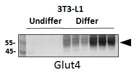
Western Blot: Glut4 Antibody [NBP1-49533] - Total protein from 3T3-L1 mouse embryonic fibroblast adipose-like cell line, separated by 4-12% SDS-PAGE, transferred to nitrocellulose membrane and blocked in 5% non-fat milk for 1h at room temperature. The membrane was probed with anti-Glut4 0.5 ug/ml in non-fat milk. Undiffer: undifferentiated; Differ: Differentiated. Image from verified customer review.![Flow Cytometry: Glut4 Antibody - BSA Free [NBP1-49533]](https://resources.rndsystems.com/images/products/Glut4-Antibody-Flow-Cytometry-NBP1-49533-img0008.jpg "Flow Cytometry: Glut4 Antibody - BSA Free [NBP1-49533]")
Flow Cytometry: Glut4 Antibody - BSA Free [NBP1-49533]
Flow Cytometry: Glut4 Antibody [NBP1-49533] - GLUT4 Biotin/APC vs. [X] PE. Image from verified customer review.![Flow Cytometry: Glut4 Antibody - BSA Free [NBP1-49533]](https://resources.rndsystems.com/images/products/Glut4-Antibody-Flow-Cytometry-NBP1-49533-img0012.jpg "Flow Cytometry: Glut4 Antibody - BSA Free [NBP1-49533]")
Flow Cytometry: Glut4 Antibody - BSA Free [NBP1-49533]
Flow Cytometry: Glut4 Antibody [NBP1-49533] - Analysis using the Alexa Fluor (R) 647 conjugate of NBP1-49533. Staining of Glut 4 expression on Murine CD4+ T cells stimulated with anti-CD3/CD28 beads and insulin (1ug/mL) for 5 days in culture media with additional glucose provided. This Alexa Fluor (R) 647 conjugated Glut 4 antibody (orange) positively stained mouse CD4+ T cells compared to Isotype Control (Rb IgG AF647, Novus NBP2-36463AF647, blue) and fluorescence minus one/FMO control (red). Image from verified customer review.![Flow (Intracellular): Glut4 Antibody - BSA Free [NBP1-49533]](https://resources.rndsystems.com/images/products/Glut4-Antibody-Flow-Intracellular-NBP1-49533-img0013.jpg "Flow (Intracellular): Glut4 Antibody - BSA Free [NBP1-49533]")
Flow (Intracellular): Glut4 Antibody - BSA Free [NBP1-49533]
Flow (Intracellular): Glut4 Antibody [NBP1-49533] - An intracellular stain was performed on HepG2 with NBP1-49533 and a matched isotype control. Cells were fixed with 4% PFA and then permeablized with 0.1% saponin. Cells were incubated in an antibody dilution of 5 ug/mL for 30 minutes at room temperature, followed by Rabbit IgG (H+L) Cross-Adsorbed Secondary Antibody.![Glut4 Antibody - BSA Free Immunohistochemistry: Glut4 Antibody - BSA Free [NBP1-49533]](https://resources.rndsystems.com/images/products/antibody/nbp1-49533_rabbit-polyclonal-glut4-antibody-immunohistochemistry-paraffin-221020258369.jpg "Immunohistochemistry: Glut4 Antibody - BSA Free [NBP1-49533]")
Immunohistochemistry: Glut4 Antibody - BSA Free [NBP1-49533]
Immunohistochemistry: Glut4 Antibody [NBP1-49533] - Analysis of a FFPE tissue section of human kidney using 1:200 dilution of Glut4 antibody. The staining was developed using HRP labeled anti-rabbit secondary antibody and DAB reagent, and nuclei of cells were counter-stained with hematoxylin.![Glut4 Antibody - BSA Free Glut4 Antibody - BSA Free Immunohistochemistry: Glut4 Antibody - BSA Free [NBP1-49533]](https://resources.rndsystems.com/images/products/antibody/nbp1-49533_rabbit-polyclonal-glut4-antibody-immunohistochemistry-paraffin-2210202583725.jpg "Immunohistochemistry: Glut4 Antibody - BSA Free [NBP1-49533]")
Immunohistochemistry: Glut4 Antibody - BSA Free [NBP1-49533]
Immunohistochemistry: Glut4 Antibody [NBP1-49533] - Analysis of a FFPE tissue section of mouse small intestine using 1:200 dilution of Glut4 antibody. The staining was developed using HRP labeled anti-rabbit secondary antibody and DAB reagent, and nuclei of cells were counter-stained with hematoxylin.Applications for Glut4 Antibody - BSA Free
Flow Cytometry
Immunocytochemistry/ Immunofluorescence
Immunohistochemistry
Immunohistochemistry-Paraffin
Knockdown Validated
Western Blot
Reviewed Applications
Read 4 reviews rated 4 using NBP1-49533 in the following applications:
Flow Cytometry Panel Builder
Bio-Techne Knows Flow Cytometry
Save time and reduce costly mistakes by quickly finding compatible reagents using the Panel Builder Tool.
Advanced Features
- Spectra Viewer - Custom analysis of spectra from multiple fluorochromes
- Spillover Popups - Visualize the spectra of individual fluorochromes
- Antigen Density Selector - Match fluorochrome brightness with antigen density

Formulation, Preparation, and Storage
Purification
Formulation
Format
Preservative
Concentration
Shipping
Stability & Storage
Background: Glut4
Alternate Names
Gene Symbol
UniProt
Additional Glut4 Products
Product Documents for Glut4 Antibody - BSA Free
Certificate of Analysis
To download a Certificate of Analysis, please enter a lot or batch number in the search box below.
Product Specific Notices for Glut4 Antibody - BSA Free
This product is for research use only and is not approved for use in humans or in clinical diagnosis. Primary Antibodies are guaranteed for 1 year from date of receipt.
Related Research Areas
Citations for Glut4 Antibody - BSA Free
 Powered by Bioz
Customer Reviews for Glut4 Antibody - BSA Free (4)
Have you used Glut4 Antibody - BSA Free?
Submit a review and receive an Amazon gift card!
$25/€18/£15/$25CAN/¥2500 Yen for a review with an image
$10/€7/£6/$10CAN/¥1110 Yen for a review without an image
Submit a review
Customer Images
-(01-ml)_NBP1-49533_6401.jpg)
-
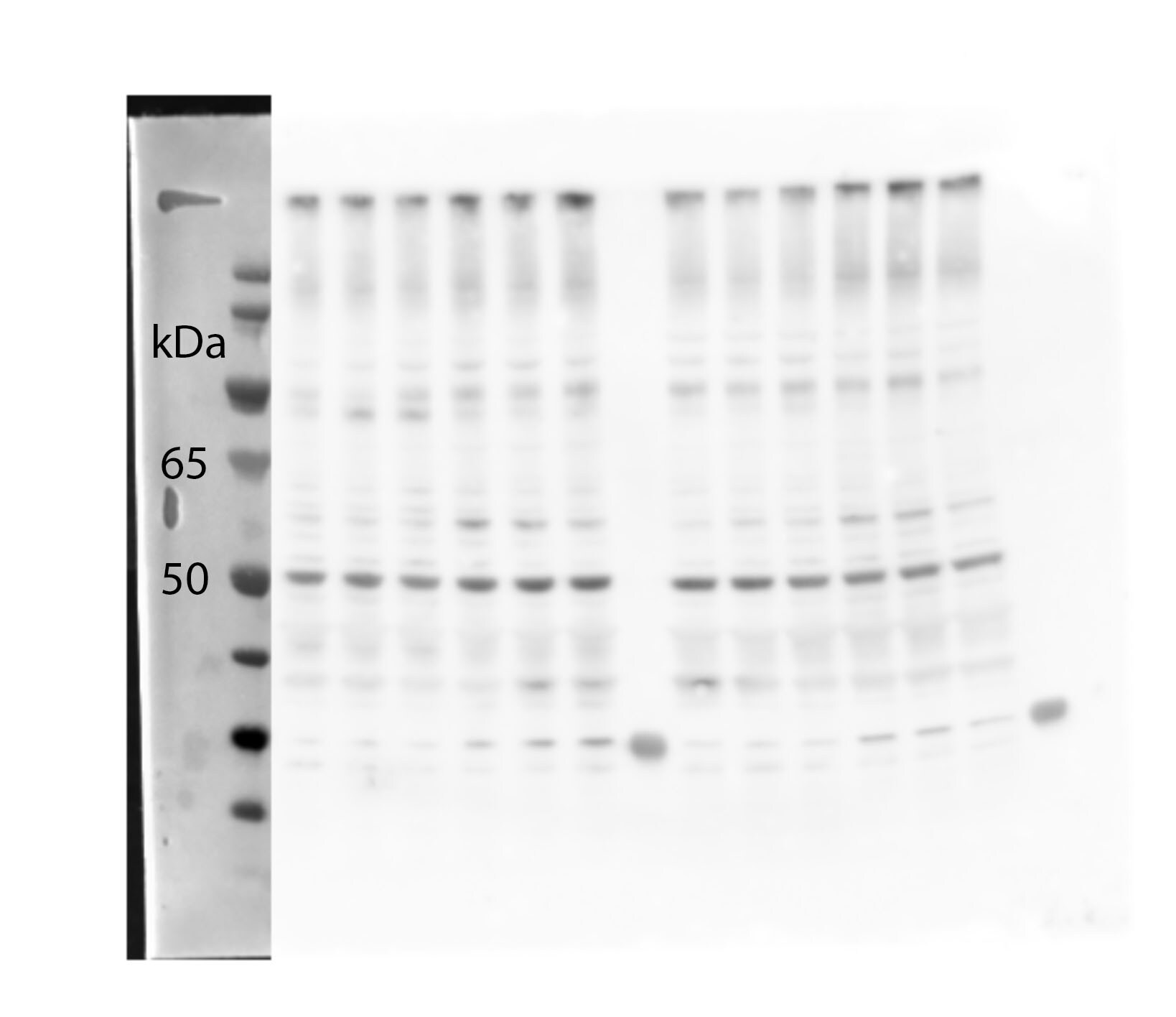
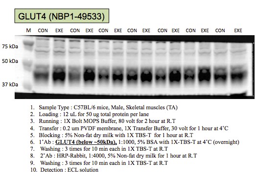
Application: Western BlotSample Tested: Mouse skeletal muscleSpecies: MouseVerified Customer | Posted 04/23/2019Total GLUT4 detection in mouse quadriceps muscle, 1:1000 dilution, ~51 kDa, a lot of non-specific bands.

-
Application: Western BlotSample Tested: 3T3-L1 mouse embryonic fibroblast adipose-like cell lineSpecies: MouseVerified Customer | Posted 03/27/2018Undiffer: undifferentiated; Differ: DifferentiatedWestern Blot: Glut4 Antibody [NBP1-49533SS] - Total protein from 3T3-L1 mouse embryonic fibroblast adipose-like cell line, separated on a 4-12% gel by SDS-PAGE, transferred to nitrocellulose membrane and blocked in 5% non-fat milk for 1h at room temperature. The membrane was probed with anti-Glut4 0.5 ug/ml in non-fat milk.
-
Application: Western BlotSample Tested: Mouse skeletal muscleSpecies: MouseVerified Customer | Posted 03/28/2017C57BL/6 Mice
-
Application: Western BlotSample Tested: lung cancer cellsSpecies: HumanVerified Customer | Posted 03/09/2014
There are no reviews that match your criteria.
Protocols
View specific protocols for Glut4 Antibody - BSA Free (NBP1-49533):
Sample Preparation.
1. Grow cells to 60-85% confluency. Flow cytometry requires between 2 x 10⁵ and 1 x 10⁶ cells for optimal performance.
2. If cells are adherent, harvest gently by washing once with staining buffer and then scraping. Avoid using trypsin as this can disrupt certain epitopes of interest. If enzymatic harvest is required, use Accutase, Collagenase, or TrypLE Express for a less damaging option.
3. Reserve 100 uL for counting, then transfer cell volume into a 50 mL conical tube and centrifuge for 8 minutes at 400 RCF.
a. Count cells using a hemocytometer and a 1:1 trypan blue exclusion stain to determine cell viability before starting the flow protocol. If cells appear blue, do not proceed.
4. Re-suspend cells to a concentration of 1 x 10⁶ cells/mL in staining buffer (NBP2-26247).
5. Aliquot out 1 mL samples in accordance with your experimental samples.
Tip: When cell surface and intracellular staining are required in the same sample, it is advisable that the cell surface staining be performed first since the fixation and permeablization steps might reduce the availability of surface antigens.
Intracellular Staining.
Tip: When performing intracellular staining, it is important to use appropriate fixation and permeabilization reagents based upon the target and its subcellular location. Generally, our Intracellular Flow Assay Kit (NBP2-29450) is a good place to start as it contains an optimized combination of reagents for intracellular staining as well as an inhibitor of intracellular protein transport (necessary if staining secreted proteins). Certain targets may require more gentle or transient permeabilization protocols such as the commonly employed methanol or saponin-based methods.
Protocol for Cytoplasmic Targets:
Optional: Perform cell surface staining as described in the previous section.
1. Fix the cells by adding 100 uL fixation solution (such as 4% PFA) to each sample for 10-15 minutes.
2. Permeabilize cells by adding 100 uL of a permeabization buffer to every 1 x 10⁶ cells present in the sample. Mix well and incubate at room temperature for 15 minutes.
a. For cytoplasmic targets, use a gentle permeabilization solution such as 1X PBS + 0.5% Saponin or 1X PBS + 0.5% Tween-20.
b. To maintain the permeabilized state throughout your experiment, use staining buffer + 0.1% of the permeabilization reagent (i.e. 0.1% Tween-20 or 0.1% Saponin).
3. Following the 15 minute incubation, add 2 mL of the staining buffer + 0.1% permeabilizer to each sample.
4. Centrifuge for 5 minutes at 400 RCF.
5. Discard supernatant and re-suspend in 1 mL of staining buffer + 0.1% permeabilizer.
6. Stain each sample at 1 uL/ 1 x 10⁶ cells of primary antibody or 1-3 uL/ 1 x 10⁶ cells for directly conjugated antibodies. Mix well and incubate at room temperature for 30 minutes- 1 hour. Gently mix samples every 10-15 minutes.
7. Following the primary/conjugate incubation, add 2 mL/sample of staining buffer +0.1% permeabilizer and centrifuge for 5 minutes at 400 RCF.
8. Remove supernatant and re-suspend each sample in 2 mL staining buffer + 0.1% permeabilizer, repeat wash for 5 minutes at 400 RCF.
9. If using a directly conjugated antibody, after the second wash, re-suspend cell pellet to a final volume of 500 uL per sample and proceed with flow analysis.
Culture cells to appropriate density in 35 mm culture dishes or 6-well plates.
1. Remove culture medium and wash the cells briefly in PBS. Add 10% formalin to the dish and fix at room temperature for 10 minutes.
2. Remove the formalin and wash the cells in PBS.
3. Permeablize the cells with 0.1% Triton X100 or other suitable detergent for 10 min.
4. Remove the permeablization buffer and wash three times for 10 minutes each in PBS. Be sure to not let the specimen dry out.
5. To block nonspecific antibody binding, incubate in 10% normal goat serum from 1 hour to overnight at room temperature.
6. Add primary antibody at appropriate dilution and incubate overnight at 4C.
7. Remove primary antibody and replace with PBS. Wash three times for 10 minutes each.
8. Add secondary antibody at appropriate dilution. Incubate for 1 hour at room temperature.
9. Remove secondary antibody and replace with PBS. Wash three times for 10 minutes each.
10. Counter stain DNA with DAPi if required.
Antigen Unmasking:
Bring slides to a boil in 10 mM sodium citrate buffer (pH 6.0) then maintain at a sub-boiling temperature for 10 minutes. Cool slides on bench-top for 30 minutes (keep slides in the sodium citrate buffer at all times).
Staining:
1. Wash sections in deionized water three times for 5 minutes each.
2. Wash sections in PBS for 5 minutes.
3. Block each section with 100-400 ul blocking solution (1% BSA in PBS) for 1 hour at room temperature.
4. Remove blocking solution and add 100-400 ul diluted primary antibody. Incubate overnight at 4 C.
5. Remove antibody solution and wash sections in wash buffer three times for 5 minutes each.
6. Add 100-400 ul HRP polymer conjugated secondary antibody. Incubate 30 minutes at room temperature.
7. Wash sections three times in wash buffer for 5 minutes each.
8. Add 100-400 ul DAB substrate to each section and monitor staining closely.
9. As soon as the sections develop, immerse slides in deionized water.
10. Counterstain sections in hematoxylin.
11. Wash sections in deionized water two times for 5 minutes each.
12. Dehydrate sections.
13. Mount coverslips.
1. Perform SDS-PAGE on samples to be analyzed, loading 10-25 ug of total protein per lane.
2. Transfer proteins to PVDF membrane according to the instructions provided by the manufacturer of the membrane and transfer apparatus.
3. Stain the membrane with Ponceau S (or similar product) to assess transfer success, and mark molecular weight standards where appropriate.
4. Rinse the blot TBS -0.05% Tween 20 (TBST).
5. Block the membrane in 5% Non-fat milk in TBST (blocking buffer) for at least 1 hour.
6. Wash the membrane in TBST three times for 10 minutes each.
7. Dilute primary antibody in blocking buffer and incubate overnight at 4C with gentle rocking.
8. Wash the membrane in TBST three times for 10 minutes each.
9. Incubate the membrane in diluted HRP conjugated secondary antibody in blocking buffer (as per manufacturer's instructions) for 1 hour at room temperature.
10. Wash the blot in TBST three times for 10 minutes each (this step can be repeated as required to reduce background).
11. Apply the detection reagent of choice in accordance with the manufacturer's instructions.
Find general support by application which include: protocols, troubleshooting, illustrated assays, videos and webinars.
- 7-Amino Actinomycin D (7-AAD) Cell Viability Flow Cytometry Protocol
- Antigen Retrieval Protocol (PIER)
- Antigen Retrieval for Frozen Sections Protocol
- Appropriate Fixation of IHC/ICC Samples
- Cellular Response to Hypoxia Protocols
- Chromogenic IHC Staining of Formalin-Fixed Paraffin-Embedded (FFPE) Tissue Protocol
- Chromogenic Immunohistochemistry Staining of Frozen Tissue
- ClariTSA™ Fluorophore Kits
- Detection & Visualization of Antibody Binding
- Extracellular Membrane Flow Cytometry Protocol
- Flow Cytometry Protocol for Cell Surface Markers
- Flow Cytometry Protocol for Staining Membrane Associated Proteins
- Flow Cytometry Staining Protocols
- Flow Cytometry Troubleshooting Guide
- Fluorescent IHC Staining of Frozen Tissue Protocol
- Graphic Protocol for Heat-induced Epitope Retrieval
- Graphic Protocol for the Preparation and Fluorescent IHC Staining of Frozen Tissue Sections
- Graphic Protocol for the Preparation and Fluorescent IHC Staining of Paraffin-embedded Tissue Sections
- Graphic Protocol for the Preparation of Gelatin-coated Slides for Histological Tissue Sections
- ICC Cell Smear Protocol for Suspension Cells
- ICC Immunocytochemistry Protocol Videos
- ICC for Adherent Cells
- IHC Sample Preparation (Frozen sections vs Paraffin)
- Immunocytochemistry (ICC) Protocol
- Immunocytochemistry Troubleshooting
- Immunofluorescence of Organoids Embedded in Cultrex Basement Membrane Extract
- Immunofluorescent IHC Staining of Formalin-Fixed Paraffin-Embedded (FFPE) Tissue Protocol
- Immunohistochemistry (IHC) and Immunocytochemistry (ICC) Protocols
- Immunohistochemistry Frozen Troubleshooting
- Immunohistochemistry Paraffin Troubleshooting
- Intracellular Flow Cytometry Protocol Using Alcohol (Methanol)
- Intracellular Flow Cytometry Protocol Using Detergents
- Intracellular Nuclear Staining Flow Cytometry Protocol Using Detergents
- Intracellular Staining Flow Cytometry Protocol Using Alcohol Permeabilization
- Intracellular Staining Flow Cytometry Protocol Using Detergents to Permeabilize Cells
- Preparing Samples for IHC/ICC Experiments
- Preventing Non-Specific Staining (Non-Specific Binding)
- Primary Antibody Selection & Optimization
- Propidium Iodide Cell Viability Flow Cytometry Protocol
- Protocol for Heat-Induced Epitope Retrieval (HIER)
- Protocol for Liperfluo
- Protocol for Making a 4% Formaldehyde Solution in PBS
- Protocol for VisUCyte™ HRP Polymer Detection Reagent
- Protocol for the Characterization of Human Th22 Cells
- Protocol for the Characterization of Human Th9 Cells
- Protocol for the Fluorescent ICC Staining of Cell Smears - Graphic
- Protocol for the Fluorescent ICC Staining of Cultured Cells on Coverslips - Graphic
- Protocol for the Preparation & Fixation of Cells on Coverslips
- Protocol for the Preparation and Chromogenic IHC Staining of Frozen Tissue Sections
- Protocol for the Preparation and Chromogenic IHC Staining of Frozen Tissue Sections - Graphic
- Protocol for the Preparation and Chromogenic IHC Staining of Paraffin-embedded Tissue Sections
- Protocol for the Preparation and Chromogenic IHC Staining of Paraffin-embedded Tissue Sections - Graphic
- Protocol for the Preparation and Fluorescent ICC Staining of Cells on Coverslips
- Protocol for the Preparation and Fluorescent ICC Staining of Non-adherent Cells
- Protocol for the Preparation and Fluorescent ICC Staining of Stem Cells on Coverslips
- Protocol for the Preparation and Fluorescent IHC Staining of Frozen Tissue Sections
- Protocol for the Preparation and Fluorescent IHC Staining of Paraffin-embedded Tissue Sections
- Protocol for the Preparation of Gelatin-coated Slides for Histological Tissue Sections
- Protocol for the Preparation of a Cell Smear for Non-adherent Cell ICC - Graphic
- Protocol: Annexin V and PI Staining by Flow Cytometry
- Protocol: Annexin V and PI Staining for Apoptosis by Flow Cytometry
- R&D Systems Quality Control Western Blot Protocol
- TUNEL and Active Caspase-3 Detection by IHC/ICC Protocol
- The Importance of IHC/ICC Controls
- Troubleshooting Guide: Fluorokine Flow Cytometry Kits
- Troubleshooting Guide: Immunohistochemistry
- Troubleshooting Guide: Western Blot Figures
- Western Blot Conditions
- Western Blot Protocol
- Western Blot Protocol for Cell Lysates
- Western Blot Troubleshooting
- Western Blot Troubleshooting Guide
- View all Protocols, Troubleshooting, Illustrated assays and Webinars
FAQs for Glut4 Antibody - BSA Free
-
Q: Do you happen to sell peptide for this antibody?
A: We do have a peptide for this particular product. The catalog number is NBP1-49533PEP.
-
Q: NBP1-49533 and NBP1-49533R are listed as being raised against a synthetic peptide made to an internal portion of the human GLUT4 protein (between residues 480-509 or 480-530 for NBP1-49533R), but since the protein is only 509aa in length, I am curious to know if the description as "internal" means that the actual sequence does not include the very c-terminal part, or if the listed sequence range is incorrect and the actual sequence in more internal?
A: We highly appreciate your feedback on the immunogen info on the datasheet of our Glucose Transporter GLUT4 antibody #NBP1-49533, and yes, the immunogen sequence is from human Glucose Transporter GLUT4 protein between residues 480-509. Similarly, the sequence for Glucose Transporter GLUT4 Antibody [DyLight 550] (NBP1-49533R) would also be the same (between residues 480-509). It is certainly C-terminal region and we apologize for the mistake committed on our end about writing it as "internal" region.
-
Q: NBP1-49533 and NBP1-49533R are listed as being raised against a synthetic peptide made to an internal portion of the human GLUT4 protein (between residues 480-509 or 480-530 for NBP1-49533R), but since the protein is only 509aa in length, I am curious to know if the description as internal means that the actual sequence does not include the very c-terminal part, or if the listed sequence range is incorrect and the actual sequence in more internal?
A: We highly appreciate your feedback on the immunogen info on the datasheet of our Glucose Transporter GLUT4 antibody #NBP1-49533, and yes, the immunogen sequence is from human Glucose Transporter GLUT4 protein between residues 480-509. Similarly, the sequence for Glucose Transporter GLUT4 Antibody [DyLight 550] (NBP1-49533R) would also be the same (between residues 480-509). It is certainly C-terminal region and we apologize for the mistake committed on our end about writing it as internal region.
-
Q: Regarding NBP1-49533AF647, Glut4-Alexa Fluor 647, the data sheet declares that the immunogen is aa480-509. Uniport says it’s a cytoplasmatic region of the protein. Is this antibody suitable for flow? Has it been validated for this application? Can you provide the suggested protocol, specifically the fixation and permeabilzation part? Any available references for this antibody and application?
A:
This antibody was validated in flow cytometry. We used our intracellular flow protocol found here. The results from this testing can be seen on the unconjugated part datasheet here
-
Q: Regarding the use of NBP1-49533, is this anti Glut4 Antibody validated for extracellular domain of Glut4 in for flow cytometry? The data sheet shows images if intracellular and additional images of flow images that are not specified.
A:
NBP1-49533 is not specific for the extracellular domain of Glut4 becuase the immunogen of the antibody pertains to the cytoplasmic C-terminus of Glut4: Immunogen: A synthetic peptide made to a C-terminal portion of the human Glucose Transporter GLUT4 protein (between residues 480-509) [UniProt P14672]. Amino acids 480-509 correspond to the cytoplasmic tail of this protein, accordingly, NBP1-49533 has only been used for labeling the intracellular staining in FLOW cytometry.
The only other option we have is MAB86541, which is made by our sister company R&D systems. They used their protocol for "Staining Membrane-associated Proteins" to product the Flow results, so if you have any other questions abotu this product I would contact them for the most accurate and proper information: https://www.rndsystems.com/resources/protocols/flow-cytometry-protocol-staining-membrane-associated-proteins-suspended-cells -
Q: Would you please help confirm if NBP1-49533 has been tested for IHC with frozen sections?
A:
NBP1-49533 has been validated in WB, ICC/IF and IHC-P, but has not been validated in IHC-Fr yet. However, because this antibody worked very well in ICC/IF, I strongly believe that it will work in IHC-Fr also. Please see the protocol for IHC-Fr which is utilized in our lab.
-
Q: Do you happen to sell peptide for this antibody?
A: We do have a peptide for this particular product. The catalog number is NBP1-49533PEP.
-
Q: NBP1-49533 and NBP1-49533R are listed as being raised against a synthetic peptide made to an internal portion of the human GLUT4 protein (between residues 480-509 or 480-530 for NBP1-49533R), but since the protein is only 509aa in length, I am curious to know if the description as "internal" means that the actual sequence does not include the very c-terminal part, or if the listed sequence range is incorrect and the actual sequence in more internal?
A: We highly appreciate your feedback on the immunogen info on the datasheet of our Glucose Transporter GLUT4 antibody #NBP1-49533, and yes, the immunogen sequence is from human Glucose Transporter GLUT4 protein between residues 480-509. Similarly, the sequence for Glucose Transporter GLUT4 Antibody [DyLight 550] (NBP1-49533R) would also be the same (between residues 480-509). It is certainly C-terminal region and we apologize for the mistake committed on our end about writing it as "internal" region.
-
Q: NBP1-49533 and NBP1-49533R are listed as being raised against a synthetic peptide made to an internal portion of the human GLUT4 protein (between residues 480-509 or 480-530 for NBP1-49533R), but since the protein is only 509aa in length, I am curious to know if the description as internal means that the actual sequence does not include the very c-terminal part, or if the listed sequence range is incorrect and the actual sequence in more internal?
A: We highly appreciate your feedback on the immunogen info on the datasheet of our Glucose Transporter GLUT4 antibody #NBP1-49533, and yes, the immunogen sequence is from human Glucose Transporter GLUT4 protein between residues 480-509. Similarly, the sequence for Glucose Transporter GLUT4 Antibody [DyLight 550] (NBP1-49533R) would also be the same (between residues 480-509). It is certainly C-terminal region and we apologize for the mistake committed on our end about writing it as internal region.
-
Q: Regarding NBP1-49533AF647, Glut4-Alexa Fluor 647, the data sheet declares that the immunogen is aa480-509. Uniport says it’s a cytoplasmatic region of the protein. Is this antibody suitable for flow? Has it been validated for this application? Can you provide the suggested protocol, specifically the fixation and permeabilzation part? Any available references for this antibody and application?
A:
This antibody was validated in flow cytometry. We used our intracellular flow protocol found here. The results from this testing can be seen on the unconjugated part datasheet here
-
Q: Regarding the use of NBP1-49533, is this anti Glut4 Antibody validated for extracellular domain of Glut4 in for flow cytometry? The data sheet shows images if intracellular and additional images of flow images that are not specified.
A:
NBP1-49533 is not specific for the extracellular domain of Glut4 becuase the immunogen of the antibody pertains to the cytoplasmic C-terminus of Glut4: Immunogen: A synthetic peptide made to a C-terminal portion of the human Glucose Transporter GLUT4 protein (between residues 480-509) [UniProt P14672]. Amino acids 480-509 correspond to the cytoplasmic tail of this protein, accordingly, NBP1-49533 has only been used for labeling the intracellular staining in FLOW cytometry.
The only other option we have is MAB86541, which is made by our sister company R&D systems. They used their protocol for "Staining Membrane-associated Proteins" to product the Flow results, so if you have any other questions abotu this product I would contact them for the most accurate and proper information: https://www.rndsystems.com/resources/protocols/flow-cytometry-protocol-staining-membrane-associated-proteins-suspended-cells -
Q: Would you please help confirm if NBP1-49533 has been tested for IHC with frozen sections?
A:
NBP1-49533 has been validated in WB, ICC/IF and IHC-P, but has not been validated in IHC-Fr yet. However, because this antibody worked very well in ICC/IF, I strongly believe that it will work in IHC-Fr also. Please see the protocol for IHC-Fr which is utilized in our lab.
Associated Pathways

