HIF-1 alpha Antibody (H1alpha67)
Novus Biologicals | Catalog # NB100-123
![Immunohistochemistry: HIF-1 alpha Antibody (H1alpha67) [NB100-123]](https://resources.rndsystems.com/images/products/HIF-1-alpha-Antibody-H1alpha67-Immunohistochemistry-NB100-123-img0033.jpg "Immunohistochemistry: HIF-1 alpha Antibody (H1alpha67) [NB100-123]")
Key Product Details
Validated by
Knockout/Knockdown, Orthogonal Validation, Biological Validation
Species Reactivity
Validated:
Human, Mouse, Rat, Porcine, Avian, Bovine, Canine, Ferret, Primate, Rabbit, Sheep
Cited:
Human, Mouse, Rat, Porcine, Ovine, Primate, Rabbit
Applications
Validated:
Knockout Validated, Immunohistochemistry, Immunohistochemistry-Paraffin, Western Blot, Immunoblotting, ELISA, Flow Cytometry, Immunocytochemistry/ Immunofluorescence, Simple Western, Immunoprecipitation, Chromatin Immunoprecipitation, Chromatin Immunoprecipitation (ChIP), Gel Super Shift Assays, Knockdown Validated
Cited:
Knockout Validated, Immunohistochemistry-Paraffin, Western Blot, ELISA, Flow Cytometry, Immunocytochemistry/ Immunofluorescence, Immunoprecipitation, Chemotaxis, Gel Supershift Assay, IF/IHC
Label
Unconjugated
Antibody Source
Monoclonal Mouse IgG2B Clone # H1alpha67
Loading...
Product Specifications
Immunogen
This HIF-1 alpha Antibody (H1alpha67) was developed against a fusion protein containing amino acids 432 - 528 of human HIF-1 alpha [Uniprot# Q16665].
Reactivity Notes
Please note that this antibody is reactive to Mouse and derived from the same host, Mouse. Additional Mouse on Mouse blocking steps may be required for IHC and ICC experiments. Please contact Technical Support for more information. Rabbit reactivity reported in scientific literature (PMID: 16738327, 26339038).
Localization
HIF-1 alpha is a nuclear protein that activates gene transcription in response to reduced cellular O2 concentration.
Clonality
Monoclonal
Host
Mouse
Isotype
IgG2B
Theoretical MW
93 kDa.
Disclaimer note: The observed molecular weight of the protein may vary from the listed predicted molecular weight due to post translational modifications, post translation cleavages, relative charges, and other experimental factors.
Disclaimer note: The observed molecular weight of the protein may vary from the listed predicted molecular weight due to post translational modifications, post translation cleavages, relative charges, and other experimental factors.
Scientific Data Images for HIF-1 alpha Antibody (H1alpha67)
![Knockdown Validated: HIF-1 alpha Antibody (H1alpha67) [NB100-123]](https://resources.rndsystems.com/images/products/HIF-1-alpha-Antibody-H1alpha67-Knockdown-Validated-NB100-123-img0028.jpg "Western Blot: HIF-1 alpha Antibody (H1alpha67) [NB100-123]")
![Western Blot: HIF-1 alpha Antibody (H1alpha67) [NB100-123]](https://resources.rndsystems.com/images/products/HIF-1-alpha-Antibody-H1alpha67-Western-Blot-NB100-123-img0030.jpg "Western Blot: HIF-1 alpha Antibody (H1alpha67) [NB100-123]")
![Immunohistochemistry: HIF-1 alpha Antibody (H1alpha67) [NB100-123]](https://resources.rndsystems.com/images/products/HIF-1-alpha-Antibody-H1alpha67-Immunohistochemistry-NB100-123-img0006.jpg "Immunohistochemistry: HIF-1 alpha Antibody (H1alpha67) [NB100-123]")
Immunohistochemistry: HIF-1 alpha Antibody (H1alpha67) [NB100-123]
Immunohistochemistry: HIF-1 alpha Antibody (H1alpha67) [NB100-123] - Analysis of HIF-1 alpha in human lung tissue. Image courtesy of product review by Aneta Gandjeva.![Immunoprecipitation: HIF-1 alpha Antibody (H1alpha67) [NB100-123]](https://resources.rndsystems.com/images/products/HIF-1-alpha-Antibody-H1alpha67-Immunoprecipitation-NB100-123-img0027.jpg "Immunoprecipitation: HIF-1 alpha Antibody (H1alpha67) [NB100-123]")
Immunoprecipitation: HIF-1 alpha Antibody (H1alpha67) [NB100-123]
HIF-1-alpha-Antibody-H1alpha67-Immunoprecipitation-NB100-123-img0027.jpg![Western Blot: HIF-1 alpha Antibody (H1alpha67) [NB100-123]](https://resources.rndsystems.com/images/products/HIF-1-alpha-Antibody-H1alpha67-Western-Blot-NB100-123-img0023.jpg "Western Blot: HIF-1 alpha Antibody (H1alpha67) [NB100-123]")
![Western Blot: HIF-1 alpha Antibody (H1alpha67) [NB100-123]](https://resources.rndsystems.com/images/products/HIF-1-alpha-Antibody-H1alpha67-Western-Blot-NB100-123-img0024.jpg "Western Blot: HIF-1 alpha Antibody (H1alpha67) [NB100-123]")
Western Blot: HIF-1 alpha Antibody (H1alpha67) [NB100-123]
Western Blot: HIF-1 alpha Antibody (H1alpha67) [NB100-123] - Analysis using the HRP conjugate of NB100-123. On day 1, MEF cells (+/+,-/-), were grown on 15cm dish (2x10 to the 6th cells). On day 2, cells were exposed to hypoxia for 4hrs. Cells were washed with ice cold PBS twice and whole cell protein was extracted extracted with RIPA buffer fortified with protease. Upon quantification, 100ug of protein was fractionated on 7% polyacralymide gel. Gel was transferred overnight onto nitrocellulose membrane. The membrane was probed with HIF-1 alpha monoclonal antibody at a 1:500 dilution (NB100-123). The secondary antibody was conjugated with HRP and was used at a 1:2500 dilution. Photo courtesy of Dr. Gregg Semenza, Johns Hopkins University.![Immunocytochemistry/ Immunofluorescence: HIF-1 alpha Antibody (H1alpha67) [NB100-123]](https://resources.rndsystems.com/images/products/HIF-1-alpha-Antibody-H1alpha67-Immunocytochemistry-Immunofluorescence-NB100-123-img0035.jpg "Immunocytochemistry/ Immunofluorescence: HIF-1 alpha Antibody (H1alpha67) [NB100-123]")
![Immunohistochemistry-Paraffin: HIF-1 alpha Antibody (H1alpha67) [NB100-123]](https://resources.rndsystems.com/images/products/HIF-1-alpha-Antibody-H1alpha67-Immunohistochemistry-Paraffin-NB100-123-img0034.jpg "Immunohistochemistry-Paraffin: HIF-1 alpha Antibody (H1alpha67) [NB100-123]")
Immunohistochemistry-Paraffin: HIF-1 alpha Antibody (H1alpha67) [NB100-123]
HIF-1-alpha-Antibody-H1alpha67-Immunohistochemistry-Paraffin-NB100-123-img0034.jpg![Immunocytochemistry/ Immunofluorescence: HIF-1 alpha Antibody (H1alpha67) [NB100-123]](https://resources.rndsystems.com/images/products/HIF-1-alpha-Antibody-H1alpha67-Immunocytochemistry-Immunofluorescence-NB100-123-img0011.jpg "Immunocytochemistry/ Immunofluorescence: HIF-1 alpha Antibody (H1alpha67) [NB100-123]")
Immunocytochemistry/ Immunofluorescence: HIF-1 alpha Antibody (H1alpha67) [NB100-123]
Immunocytochemistry/Immunofluorescence: HIF-1 alpha Antibody (H1alpha67) [NB100-123] - Staining in pig endothelial cells under hypoxia condition using NB100-123. Image from verified customer review.![Immunohistochemistry-Paraffin: HIF-1 alpha Antibody (H1alpha67) [NB100-123]](https://resources.rndsystems.com/images/products/HIF-1-alpha-Antibody-H1alpha67-Immunohistochemistry-Paraffin-NB100-123-img0012.jpg "Immunohistochemistry-Paraffin: HIF-1 alpha Antibody (H1alpha67) [NB100-123]")
Immunohistochemistry-Paraffin: HIF-1 alpha Antibody (H1alpha67) [NB100-123]
Immunohistochemistry-Paraffin: HIF-1 alpha Antibody (H1alpha67) [NB100-123] - Staining on pig tissue (brown) using NB100-123. Image from verified customer review.![Immunohistochemistry: HIF-1 alpha Antibody (H1alpha67) [NB100-123]](https://resources.rndsystems.com/images/products/HIF-1-alpha-Antibody-H1alpha67-Immunohistochemistry-NB100-123-img0026.jpg "Immunohistochemistry: HIF-1 alpha Antibody (H1alpha67) [NB100-123]")
Immunohistochemistry: HIF-1 alpha Antibody (H1alpha67) [NB100-123]
Immunohistochemistry: HIF-1 alpha Antibody (H1alpha67) [NB100-123] - Staining of biotin conjugated HIF-1 alpha (NB100-123B) in human kidney.![Immunoprecipitation: HIF-1 alpha Antibody (H1alpha67) [NB100-123]](https://resources.rndsystems.com/images/products/HIF-1-alpha-Antibody-H1alpha67-Immunoprecipitation-NB100-123-img0025.jpg "Immunoprecipitation: HIF-1 alpha Antibody (H1alpha67) [NB100-123]")
 [NB100-123] -")
Western Blot: HIF-1 alpha Antibody (H1alpha67) [NB100-123] -
Immunohistological and western blot analyses of HIF-1 alpha.(A) PAB liver stained by HIF-1 alpha (original magnification, ×20). (B) Sham-operated control liver stained by HIF-1 alpha (original magnification, ×20). (C) Enlarged view of the boxed area in the Image (A) (original magnification, ×100). (D) Western blot analysis of HIF-1 alpha (mean ± SEM), * P<0.05. HIF-1 alpha : hypoxia-inducible factor-1 alpha ; PAB: pulmonary artery banding. [NB100-123]")
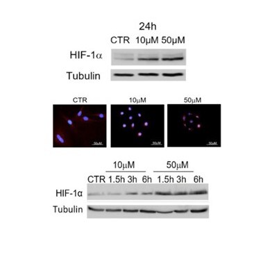
Immunocytochemistry/Immunofluorescence: Mouse Monoclonal HIF-1 alpha Antibody (H1alpha67) [NB100-123]
Immunocytochemistry/Immunofluorescence: Mouse Monoclonal HIF-1 alpha Antibody (H1alpha67) [NB100-123] - Images of HIF-1 alpha localization (red) in CTR and CoCl2-treated oAECs after 24 h of culture. Image from a verified customer review. [NB100-123]")
Western Blot: Mouse Monoclonal HIF-1 alpha Antibody (H1alpha67) [NB100-123]
Western Blot: Mouse Monoclonal HIF-1 alpha Antibody (H1alpha67) [NB100-123] - Image of HIF-1 alpha activation in AECs control cells (CTR) and after 24h of 10 and 50µM of CoCl2-treatment; HIF-1 alpha time course at shorter CoCl2 incubation times. Image from a verified customer review. [NB100-123]")
Western Blot: Mouse Monoclonal HIF-1 alpha Antibody (H1alpha67) [NB100-123]
Western Blot: Mouse Monoclonal HIF-1 alpha Antibody (H1alpha67) [NB100-123] - Image of HIF-1 alpha activation in AECs control cells (CTR) and after 24h of 10 and 50µM of CoCl2-treatment; HIF-1 alpha time course at shorter CoCl2 incubation times. Image from a verified customer review. [NB100-123] -")
Immunohistochemistry: HIF-1 alpha Antibody (H1alpha67) [NB100-123] -
Immunohistochemistry: HIF-1 alpha Antibody (H1alpha67) [NB100-123] - Immunohistochemical detection of HIF-1 alpha protein in human tissues. The nature of the tissue is indicated on top of each figure. Original magnifications are as follows: A, ×400; B, ×400; C, ×100; D, ×100; E, ×100; F, ×100; G, ×40; H, ×40. Image collected & cropped by CiteAb from the following publication (http://bmcgenet.biomedcentral.com/articles/10.1186/1471-2156-5-27), licensed under a CC-BY license. Not internally tested by Novus Biologicals. [NB100-123] -")
Western Blot: HIF-1 alpha Antibody (H1alpha67) [NB100-123] -
Western Blot: HIF-1 alpha Antibody (H1alpha67) [NB100-123] - Immunohistological & western blot analyses of HIF-1 alpha.(A) PAB liver stained by HIF-1 alpha (original magnification, ×20). (B) Sham-operated control liver stained by HIF-1 alpha (original magnification, ×20). (C) Enlarged view of the boxed area in the Image (A) (original magnification, ×100). (D) Western blot analysis of HIF-1 alpha (mean ± SEM), * P<0.05. HIF-1 alpha : hypoxia-inducible factor-1 alpha ; PAB: pulmonary artery banding. Image collected & cropped by CiteAb from the following publication (https://pubmed.ncbi.nlm.nih.gov/26863419), licensed under a CC-BY license. Not internally tested by Novus Biologicals. [NB100-123] -")
Immunocytochemistry/ Immunofluorescence: HIF-1 alpha Antibody (H1alpha67) [NB100-123] -
Immunocytochemistry/ Immunofluorescence: HIF-1 alpha Antibody (H1alpha67) [NB100-123] - Analysis of CBF treatment in HCT116 implanted mouse models & tumour tissue samples. (a) Tumour size versus CBF treatment. The lowest tumour growth rate was found in i.p. group during CBF treatment. (b) Analysis of HIF-1 alpha mRNA level in tumour tissues. There was a significant upregulated HIF-1 alpha level in i.p. group. Results are means with standard errors from 4 replicates. (c) Inhibition of HIF-1 alpha nuclear translocation in the i.t. sample. CBF strongly inhibited HIF-1 alpha nuclear accumulation in the tumour of xenografts implanted with HCT116 cells. Scale bars equal 10 μm. White arrows: HIF-1 alpha in the cytosol. Image collected & cropped by CiteAb from the following publication (https://pubmed.ncbi.nlm.nih.gov/23818933), licensed under a CC-BY license. Not internally tested by Novus Biologicals. [NB100-123] -")
Immunohistochemistry: HIF-1 alpha Antibody (H1alpha67) [NB100-123] -
Immunohistochemistry: HIF-1 alpha Antibody (H1alpha67) [NB100-123] - Immunohistochemical detection of HIF-1 alpha protein in human tissues. The nature of the tissue is indicated on top of each figure. Original magnifications are as follows: A, ×400; B, ×400; C, ×100; D, ×100; E, ×100; F, ×100; G, ×40; H, ×40. Image collected & cropped by CiteAb from the following publication (http://bmcgenet.biomedcentral.com/articles/10.1186/1471-2156-5-27), licensed under a CC-BY license. Not internally tested by Novus Biologicals. [NB100-123] -")
Immunohistochemistry: HIF-1 alpha Antibody (H1alpha67) [NB100-123] -
Immunohistochemistry: HIF-1 alpha Antibody (H1alpha67) [NB100-123] - CycT alleviates tumor hypoxia. The effects of CycT on the levels of exogenous hypoxia-marker pimonidazole labeling (A), the levels of hypoxia-inducible CA9 enzyme (B), & the levels of hypoxia-inducible factor HIF1 alpha (C) in orthotopic tumor xenografts. Scale bar: Montage, 1 mm; 10X, 20 μm. Data are plotted as mean ± SEM. For statistical analysis, the levels in treated tumors were compared to the levels in control tumors with a Welch 2-sample t-test. **p-value < 0.005. IHC images are representative of 3 independent experiments. Image collected & cropped by CiteAb from the following publication (https://pubmed.ncbi.nlm.nih.gov/30723259), licensed under a CC-BY license. Not internally tested by Novus Biologicals. [NB100-123] -")
Immunohistochemistry: HIF-1 alpha Antibody (H1alpha67) [NB100-123] -
Immunohistochemistry: HIF-1 alpha Antibody (H1alpha67) [NB100-123] - Immunohistochemical detection of HIF-1 alpha protein in human tissues. The nature of the tissue is indicated on top of each figure. Original magnifications are as follows: A, ×400; B, ×400; C, ×100; D, ×100; E, ×100; F, ×100; G, ×40; H, ×40. Image collected & cropped by CiteAb from the following publication (http://bmcgenet.biomedcentral.com/articles/10.1186/1471-2156-5-27), licensed under a CC-BY license. Not internally tested by Novus Biologicals. [NB100-123] -")
Immunohistochemistry: HIF-1 alpha Antibody (H1alpha67) [NB100-123] -
Immunohistochemistry: HIF-1 alpha Antibody (H1alpha67) [NB100-123] - Schematic demonstration of the three HIF1 alpha antibodies used in this study & their epitopes (Figs. 2a & b). Ab1 is a polyclonal antibody raised against N-domain of wild type HIF1 alpha & does not recognize HIF1 alpha 1.2 due to it's different N-terminal part (Fig. 2a). Ab2 & Ab3 are monoclonal & polyclonal antibodies, respectively, with epitopes in the common parts of HIF1 alpha & HIF1 alpha 1.2 (Fig.2a & b). Immunohistochemical analysis performed on thin adjacent sections of NE-differentiated prostate cancer using the HIF1 alpha antibodies Ab1 (Fig. 2c), Ab 2 (Fig. 2d), Ab 3 (Fig. 2e) & HIF1 beta antibody (Fig. 2f). Immunopositivity was detected for HIF1 alpha with Ab2 & Ab3 while HIF1 alpha Ab1 produced no detectable staining. HIF1 beta was also positive in adjacent section (Fig. 2f). Double staining of chromogranin A & androgen receptor antigens on adjacent sections (Fig. 2g) showed immunopositivity for chromogranin A (Fast red). Androgen receptor antibody (DAB, brown) produced no staining. Immunostaining of benign prostate tissue with HIF1 alpha Ab3 showed immunopositivity in NE-like cells of benign prostate tissue (Fig. 2h). In addition, HIF1 beta antibody recognized benign NE-like cells in benign prostate hyperplasia (Fig. 2i). Double staining with HIF1 alpha Ab3 & HIF1 beta (Fig. 2j) shows co-localization of the two proteins in NE-like cells of benign prostate hyperplasia (HIF1 alpha Ab3 red stain, HIF1 beta brown stain). Panels a, b, c, d, e, f & g: 40× objective. Panels h, i & j: 60× objective. Image collected & cropped by CiteAb from the following publication (https://pubmed.ncbi.nlm.nih.gov/20663134), licensed under a CC-BY license. Not internally tested by Novus Biologicals. [NB100-123] -")
Immunocytochemistry/ Immunofluorescence: HIF-1 alpha Antibody (H1alpha67) [NB100-123] -
Immunocytochemistry/ Immunofluorescence: HIF-1 alpha Antibody (H1alpha67) [NB100-123] - GAD65/67-positive neurons expressed HIF-1 alpha under hypoxic conditions. A) HIF-1 alpha expression co-localized with GAD65/67-ir in neurons exposed to hypoxia when compared to normoxia (upper panel) or GAD65/67-negative neurons in hypoxia (bottom panel, open arrow). B) Quantification shows the percentage of HIF-1 alpha -expressing GAD65/67-positive neurons after hypoxia in vitro (mean ± SD; from n = 6 cultures). C)In vivo immunostaining illustrates HIF-1 alpha -positive (bottom panel, solid arrow) & HIF-1 alpha -negative (bottom panel, open arrow) in GAD65/67-ir neurons in the ipsilateral region, whereas the contralateral region shows no HIF-1 alpha staining in GAD65/67-ir neurons (see Figure 1 for region selection). Scale bars, 10 μm (A); 20 μm (B). Image collected & cropped by CiteAb from the following publication (https://actaneurocomms.biomedcentral.com/articles/10.1186/2051-5960-2-51), licensed under a CC-BY license. Not internally tested by Novus Biologicals. [NB100-123] -")
Immunocytochemistry/ Immunofluorescence: HIF-1 alpha Antibody (H1alpha67) [NB100-123] -
Immunocytochemistry/ Immunofluorescence: HIF-1 alpha Antibody (H1alpha67) [NB100-123] - Levels of GSH in cortical neurons exposed to hypoxia. A) During hypoxia, GSH increased in MAP2-ir neurons with round somata (bottom panel, open arrow) & decreased in neurons with pyramidal-like morphology (upper panel, solid arrow). B) GSH increased in a subset of GAD65/67–ir neurons exposed to hypoxia (bottom panel, open arrow). C) Elevated HIF-1 alpha expression in GAD65/67–ir neurons containing high levels of GSH when exposed to hypoxia (middle panel). BSO treatment decreased HIF-1 alpha expression in GAD65/67-positive neurons during hypoxia. D) Percentages of HIF-1 alpha -expressing GAD65/67-ir neurons expressing a high level of GSH & GAD65/67-ir neurons expressing a low levels of GSH & HIF-1 alpha (mean ± SD; n = 3). The “low” level referred to the level of GSH in normoxic GAD65/67 neurons that was normalized to 1. The “high” level referred to the elevated GSH level in GAD65/67 neurons with a mean of 1.40 ± 0.10. E) Total MCB-GSH intensity in GAD65/67-positive neurons with & without BSO after hypoxia (mean ± SD; 8-10 neurons quantified from each experiment, n = 3 independent cultures). Scale bars, 20 μm. Image collected & cropped by CiteAb from the following publication (https://actaneurocomms.biomedcentral.com/articles/10.1186/2051-5960-2-51), licensed under a CC-BY license. Not internally tested by Novus Biologicals. [NB100-123] -")
Immunohistochemistry: HIF-1 alpha Antibody (H1alpha67) [NB100-123] -
Immunohistochemistry: HIF-1 alpha Antibody (H1alpha67) [NB100-123] - Results of in situ hybridization & immunohistochemistry on thin adjacent section to detect expression of HIF-1 alpha 1.2 mRNA & HIF1 alpha protein in malignant & benign prostate tissue. In situ hybridization (antisense probe, Fig. 3a) & immunostaining with HIF1 alpha Ab2 (Fig. 3c) on thin adjacent sections of NE-differentiated prostate adenocarcinoma showed co-localization of HIF1 alpha 1.2 transcript & HIF-1 alpha protein. Incubation with sense probe did not generate any detectable hybridization signals (Fig. 3b). Both In situ hybridization (Fig. 3d antisense) & HIF-1 alpha Ab2 immunostaining (Fig. 3f) were negative in non-NE-differentiated prostate adenocarcinoma. In situ hybridization with sense probe performed on non-NE-differentiated prostate cancer (Fig. 3e) was negative. In situ hybridizering on benign prostate tissue showed HIF1 alpha 1.2 transcript in NE-like cells of benign prostate tissue (Fig. 3g, ↗). The sense probe on thin adjacent section generated no signals (Fig. 3h,↗). Furthermore, co-localization of HIF1 alpha 1.2 transcript (Fig. 3i,↗,◢,↖) & HIF1 alpha protein, detected with HIF1 alpha Ab3 (Fig. 3j,↗,◢,↖) was also shown in NE-like cells of benign prostate tissue. Panels a, b, c, d, e, f, g, h, i, j: 40× objective. Image collected & cropped by CiteAb from the following publication (https://pubmed.ncbi.nlm.nih.gov/20663134), licensed under a CC-BY license. Not internally tested by Novus Biologicals. [NB100-123] -")
Western Blot: HIF-1 alpha Antibody (H1alpha67) [NB100-123] -
Western Blot: HIF-1 alpha Antibody (H1alpha67) [NB100-123] - HX-induced Sirt1 expression in white matter OPCs requires HIF1 alpha.(a) HIF1 alpha stabilization of Sirt1 transcript expression in OPCs as revealed by representative RT–PCR represents higher level of Sirt1 mRNA in VHL cKO mice. GDPDH mRNA serves as a control. Mean±s.e.m., n=3 brains for each group. (b,c) Representative western blot demonstrates a transient increase of HIF1 alpha expression in HX white matter at P18 with no significant effect at P11 (P=0.7955) & P45 (P=0.7333). Histograms show mean±s.e.m. (d,e) Graphs represent the percentages of Sirt1+Ki67+ & NG2+Ki67+ cells after HX white matter in WT & HIF1 alpha KO mice. Number in parentheses within bar indicates number of samples (n=4 brains per group & per genotype; ****P<0.0001, one-way analysis of variance, Bonferroni post hoc test, mean±s.e.m.). (f,g) Western blot demonstrates no increase (P=0.8231) in Sirt1 & HIF1 alpha expression in white matter of Hif1 alpha KO mice. Actin was used as loading control (mean±s.e.m.; n=3 brains for each experiment, group & genotype). Image collected & cropped by CiteAb from the following publication (https://www.nature.com/articles/ncomms13866), licensed under a CC-BY license. Not internally tested by Novus Biologicals. [NB100-123] -")
Immunohistochemistry: HIF-1 alpha Antibody (H1alpha67) [NB100-123] -
Immunohistochemistry: HIF-1 alpha Antibody (H1alpha67) [NB100-123] - Results of in situ hybridization & immunohistochemistry on thin adjacent section to detect expression of HIF-1 alpha 1.2 mRNA & HIF1 alpha protein in malignant & benign prostate tissue. In situ hybridization (antisense probe, Fig. 3a) & immunostaining with HIF1 alpha Ab2 (Fig. 3c) on thin adjacent sections of NE-differentiated prostate adenocarcinoma showed co-localization of HIF1 alpha 1.2 transcript & HIF-1 alpha protein. Incubation with sense probe did not generate any detectable hybridization signals (Fig. 3b). Both In situ hybridization (Fig. 3d antisense) & HIF-1 alpha Ab2 immunostaining (Fig. 3f) were negative in non-NE-differentiated prostate adenocarcinoma. In situ hybridization with sense probe performed on non-NE-differentiated prostate cancer (Fig. 3e) was negative. In situ hybridizering on benign prostate tissue showed HIF1 alpha 1.2 transcript in NE-like cells of benign prostate tissue (Fig. 3g, ↗). The sense probe on thin adjacent section generated no signals (Fig. 3h,↗). Furthermore, co-localization of HIF1 alpha 1.2 transcript (Fig. 3i,↗,◢,↖) & HIF1 alpha protein, detected with HIF1 alpha Ab3 (Fig. 3j,↗,◢,↖) was also shown in NE-like cells of benign prostate tissue. Panels a, b, c, d, e, f, g, h, i, j: 40× objective. Image collected & cropped by CiteAb from the following publication (https://pubmed.ncbi.nlm.nih.gov/20663134), licensed under a CC-BY license. Not internally tested by Novus Biologicals. [NB100-123] -")
Immunocytochemistry/ Immunofluorescence: HIF-1 alpha Antibody (H1alpha67) [NB100-123] -
Immunocytochemistry/ Immunofluorescence: HIF-1 alpha Antibody (H1alpha67) [NB100-123] - HIF-1 alpha expression in primary cortical neurons exposed to hypoxia/ischemia. A & A’) Neurons were double-stained for HIF-1 alpha & MAP2 in the presence of 5 & 25 mM glucose with & without hypoxia. HIF-1 alpha expression in the somata was observed in cells with interneuron-like morphology after hypoxia. B) CoCl2 (0.3 mM) induced HIF-1 alpha expression in cells with interneuron-like morphology. C) Quantification represents the increase in HIF-1 alpha –ir staining (mean ± SD; 5-10 neurons quantified from each experiment, n = 3 experiments). D)In vivo brain slice shows a similar pattern of positive HIF-1 alpha -ir in round soma (open arrow) & negative in neurons with pyramidal-like morphology (solid arrow) in the ipsilateral side. *p < 0.05, compared with normoxia (25 mM glucose), #p < 0.05, compared with hypoxia (25 mM glucose). Scale bars, 20 μm (A, A’, B); 10 μm (D). Image collected & cropped by CiteAb from the following publication (https://actaneurocomms.biomedcentral.com/articles/10.1186/2051-5960-2-51), licensed under a CC-BY license. Not internally tested by Novus Biologicals. [NB100-123] -")
Western Blot: HIF-1 alpha Antibody (H1alpha67) [NB100-123] -
Western Blot: HIF-1 alpha Antibody (H1alpha67) [NB100-123] - HX-induced Sirt1 expression in white matter OPCs requires HIF1 alpha.(a) HIF1 alpha stabilization of Sirt1 transcript expression in OPCs as revealed by representative RT–PCR represents higher level of Sirt1 mRNA in VHL cKO mice. GDPDH mRNA serves as a control. Mean±s.e.m., n=3 brains for each group. (b,c) Representative western blot demonstrates a transient increase of HIF1 alpha expression in HX white matter at P18 with no significant effect at P11 (P=0.7955) & P45 (P=0.7333). Histograms show mean±s.e.m. (d,e) Graphs represent the percentages of Sirt1+Ki67+ & NG2+Ki67+ cells after HX white matter in WT & HIF1 alpha KO mice. Number in parentheses within bar indicates number of samples (n=4 brains per group & per genotype; ****P<0.0001, one-way analysis of variance, Bonferroni post hoc test, mean±s.e.m.). (f,g) Western blot demonstrates no increase (P=0.8231) in Sirt1 & HIF1 alpha expression in white matter of Hif1 alpha KO mice. Actin was used as loading control (mean±s.e.m.; n=3 brains for each experiment, group & genotype). Image collected & cropped by CiteAb from the following publication (https://www.nature.com/articles/ncomms13866), licensed under a CC-BY license. Not internally tested by Novus Biologicals.Applications for HIF-1 alpha Antibody (H1alpha67)
Application
Recommended Usage
Chromatin Immunoprecipitation
1:10 - 1:500. Use reported in scientific literature
Chromatin Immunoprecipitation (ChIP)
1:10-1:500
ELISA
1:100 - 1:2000. Use reported in scientific literature
Flow Cytometry
1:10 - 1:1000
Gel Super Shift Assays
1:1 - 1:100. Use reported in scientific literature
Immunoblotting
reported in multiple pieces of scientific literature
Immunohistochemistry
1:100 - 1:300
Immunohistochemistry-Paraffin
1:100 - 1:300
Immunoprecipitation
1:10
Knockout Validated
reported in scientific literature (PMID 27991597)
Simple Western
1:50 - 1:1000
Western Blot
1:500 - 1:1000
Application Notes
By WB, this antibody recognizes bands at 120kDa representing HIF-1 alpha in induced tissues and cells. Multiple bands may be seen at 120kDa representing post-translational modifications. Nuclear extracts are recommended for WB.
Reviewed Applications
Read 10 reviews rated 4.2 using NB100-123 in the following applications:
Flow Cytometry Panel Builder
Bio-Techne Knows Flow Cytometry
Save time and reduce costly mistakes by quickly finding compatible reagents using the Panel Builder Tool.
Advanced Features
- Spectra Viewer - Custom analysis of spectra from multiple fluorochromes
- Spillover Popups - Visualize the spectra of individual fluorochromes
- Antigen Density Selector - Match fluorochrome brightness with antigen density

Formulation, Preparation, and Storage
Purification
Protein A purified
Formulation
PBS with 1% BSA
Preservative
0.05% Sodium Azide
Concentration
1.0 mg/ml
Shipping
The product is shipped with polar packs. Upon receipt, store it immediately at the temperature recommended below.
Stability & Storage
Store at 4C short term. Aliquot and store at -20C long term. Avoid freeze-thaw cycles.
Background: HIF-1 alpha/HIF1A
HIF-1 or hypoxia inducible factor 1 (predicted molecular weight 93kDa), is a transcription factor commonly referred to as a "master regulator of the hypoxic response" for its central role in the regulation of cellular adaptations to hypoxia. In its active form under hypoxic conditions, HIF-1 is stabilized by the formation of a heterodimer of HIF-1 alpha and ARNT/HIF-1 beta subunits. Nuclear HIF-1 engages p300/CBP for binding to hypoxic response elements (HREs). This process induces transcription and regulation of genes including EPO, VEGF, iNOS2, ANGPT1 and OCT4 (4,5).
Under normoxic conditions, the HIF-1 alpha subunit is rapidly targeted and degraded by the ubiquitin proteasome system. This process is mediated by prolyl hydroxylase domain enzymes (PHDs), which catalyze the hydroxylation of key proline residues (Pro-402 and Pro-564) within the oxygen-dependent degradation domain of HIF-1 alpha. Once hydroxylated, HIF-1 alpha binds the von Hippel-Lindau tumor suppressor protein (pVHL) for subsequent ubiquitination and proteasomal degradation (4). pVHL dependent regulation of HIF-1 alpha plays a role in normal physiology and disease states. Regulation of HIF-1 alpha by pVHL is critical for the suppressive function of FoxP3+ regulatory Tcells (6). Repression of pVHL expression in chronic lymphocytic leukemia (CLL) B cells leads to HIF-1 alpha stabilization and increased VEGF secretion (7).
References
1. Semenza, G. L., Agani, F., Feldser, D., Iyer, N., Kotch, L., Laughner, E., & Yu, A. (2000). Hypoxia, HIF-1, and the pathophysiology of common human diseases. Advances in Experimental Medicine and Biology.
2. Muz, B., de la Puente, P., Azab, F., & Azab, A. K. (2015). The role of hypoxia in cancer progression, angiogenesis, metastasis, and resistance to therapy. Hypoxia. https://doi.org/10.2147/hp.s93413
3. Huang, Y., Lin, D., & Taniguchi, C. M. (2017). Hypoxia inducible factor (HIF) in the tumor microenvironment: friend or foe? Science China Life Sciences. https://doi.org/10.1007/s11427-017-9178-y
4. Koyasu, S., Kobayashi, M., Goto, Y., Hiraoka, M., & Harada, H. (2018). Regulatory mechanisms of hypoxia-inducible factor 1 activity: Two decades of knowledge. Cancer Science. https://doi.org/10.1111/cas.13483
5. Dengler, V. L., Galbraith, M. D., & Espinosa, J. M. (2014). Transcriptional regulation by hypoxia inducible factors. Critical Reviews in Biochemistry and Molecular Biology. https://doi.org/10.3109/10409238.2013.838205
6. Lee, J. H., Elly, C., Park, Y., & Liu, Y. C. (2015). E3Ubiquitin Ligase VHL Regulates Hypoxia-Inducible Factor-1 alpha to Maintain Regulatory T Cell Stability and Suppressive Capacity. Immunity. https://doi.org/10.1016/j.immuni.2015.05.016
7. Ghosh, A. K., Shanafelt, T. D., Cimmino, A., Taccioli, C., Volinia, S., Liu, C. G.,... Kay, N. E. (2009). Aberrant regulation of pVHL levels by microRNA promotes the HIF/VEGF axis in CLL B cells. Blood. https://doi.org/10.1182/blood-2008-10-185686
Long Name
Hypoxia Inducible Factor 1 Subunit Alpha
Alternate Names
BHLHE78, HIF 1A, HIF-1a, HIF1 alpha, HIF1A, MOP1, PASD8, H1alpha67
Gene Symbol
HIF1A
Additional HIF-1 alpha/HIF1A Products
Product Documents for HIF-1 alpha Antibody (H1alpha67)
Certificate of Analysis
To download a Certificate of Analysis, please enter a lot or batch number in the search box below.
Product Specific Notices for HIF-1 alpha Antibody (H1alpha67)
This product is for research use only and is not approved for use in humans or in clinical diagnosis. Primary Antibodies are guaranteed for 1 year from date of receipt.
Citations for HIF-1 alpha Antibody (H1alpha67)
 Powered by Bioz
Customer Reviews for HIF-1 alpha Antibody (H1alpha67) (10)
4.2 out of 5
10 Customer Ratings
Have you used HIF-1 alpha Antibody (H1alpha67)?
Submit a review and receive an Amazon gift card!
$25/€18/£15/$25CAN/¥2500 Yen for a review with an image
$10/€7/£6/$10CAN/¥1110 Yen for a review without an image
Submit a review
Customer Images
-(01-ml)_NB100-123_7131.jpg)
-(01-ml)_NB100-123_6856.jpg)
Showing
1
-
5 of
10 reviews
Showing All
Filter By:
-
Application: Western BlotSample Tested: Amniotic-derived Epithelial CellsSpecies: SheepVerified Customer | Posted 07/29/2024WB images of HIF-1 alpha activation in AECs control cells (CTR) and after 24h of 10 and 50µM of CoCl2-treatment; HIF-1 alpha time course at shorter CoCl2 incubation times. IF images of HIF-1 alpha localization (red) in CTR and CoCl2-treated oAECs after 24 h of culture.The antibody has been tested following the product-associated general protocols using a concentration of 1:500 for WB assay and 1:100 for immunofluorescence.

-
Application: Western BlotSample Tested: See PMID:23091063Species: HumanVerified Customer | Posted 12/12/2014
-
Application: Western BlotSample Tested: Rat liver total lysateSpecies: OtherVerified Customer | Posted 07/15/2014
-
Application: ImmunoprecipitationSample Tested:Species: HumanVerified Customer | Posted 04/28/2014HIF-1a immunoprecipitation
-
Application: Western BlotSample Tested: Mouse brain cortex whole lysateSpecies: OtherVerified Customer | Posted 04/25/2014
-
Application: ImmunohistochemistrySample Tested:Species: OtherVerified Customer | Posted 04/14/2014HIF-1 staining on pig tissue (brown)
-
Application: ImmunofluorescenceSample Tested:Species: OtherVerified Customer | Posted 04/03/2014HIF staining in pig endothelial cells under hipoxia condition.
-
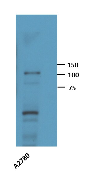
Application: Western BlotSample Tested: A2780 CELL LYSATESpecies: HumanVerified Customer | Posted 01/24/2014Western Blot: HIF-1 alpha Antibody [NB100-123]
-
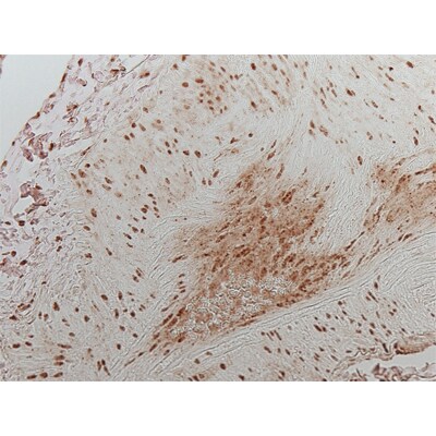
Application: Immunohistochemistry-ParaffinSample Tested: human lungSpecies: HumanVerified Customer | Posted 05/15/2012HIF-1a - APAH human lung
-
Application: ImmunocytochemistrySample Tested: Rat Primary Motor NeuronsSpecies: RatVerified Customer | Posted 03/24/2009
There are no reviews that match your criteria.
Protocols
View specific protocols for HIF-1 alpha Antibody (H1alpha67) (NB100-123):
1. Fix cells in 3% Paraformaldehyde in PBS for 15 minutes at room temperature, gently rocking.
2. Rinse cells 3 times for 5 minutes in PBS.
3. Block and permeabilize cells in 2% non-fat dry milk (NFDM) dissolved in PBS with 0.1% TX-100 overnight at 4C (covered to prevent evaporation).
4. Rinse cells 3 times for 5 minutes in PBS.
5. Dilute NB100-123 1:500 in dilution buffer [2% BSA in PBS with 0.01% TX-100].
6. Place cover slip upside down on a 50 ul drop of diluted antibody on parafilm, in humidity box.
7. Incubate for 1 hour at 37C.
8. Flip slips right side up in wells and rinse 3 times for 5 minutes each, in PBS.
9. In an amber microfuge tube, dilute secondary antibody (Cy3 anti-ms IgG) 1:500 in dilution buffer [2% BSA in PBS with 0.01% TX-100].
10. Place 800 ul of diluted secondary antibody in each well and make sure the fluid film covers over the cells on the slip. Alternatively, secondary antibody can be applied in the same manner as the primary (slip upside-down on drop of secondary that has been placed on a sheet of parafilm that is inside of a humidity box).
11. Incubate for 1 hour at 37C, in the dark.
12. Rinse cells at room temperature 4 times for 15 minutes each, in PBS, gently rocking.
13. Mount on frosted slides with AquaPoly Mount (Polysciences).
14. Refrigerate flat and covered.
Please see:
Primary Reference: Zhong, H., et al. Overexpression of Hypoxia-inducible Factor 1alpha in Common Human Cancers and Their Metastases. Cancer Research. 59: 5830-5835, 1999.
Western Blot Protocol
1. Resolve aliquots (25-30 ug) of induced nuclear protein extracts on a 4-20% Tris-HCl gel.
2. Transfer proteins to nitrocellulose membrane in 20 mM Tris-HCL (pH 8.0)/150 mM glycine/20% (vol/vol) methanol.
3. Block membrane for 1 hour with 1X western wash buffer containing 5% non-fat dry milk (NFDM).
4. Incubate membrane overnight at 4C in NB 100-123 diluted in 1X western wash/5% NFDM.
5. Wash with 1X western wash for 35 minutes at RT (1 X 15 minutes, 2 X 10 minutes).
6. Incubate membrane with HRP conjugated anti-mouse IgG for 1 hour (RT) in 1X western wash/5% NFDM.
7. Wash with 1X western wash for 35 minutes at RT (1 X 15 minutes, 2 X 10 minutes).
8. Drain membrane and place on saran wrap.
9. Using Amersham ECL Kit, mix equal volumes of two reagents. Pour over membrane (protein side facing up). Let solution sit on membrane for 15-20 seconds.
10. Drain membrane and place on new saran wrap.
11. Wrap up membrane and expose to film.
12. Develop accordingly.
10X Western wash: 24.2 g Tris, 80g NaCl, Tween-20 to 1%, pH 7.6 and QS to 4L.
Stripping buffer: 100 mM BME 2% SDS 62.5 mM Tris (pH 6.7)
To strip membrane: Incubate membrane in stripping buffer for 30 minutes at 56C. Wash membrane for 15 minutes with several change of 1X western wash.
Notes: If hypoxia treatment is not hypoxic enough (less than 2% oxygen to get an induction), signal will be absent. Also, if the harvest time is too slow or there are not enough protease inhibitors, etc., the induced protein will be rapidly lost as HIF-1alpha has a very short half-life.
Nuclear Extract Preparation Reference: Wang and Semenza. Purification and Characterization of Hypoxia-Inducible Factor. Journal of Biological Chemistry. 270(3): 1230-1237, 1995.
**This antibody has demonstrated varying results in Western blot applications. Product NB100-105 is recommended for most Western blot experiments.
Find general support by application which include: protocols, troubleshooting, illustrated assays, videos and webinars.
- 7-Amino Actinomycin D (7-AAD) Cell Viability Flow Cytometry Protocol
- Antigen Retrieval Protocol (PIER)
- Antigen Retrieval for Frozen Sections Protocol
- Appropriate Fixation of IHC/ICC Samples
- Cellular Response to Hypoxia Protocols
- ChIP Protocol Video
- Chromatin Immunoprecipitation (ChIP) Protocol
- Chromatin Immunoprecipitation Protocol
- Chromogenic IHC Staining of Formalin-Fixed Paraffin-Embedded (FFPE) Tissue Protocol
- Chromogenic Immunohistochemistry Staining of Frozen Tissue
- ClariTSA™ Fluorophore Kits
- Detection & Visualization of Antibody Binding
- ELISA Sample Preparation & Collection Guide
- ELISA Troubleshooting Guide
- Extracellular Membrane Flow Cytometry Protocol
- Flow Cytometry Protocol for Cell Surface Markers
- Flow Cytometry Protocol for Staining Membrane Associated Proteins
- Flow Cytometry Staining Protocols
- Flow Cytometry Troubleshooting Guide
- Fluorescent IHC Staining of Frozen Tissue Protocol
- Graphic Protocol for Heat-induced Epitope Retrieval
- Graphic Protocol for the Preparation and Fluorescent IHC Staining of Frozen Tissue Sections
- Graphic Protocol for the Preparation and Fluorescent IHC Staining of Paraffin-embedded Tissue Sections
- Graphic Protocol for the Preparation of Gelatin-coated Slides for Histological Tissue Sections
- How to Run an R&D Systems DuoSet ELISA
- How to Run an R&D Systems Quantikine ELISA
- How to Run an R&D Systems Quantikine™ QuicKit™ ELISA
- ICC Cell Smear Protocol for Suspension Cells
- ICC Immunocytochemistry Protocol Videos
- ICC for Adherent Cells
- IHC Sample Preparation (Frozen sections vs Paraffin)
- Immunocytochemistry (ICC) Protocol
- Immunocytochemistry Troubleshooting
- Immunofluorescence of Organoids Embedded in Cultrex Basement Membrane Extract
- Immunofluorescent IHC Staining of Formalin-Fixed Paraffin-Embedded (FFPE) Tissue Protocol
- Immunohistochemistry (IHC) and Immunocytochemistry (ICC) Protocols
- Immunohistochemistry Frozen Troubleshooting
- Immunohistochemistry Paraffin Troubleshooting
- Immunoprecipitation Protocol
- Intracellular Flow Cytometry Protocol Using Alcohol (Methanol)
- Intracellular Flow Cytometry Protocol Using Detergents
- Intracellular Nuclear Staining Flow Cytometry Protocol Using Detergents
- Intracellular Staining Flow Cytometry Protocol Using Alcohol Permeabilization
- Intracellular Staining Flow Cytometry Protocol Using Detergents to Permeabilize Cells
- Preparing Samples for IHC/ICC Experiments
- Preventing Non-Specific Staining (Non-Specific Binding)
- Primary Antibody Selection & Optimization
- Propidium Iodide Cell Viability Flow Cytometry Protocol
- Protocol for Heat-Induced Epitope Retrieval (HIER)
- Protocol for Liperfluo
- Protocol for Making a 4% Formaldehyde Solution in PBS
- Protocol for VisUCyte™ HRP Polymer Detection Reagent
- Protocol for the Characterization of Human Th22 Cells
- Protocol for the Characterization of Human Th9 Cells
- Protocol for the Fluorescent ICC Staining of Cell Smears - Graphic
- Protocol for the Fluorescent ICC Staining of Cultured Cells on Coverslips - Graphic
- Protocol for the Preparation & Fixation of Cells on Coverslips
- Protocol for the Preparation and Chromogenic IHC Staining of Frozen Tissue Sections
- Protocol for the Preparation and Chromogenic IHC Staining of Frozen Tissue Sections - Graphic
- Protocol for the Preparation and Chromogenic IHC Staining of Paraffin-embedded Tissue Sections
- Protocol for the Preparation and Chromogenic IHC Staining of Paraffin-embedded Tissue Sections - Graphic
- Protocol for the Preparation and Fluorescent ICC Staining of Cells on Coverslips
- Protocol for the Preparation and Fluorescent ICC Staining of Non-adherent Cells
- Protocol for the Preparation and Fluorescent ICC Staining of Stem Cells on Coverslips
- Protocol for the Preparation and Fluorescent IHC Staining of Frozen Tissue Sections
- Protocol for the Preparation and Fluorescent IHC Staining of Paraffin-embedded Tissue Sections
- Protocol for the Preparation of Gelatin-coated Slides for Histological Tissue Sections
- Protocol for the Preparation of a Cell Smear for Non-adherent Cell ICC - Graphic
- Protocol: Annexin V and PI Staining by Flow Cytometry
- Protocol: Annexin V and PI Staining for Apoptosis by Flow Cytometry
- Quantikine HS ELISA Kit Assay Principle, Alkaline Phosphatase
- Quantikine HS ELISA Kit Principle, Streptavidin-HRP Polymer
- R&D Systems Quality Control Western Blot Protocol
- Sandwich ELISA (Colorimetric) – Biotin/Streptavidin Detection Protocol
- Sandwich ELISA (Colorimetric) – Direct Detection Protocol
- TUNEL and Active Caspase-3 Detection by IHC/ICC Protocol
- The Importance of IHC/ICC Controls
- Troubleshooting Guide: ELISA
- Troubleshooting Guide: Fluorokine Flow Cytometry Kits
- Troubleshooting Guide: Immunohistochemistry
- Troubleshooting Guide: Western Blot Figures
- Western Blot Conditions
- Western Blot Protocol
- Western Blot Protocol for Cell Lysates
- Western Blot Troubleshooting
- Western Blot Troubleshooting Guide
- View all Protocols, Troubleshooting, Illustrated assays and Webinars
FAQs for HIF-1 alpha Antibody (H1alpha67)
Showing
1
-
5 of
17 FAQs
Showing All
-
Q: Can HIF-1 alpha Antibody (Dylight 488), product NB100-479G, react with goat species? Does this product have preservatives in it?
A:
NB100-479G has not been tested in goat species. Only the listed species on the product page and datasheet will be guaranteed.
Reactivity: Hu, Mu, Rt, Ca, Fi, Ha, Pm, Rb
The immunogen for this antibody corresponds to amino acids 530-825 of mouse Hifa. Running a sequence alignment of this sequence with the goat sequence found on UniProt yields around 81% homology.Mouse Hif1a: https://www.uniprot.org/uniprot/Q61221#sequences
Goat Hif1a: https://www.uniprot.org/uniprot/A0A023R978#sequencesLastly, there is 0.05% Sodium Azide present in the formulation of the product. This is also listed on the product page and datasheet.
Buffer: 50mM Sodium Borate
Preservative: 0.05% Sodium Azide -
Q: Could you clarify if Yeast is a reactive species for HIF1a product #NB100-105?
A: This HIF-1 alpha antibody has not been tested in yeast. The homology is not significantly homologous so we do not believe there will be cross reactivity to the yeast protein.
-
Q: I am curious to know the biochemical reactions of CoCl2 that mimic hypoxia. Is it that CoCl2 can bind any ubiquitin enzyme which regulates their degradation?
A:
CoCl2 inhibits PHD enzymes (the body’s “oxygen sensors”) by replacing the Fe ion with Co, preventing these enzymes from marking HIF-1 alpha for degradation. CoCl2-based hypoxia mimetic samples are often used as positive control in HIF analysis. For more troubleshooting tips and frequently asked questions regarding hypoxia/HIFs, you can refer to our hypoxia-related FAQs.
-
Q: I am doing HIF1 westerns in HIF-overexpressing mouse liver and adipose tissue using Novus antirabbit HIF1a antibody with overnight incubation. I am getting strong bands around 90kDa. I am aware that HIF theoretical molecular weight is 93kDa, but in westerns, the HIF band is usually around 120kDa according to my internet research. Can someone let me know if I’m getting the right HIF band or just some non-specific bands? Thanks.
A:
(1) HIF-1 alpha’s theoretical molecular weight is 93kDa. The post translationally modified/ubiquitinated form of HIF-1 alpha protein (fails to undergo proteasomal degradation) shows up as a band in the 110-130 kDa range on a Western blot.
(2) The dimeric protein may appear at a position above 200 kDa on non-reducing gels.
(3) Importantly, HIFs are among the most rapidly degradable proteins; therefore, sample preparation is highly important when analyzing HIF1 alpha or HIF2 alpha. When degraded, HIF-1 alpha may show up between 40-80 kDa position on Western blot. Degradation may be avoided by preparing the samples as soon as possible after collection of cells/tissues in hypoxic chamber. Notably, the tissues/cells should be kept on ice during lysate preparation and the lysates should be analyzed as soon as possible.
(4) For troubleshooting suggestions/feedback on more than 25 similar frequently asked questions, I would recommend visiting Novus page: FAQs - Hypoxia and HIFs
(5) Last but not the least, Novus technical support team may be contacted via email -
Q: I have Hif1a nuclear protein extract at -80C. I am wondering if anyone knows how long it would be good for at that temperature since HIf1a is known to be degraded easily.
Thank you!A: You could try a few things to further inhibit the degradation.
1) Use the protease inhibitors (if you are not already using them).
2) Lyse cells into a buffer that contains SDS or LDS (eg: Laemmli's buffer), since SDS and LDS denature and inhibit proteases. Lysis may even be performed with reducing agents in the buffer (eg. DTT), but this will make your lysates unsuitable for BCA assay.
3) Lysing samples rapidly ensures that the samples are instantly homogenized (it also shears DNA released by the SDS).
5) Flash-freezing samples in liquid nitrogen rather than freezing at -80*C reduces the window of time for protease activity.
6) Freeze samples in individual aliquots, instead of thawing the same vial multiple times. -
Q: I performed several Western Blots of HIF-1 alpha with different lysis buffers, whole lysates, and cytoplasm/nuclei extractions. I can’t seem to get a good western blot (poor signal, band much lower than expected, etc.). Can someone suggest some technical considerations/tricks I should consider using?
A:
A major issue that researchers working with HIF-1 alpha is degradation due to exposure to oxygen. In western blot, this results in a weaker band and/or the appearance of multiple low molecular weight bands (40-80 kDa). We recommend preparing the lysates after collection of cells/tissues as quickly as possible (on ice), preferably in a hypoxic chamber. We also recommend including a true hypoxia mimetic control (eg: cells treated with CoCl2, DMOG… etc.). The controls help distinguish your band of interest from potential degradation/dimer bands.
For more troubleshooting tips and frequently asked questions regarding hypoxia/HIFs, you can refer to our hypoxia-related FAQs. -
Q: I would like to know, does a path exist for detection of HIF 1 in venous blood before and after revascularization of the leg?
A: We are not entirely sure if HIF-1 alpha will be present in the leg after revascularization. It may be present, but you may want to search the literature to see if this has been looked at before. If not, then this would certainly be an experiment worth doing.
-
Q: Is cross-reactivity with HIF-2 alpha tested/predicted?
A: Although we don’t have cross-reactivity data with regards to HIF-2 alpha, we predict minimal cross-reactivity based on low sequence similarity observed from BLAST analysis between HIF-1 alpha and HIF-2 alpha.
-
Q: NB100-105 has the same clone name (H1alpha67) as NB100-123.
Are these the same clone?A:
These are the same clones, however their purifications are slightly different (and thus their recommended dilutions for certain applications are a bit different, please see the section "Applications/Dilutions" on the specific product pages for differences).
NB100-123: Protein A purified
The basis for purification of IgG, IgG fragments and IgG subclasses is the high affinity of protein A and protein G for the Fc region of polyclonal and monoclonal IgG-type antibodies. Protein A and protein G are bacterial proteins, which, when coupled to chromatography matrix such as Agarose or Sepharose, generate exceptionally useful, easy to use media (resin) for many routine applications. Protein A/G is a recombinant of Protein A and Protein G that has the additive binding properties of both proteins. Protein A, protein G and protein A/G can be used for purification of monoclonal IgG-type antibodies, purification of polyclonal IgG subclasses, and the adsorption and purification of immune complexes involving IgG. IgG subclasses can be isolated from cell culture supernatants and serum and from ascites fluid.
Lastly, we have been producing NB100-105 for a longer time period, and thus why it has considerably more publication references than NB100-123. -
Q: Our lab recently ordered NB100-449, HIF-1 alpha antibody. Unfortunately an inexperienced technician stored it at -20C rather than 4C for approximately 2 days. Have you done any tests to determine antibody functionality if frozen?
A: The recommended storage condition of HIF-1 alpha antibody NB100-449 is 4C and we highly recommended not storing the product lower than the freezing point, as it may potentially disrupt the protein folding and destroy the antigen binding site of the antibody. Since we likely have not tested a storage condition of -20C for this antibody, we cannot really say if this antibody has been impaired by the storage condition. Our recommendation would be to test the antibody in a small portion of your treated cell line and see if the antibody is still reactive to the HIF-1 alpha protein.
-
Q: We got the Hif1a (NB100-105) antibody from you guys. I used the concentration that is mentioned on your website, but I am getting a band of a completely different size (~70kDa) and not the 120 kDa mentioned.
A:
HIF-1 alpha is a notoriously difficult protein to work with due to its rapid degradation. Therefore, the ~70kDa bands are most likely degradation products. It is very important to lyse the cells in hypoxic conditions. We strongly recommend lysing the cells directly into the Laemmli buffer and doing that quickly, so that the exposure to oxygen is minimized.
Please go through our hypoxia related FAQs, you should find them very informative.Also, running a positive control may help confirm the band specificity in your samples. You may prepare them yourself or choose some from our catalog, for example:
1) HeLa Hypoxic / Normoxic Cell Lysate (NBP2-36452)
2) HeLa Hypoxic (CoCl2) / Normoxic Cell Lysate (NBP2-36450) -
Q: We ordered and received the HIF-1 alpha antibody NB100-449 and on the packing slip it says that is prepared in TBS+0.1% BSA. I will be using it for western blots. DO you think my choice of blocking buffer (milk or Blotto) could interfere with the activity of this antibody or should it be necessarily BSA based?
A:
Choice of blocking buffer is entirely within your discretion; it will not affect the antibody binding activity. Please note, some blocking buffers may work better than others and sometimes optimization is needed.
When working with hypoxia there are other important factors to consider, as HIF-alpha is very easily degraded. The lysates should be freshly prepared. Also lysate preparation should be as quick as possible to avoid any exposure to oxygen - we recommend lysing cells directly into the SDS sample loading buffer (Laemmli buffer). We also highly recommend using positive control (you can prepare them yourself or choose some of those we have for sale).
I have attached some additional information that you may find quite useful. Also here are some hypoxia related FAQ addressing common concerns. -
Q: What is the difference between NB100-105 and NB100-123?
A: NB100-105 and NB100-123 are similar as far as immunogens and clones are concerned. However, they are different with respect to their purification process, concentrations and recommended dilutions. NB100-123 is Protein A purified IgG2b with a concentration of 1.5 mg/ml, whereas NB100-105 is Protein G purified IgG2b with 4.0 mg/ml concentration. Another difference is the recommended dilutions (because of the differences in their concentration).
-
Q: What is the molecular weight (kDa) of protein HIF 1 alpha in western blot?
A: The theoretical molecular weight of HIF 1-alpha is ~93kDa. However, you will likely see a band between 100-120kDa due to phosphorylation.
-
Q: What's the difference between NBP2-75977 vs NBP2-75978?
A: While the same immunogen was used to make both HIF-1 alpha antibodies, they are different clones, meaning they recognize a different epitope on the immunogen.
-
Q: Which antibody(ies) do you recommend for the detection of HIF-1a by immunohistochemistry in the sections of paraffin-embedded mouse liver samples? I would appreciate if you can give me several choices and rank them in the order of performance. My goal is to distinguish HIF upregulation by prolyl hydroxylase inhibitor in different liver cells.
A: All of our antibodies are of high quality and are well tested/validated in species/applications we list on the datasheet. However, we suggest the following four HIF-1 alpha antibodies based upon customer reviews, as well as the number of peer reviewed publications in which these products have been cited by researchers from reputed institutes. (1) HIF-1 alpha Antibody (H1alpha67) (cat# NB100-105) (cited in at least 218 peer reviewed publications) (2) HIF-1 alpha Antibody (cat# NB100-479) (cited in at least 51 peer reviewed publications) (3) HIF-1 alpha Antibody (H1alpha67) (cat# NB100-123 ) (cited in at least 38 peer reviewed publications) (4) HIF-1 alpha Antibody (cat# NB100-449) (cited in at least 31 peer reviewed publications).
-
Q: Why is there a difference between the theoretical MW for HIF1A and the observed MW for HIF-1 alpha?
A: HIF1A, like many other proteins, has post-translational modifications. Depending on the size, amount and nature of the post-translational modifications, it can cause subtle to very large changes in molecular weight.
-
Q: Can HIF-1 alpha Antibody (Dylight 488), product NB100-479G, react with goat species? Does this product have preservatives in it?
A:
NB100-479G has not been tested in goat species. Only the listed species on the product page and datasheet will be guaranteed.
Reactivity: Hu, Mu, Rt, Ca, Fi, Ha, Pm, Rb
The immunogen for this antibody corresponds to amino acids 530-825 of mouse Hifa. Running a sequence alignment of this sequence with the goat sequence found on UniProt yields around 81% homology.Mouse Hif1a: https://www.uniprot.org/uniprot/Q61221#sequences
Goat Hif1a: https://www.uniprot.org/uniprot/A0A023R978#sequencesLastly, there is 0.05% Sodium Azide present in the formulation of the product. This is also listed on the product page and datasheet.
Buffer: 50mM Sodium Borate
Preservative: 0.05% Sodium Azide -
Q: Could you clarify if Yeast is a reactive species for HIF1a product #NB100-105?
A: This HIF-1 alpha antibody has not been tested in yeast. The homology is not significantly homologous so we do not believe there will be cross reactivity to the yeast protein.
-
Q: I am curious to know the biochemical reactions of CoCl2 that mimic hypoxia. Is it that CoCl2 can bind any ubiquitin enzyme which regulates their degradation?
A:
CoCl2 inhibits PHD enzymes (the body’s “oxygen sensors”) by replacing the Fe ion with Co, preventing these enzymes from marking HIF-1 alpha for degradation. CoCl2-based hypoxia mimetic samples are often used as positive control in HIF analysis. For more troubleshooting tips and frequently asked questions regarding hypoxia/HIFs, you can refer to our hypoxia-related FAQs.
-
Q: I am doing HIF1 westerns in HIF-overexpressing mouse liver and adipose tissue using Novus antirabbit HIF1a antibody with overnight incubation. I am getting strong bands around 90kDa. I am aware that HIF theoretical molecular weight is 93kDa, but in westerns, the HIF band is usually around 120kDa according to my internet research. Can someone let me know if I’m getting the right HIF band or just some non-specific bands? Thanks.
A:
(1) HIF-1 alpha’s theoretical molecular weight is 93kDa. The post translationally modified/ubiquitinated form of HIF-1 alpha protein (fails to undergo proteasomal degradation) shows up as a band in the 110-130 kDa range on a Western blot.
(2) The dimeric protein may appear at a position above 200 kDa on non-reducing gels.
(3) Importantly, HIFs are among the most rapidly degradable proteins; therefore, sample preparation is highly important when analyzing HIF1 alpha or HIF2 alpha. When degraded, HIF-1 alpha may show up between 40-80 kDa position on Western blot. Degradation may be avoided by preparing the samples as soon as possible after collection of cells/tissues in hypoxic chamber. Notably, the tissues/cells should be kept on ice during lysate preparation and the lysates should be analyzed as soon as possible.
(4) For troubleshooting suggestions/feedback on more than 25 similar frequently asked questions, I would recommend visiting Novus page: FAQs - Hypoxia and HIFs
(5) Last but not the least, Novus technical support team may be contacted via email -
Q: I have Hif1a nuclear protein extract at -80C. I am wondering if anyone knows how long it would be good for at that temperature since HIf1a is known to be degraded easily.
Thank you!A: You could try a few things to further inhibit the degradation.
1) Use the protease inhibitors (if you are not already using them).
2) Lyse cells into a buffer that contains SDS or LDS (eg: Laemmli's buffer), since SDS and LDS denature and inhibit proteases. Lysis may even be performed with reducing agents in the buffer (eg. DTT), but this will make your lysates unsuitable for BCA assay.
3) Lysing samples rapidly ensures that the samples are instantly homogenized (it also shears DNA released by the SDS).
5) Flash-freezing samples in liquid nitrogen rather than freezing at -80*C reduces the window of time for protease activity.
6) Freeze samples in individual aliquots, instead of thawing the same vial multiple times. -
Q: I performed several Western Blots of HIF-1 alpha with different lysis buffers, whole lysates, and cytoplasm/nuclei extractions. I can’t seem to get a good western blot (poor signal, band much lower than expected, etc.). Can someone suggest some technical considerations/tricks I should consider using?
A:
A major issue that researchers working with HIF-1 alpha is degradation due to exposure to oxygen. In western blot, this results in a weaker band and/or the appearance of multiple low molecular weight bands (40-80 kDa). We recommend preparing the lysates after collection of cells/tissues as quickly as possible (on ice), preferably in a hypoxic chamber. We also recommend including a true hypoxia mimetic control (eg: cells treated with CoCl2, DMOG… etc.). The controls help distinguish your band of interest from potential degradation/dimer bands.
For more troubleshooting tips and frequently asked questions regarding hypoxia/HIFs, you can refer to our hypoxia-related FAQs. -
Q: I would like to know, does a path exist for detection of HIF 1 in venous blood before and after revascularization of the leg?
A: We are not entirely sure if HIF-1 alpha will be present in the leg after revascularization. It may be present, but you may want to search the literature to see if this has been looked at before. If not, then this would certainly be an experiment worth doing.
-
Q: Is cross-reactivity with HIF-2 alpha tested/predicted?
A: Although we don’t have cross-reactivity data with regards to HIF-2 alpha, we predict minimal cross-reactivity based on low sequence similarity observed from BLAST analysis between HIF-1 alpha and HIF-2 alpha.
-
Q: NB100-105 has the same clone name (H1alpha67) as NB100-123.
Are these the same clone?A:
These are the same clones, however their purifications are slightly different (and thus their recommended dilutions for certain applications are a bit different, please see the section "Applications/Dilutions" on the specific product pages for differences).
NB100-123: Protein A purified
The basis for purification of IgG, IgG fragments and IgG subclasses is the high affinity of protein A and protein G for the Fc region of polyclonal and monoclonal IgG-type antibodies. Protein A and protein G are bacterial proteins, which, when coupled to chromatography matrix such as Agarose or Sepharose, generate exceptionally useful, easy to use media (resin) for many routine applications. Protein A/G is a recombinant of Protein A and Protein G that has the additive binding properties of both proteins. Protein A, protein G and protein A/G can be used for purification of monoclonal IgG-type antibodies, purification of polyclonal IgG subclasses, and the adsorption and purification of immune complexes involving IgG. IgG subclasses can be isolated from cell culture supernatants and serum and from ascites fluid.
Lastly, we have been producing NB100-105 for a longer time period, and thus why it has considerably more publication references than NB100-123. -
Q: Our lab recently ordered NB100-449, HIF-1 alpha antibody. Unfortunately an inexperienced technician stored it at -20C rather than 4C for approximately 2 days. Have you done any tests to determine antibody functionality if frozen?
A: The recommended storage condition of HIF-1 alpha antibody NB100-449 is 4C and we highly recommended not storing the product lower than the freezing point, as it may potentially disrupt the protein folding and destroy the antigen binding site of the antibody. Since we likely have not tested a storage condition of -20C for this antibody, we cannot really say if this antibody has been impaired by the storage condition. Our recommendation would be to test the antibody in a small portion of your treated cell line and see if the antibody is still reactive to the HIF-1 alpha protein.
-
Q: We got the Hif1a (NB100-105) antibody from you guys. I used the concentration that is mentioned on your website, but I am getting a band of a completely different size (~70kDa) and not the 120 kDa mentioned.
A:
HIF-1 alpha is a notoriously difficult protein to work with due to its rapid degradation. Therefore, the ~70kDa bands are most likely degradation products. It is very important to lyse the cells in hypoxic conditions. We strongly recommend lysing the cells directly into the Laemmli buffer and doing that quickly, so that the exposure to oxygen is minimized.
Please go through our hypoxia related FAQs, you should find them very informative.Also, running a positive control may help confirm the band specificity in your samples. You may prepare them yourself or choose some from our catalog, for example:
1) HeLa Hypoxic / Normoxic Cell Lysate (NBP2-36452)
2) HeLa Hypoxic (CoCl2) / Normoxic Cell Lysate (NBP2-36450) -
Q: We ordered and received the HIF-1 alpha antibody NB100-449 and on the packing slip it says that is prepared in TBS+0.1% BSA. I will be using it for western blots. DO you think my choice of blocking buffer (milk or Blotto) could interfere with the activity of this antibody or should it be necessarily BSA based?
A:
Choice of blocking buffer is entirely within your discretion; it will not affect the antibody binding activity. Please note, some blocking buffers may work better than others and sometimes optimization is needed.
When working with hypoxia there are other important factors to consider, as HIF-alpha is very easily degraded. The lysates should be freshly prepared. Also lysate preparation should be as quick as possible to avoid any exposure to oxygen - we recommend lysing cells directly into the SDS sample loading buffer (Laemmli buffer). We also highly recommend using positive control (you can prepare them yourself or choose some of those we have for sale).
I have attached some additional information that you may find quite useful. Also here are some hypoxia related FAQ addressing common concerns. -
Q: What is the difference between NB100-105 and NB100-123?
A: NB100-105 and NB100-123 are similar as far as immunogens and clones are concerned. However, they are different with respect to their purification process, concentrations and recommended dilutions. NB100-123 is Protein A purified IgG2b with a concentration of 1.5 mg/ml, whereas NB100-105 is Protein G purified IgG2b with 4.0 mg/ml concentration. Another difference is the recommended dilutions (because of the differences in their concentration).
-
Q: What is the molecular weight (kDa) of protein HIF 1 alpha in western blot?
A: The theoretical molecular weight of HIF 1-alpha is ~93kDa. However, you will likely see a band between 100-120kDa due to phosphorylation.
-
Q: What's the difference between NBP2-75977 vs NBP2-75978?
A: While the same immunogen was used to make both HIF-1 alpha antibodies, they are different clones, meaning they recognize a different epitope on the immunogen.
-
Q: Which antibody(ies) do you recommend for the detection of HIF-1a by immunohistochemistry in the sections of paraffin-embedded mouse liver samples? I would appreciate if you can give me several choices and rank them in the order of performance. My goal is to distinguish HIF upregulation by prolyl hydroxylase inhibitor in different liver cells.
A: All of our antibodies are of high quality and are well tested/validated in species/applications we list on the datasheet. However, we suggest the following four HIF-1 alpha antibodies based upon customer reviews, as well as the number of peer reviewed publications in which these products have been cited by researchers from reputed institutes. (1) HIF-1 alpha Antibody (H1alpha67) (cat# NB100-105) (cited in at least 218 peer reviewed publications) (2) HIF-1 alpha Antibody (cat# NB100-479) (cited in at least 51 peer reviewed publications) (3) HIF-1 alpha Antibody (H1alpha67) (cat# NB100-123 ) (cited in at least 38 peer reviewed publications) (4) HIF-1 alpha Antibody (cat# NB100-449) (cited in at least 31 peer reviewed publications).
-
Q: Why is there a difference between the theoretical MW for HIF1A and the observed MW for HIF-1 alpha?
A: HIF1A, like many other proteins, has post-translational modifications. Depending on the size, amount and nature of the post-translational modifications, it can cause subtle to very large changes in molecular weight.
Loading...
Associated Pathways


