
Loading...
Key Product Details
Species Reactivity
Validated:
Human
Cited:
Human
Applications
Validated:
Western Blot, Simple Western
Cited:
Western Blot
Label
Unconjugated
Antibody Source
Polyclonal Rabbit IgG
Loading...
Product Specifications
Immunogen
Akt synthetic peptide
IRCLQWTTVIERTFHV
Accession # P31749
IRCLQWTTVIERTFHV
Accession # P31749
Specificity
Detects human Akt1, Akt2, and Akt3 in Western blots.
Clonality
Polyclonal
Host
Rabbit
Isotype
IgG
Scientific Data Images for Human Akt Pan Specific Antibody
Detection of Human Akt by Western Blot.
Western blot shows lysates of HeLa human cervical epithelial carcinoma cell line, Jurkat human acute T cell leukemia cell line, HepG2 human hepatocellular carcinoma cell line, MCF-7 human breast cancer cell line, and KG-1 human myeloid leukemia cell lines. PVDF membrane was probed with 0.1 µg/mL of Human Akt Pan Specific, Antigen Affinity-purified Polyclonal Antibody (Catalog # AF2055) followed by HRP-conjugated Anti-Rabbit IgG Secondary Antibody (Catalog # HAF008). A specific band was detected for Akt Pan Specific, at approximately 60 kDa (as indicated). This experiment was conducted under reducing conditions and using Immunoblot Buffer Group 1.
Detection of Human Akt by Simple WesternTM.
Simple Western lane view shows lysates of HDLM‑2 human Hodgkin’s lymphoma cell line, loaded at 0.2 mg/mL. A specific band was detected for Akt at approximately 60 kDa (as indicated) using 20 µg/mL of Rabbit Anti-Human Akt Pan Specific Antigen Affinity-purified Polyclonal Antibody (Catalog # AF2055). This experiment was conducted under reducing conditions and using the 12-230 kDa separation system.Applications for Human Akt Pan Specific Antibody
Application
Recommended Usage
Simple Western
20 µg/mL
Sample: HDLM‑2 human Hodgkin's lymphoma cell line
Sample: HDLM‑2 human Hodgkin's lymphoma cell line
Western Blot
0.1 µg/mL
Sample: HeLa human cervical epithelial carcinoma cell line, Jurkat human acute T cell leukemia cell line, HepG2 human hepatocellular carcinoma cell line, MCF-7 human breast cancer cell line, and KG-1 human myeloid leukemia cell lines
Sample: HeLa human cervical epithelial carcinoma cell line, Jurkat human acute T cell leukemia cell line, HepG2 human hepatocellular carcinoma cell line, MCF-7 human breast cancer cell line, and KG-1 human myeloid leukemia cell lines
Reviewed Applications
Read 1 review rated 4 using AF2055 in the following applications:
Formulation, Preparation, and Storage
Purification
Antigen Affinity-purified
Reconstitution
Reconstitute at 0.2 mg/mL in sterile PBS. For liquid material, refer to CoA for concentration.
Loading...
Formulation
Lyophilized from a 0.2 μm filtered solution in PBS with Trehalose. *Small pack size (SP) is supplied either lyophilized or as a 0.2 µm filtered solution in PBS.
Shipping
Lyophilized product is shipped at ambient temperature. Liquid small pack size (-SP) is shipped with polar packs. Upon receipt, store immediately at the temperature recommended below.
Stability & Storage
Use a manual defrost freezer and avoid repeated freeze-thaw cycles.
- 12 months from date of receipt, -20 to -70 °C as supplied.
- 1 month, 2 to 8 °C under sterile conditions after reconstitution.
- 6 months, -20 to -70 °C under sterile conditions after reconstitution.
Calculators
Background: Akt
Additional Akt Products
Product Documents for Human Akt Pan Specific Antibody
Certificate of Analysis
To download a Certificate of Analysis, please enter a lot or batch number in the search box below.
Note: Certificate of Analysis not available for kit components.
Product Specific Notices for Human Akt Pan Specific Antibody
For research use only
Citations for Human Akt Pan Specific Antibody
 Powered by Bioz
Customer Reviews for Human Akt Pan Specific Antibody (1)
4 out of 5
1 Customer Rating
Have you used Human Akt Pan Specific Antibody?
Submit a review and receive an Amazon gift card!
$25/€18/£15/$25CAN/¥2500 Yen for a review with an image
$10/€7/£6/$10CAN/¥1110 Yen for a review without an image
Submit a review
Customer Images
Showing
1
-
1 of
1 review
Showing All
Filter By:
-
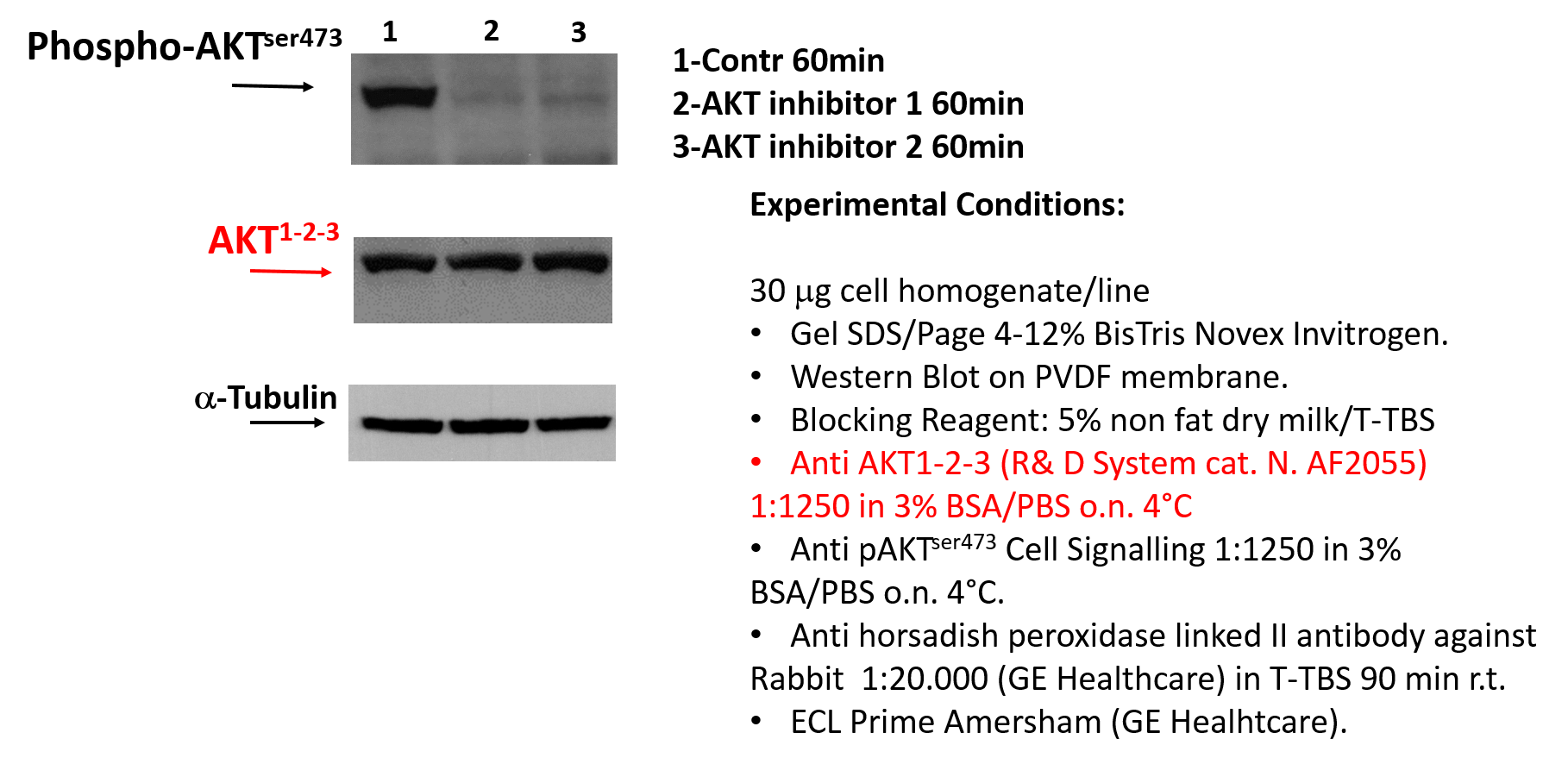
Application: Western BlotSample Tested: Human Chronic Lymphocytic Cell LineSpecies: HumanVerified Customer | Posted 04/28/201730 microg cell homogenate/line Gel SDS/Page 4-12% BisTris Novex Invitrogen. Western Blot on PVDF membrane. Blocking Reagent: 5% non fat dry milk/T-TBS Anti AKT1-2-3 (R& D System cat. N. AF2055) 1:1250 in 3% BSA/PBS o.n. 4°C Anti pAKTser473 Cell Signalling 1:1250 in 3% BSA/PBS o.n. 4°C. Anti horsadish peroxidase linked II antibody against Rabbit 1:20.000 (GE Healthcare) in T-TBS 90 min r.t. ECL Prime Amersham (GE Healhtcare).

There are no reviews that match your criteria.
Protocols
Find general support by application which include: protocols, troubleshooting, illustrated assays, videos and webinars.
- Cellular Response to Hypoxia Protocols
- R&D Systems Quality Control Western Blot Protocol
- Troubleshooting Guide: Western Blot Figures
- Western Blot Conditions
- Western Blot Protocol
- Western Blot Protocol for Cell Lysates
- Western Blot Troubleshooting
- Western Blot Troubleshooting Guide
- View all Protocols, Troubleshooting, Illustrated assays and Webinars
Loading...