CCR10 is a G protein-linked seven transmembrane domain protein expressed by T cells and B cell subsets that function as a receptor for CCL27. CCR10 mediates lymphocyte migration to the skin and mucosa, and its expression correlates with the metastatic capacity of melanomas.

Key Product Details
Validated by
Biological Validation
Species Reactivity
Validated:
Human
Cited:
Human
Applications
Validated:
Flow Cytometry, CyTOF-reported
Cited:
Neutralization, Flow Cytometry
Label
Unconjugated
Antibody Source
Monoclonal Rat IgG2A Clone # 314305
Loading...
Product Specifications
Immunogen
Y3 rat myeloid cell line transfected with human CCR10
Gln8-Asn362
Accession # P46092
Gln8-Asn362
Accession # P46092
Specificity
Detect human CCR10. Stains human CCR10-transfected cells but not irrelevant transfectants.
Clonality
Monoclonal
Host
Rat
Isotype
IgG2A
Scientific Data Images for Human CCR10 Antibody (314305)
Detection of CCR10 in Human Blood Lymphocytes by Flow Cytometry.
Human peripheral blood lymphocytes were stained with Mouse Anti-Human CD3e APC-conjugated Monoclonal Antibody (Catalog # FAB100A) and either (A) Rat Anti-Human CCR10 Monoclonal Antibody (Catalog # MAB3478) or (B) Rat IgG2AIsotype Control (Catalog # MAB006) followed by Phycoerythrin-conjugated Anti-Rat IgG Secondary Antibody (Catalog # F0105B). View our protocol for Staining Membrane-associated Proteins.
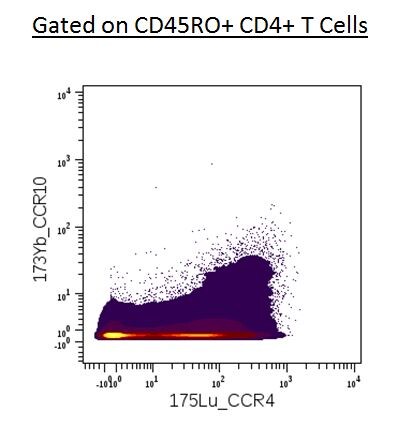
Detection of Human CCR10/GPR2 by Flow Cytometry
The effect of IL-21 and CD40L exposure on MEC1 and MEC2 cells.Expression of EBNA-2 and LMP-1 in IL-21 treated cells (A, B). (A) Simultaneous immunofluorescence staining of EBNA-2 (Green) and LMP-1 (Red); magnification (×100), scale bar 25 µm. Note the downregulation of EBNA-2 and upregultion of LMP-1 after IL-21 treatment. (B) Expression of EBNA-2, LMP-1 and Blimp-1 by immunoblotting; positive control: CBM1-Ral-STO, negative control: Ramos. 1.5×10⁵ cells were loaded in the control lanes and 5×10⁵ were loaded in both untreated and IL-21 treated MEC1 and MEC2 lanes. Note low expression of EBNA-2 and high expression of LMP-1 after IL-21 treatment and induction of Blimp-1 after IL-21 treatment. (C) Activity of the W and C promoters that regulate EBNA-2 expression and LMP-1 mRNA expression by Q-PCR. Note the difference in EBNA-2 regulation; the MEC2 cell uses both Wp and Cp while in MEC1 only Wp is active. (D) Expression of EBNA-2 and LMP-1 in cells exposed to CD40L. Simultaneous immunofluorescence staining; for details see (A). Note: EBNA-2 and LMP-1 are downregulated by CD40L in both lines. (E) CD40L induced modulation of surface marker by FACS analysis. Image collected and cropped by CiteAb from the following publication (https://dx.plos.org/10.1371/journal.pone.0106008), licensed under a CC-BY license. Not internally tested by R&D Systems.
Detection of Human CCR10/GPR2 by Flow Cytometry
Comparison of the MEC1 and MEC2 cells.(A) Expression of EBV encoded proteins EBNA-2 and LMP-1 by immunofluorescence; magnification (×100), scale bar 25 µm. Note: the MEC2 cells are larger. (B) Expression of EBNA-2 and LMP-1 by immunoblotting; positive control: CBM1-Ral-STO, negative control: Ramos. 1.5×10⁵ cells were loaded in control lanes and 5×10⁵ were loaded in MEC1 and MEC2 lanes. Note MEC2 expresses higher amount of EBNA-2. (C) Expression of Bright and BARF1 by Q-PCR. (D) FACS analysis of surface markers that are differently expressed in the 2 lines. Image collected and cropped by CiteAb from the following publication (https://dx.plos.org/10.1371/journal.pone.0106008), licensed under a CC-BY license. Not internally tested by R&D Systems.Applications for Human CCR10 Antibody (314305)
Application
Recommended Usage
CyTOF-reported
This clone has been commercially reported for use in CyTOF®. Ready to be labeled using established conjugation methods. No BSA or other carrier proteins that could interfere with conjugation.
Flow Cytometry
0.25 µg/106 cells
Sample:
Sample:
Human peripheral blood lymphocytes
Reviewed Applications
Read 1 review rated 4 using MAB3478 in the following applications:
Flow Cytometry Panel Builder
Bio-Techne Knows Flow Cytometry
Save time and reduce costly mistakes by quickly finding compatible reagents using the Panel Builder Tool.
Advanced Features
- Spectra Viewer - Custom analysis of spectra from multiple fluorochromes
- Spillover Popups - Visualize the spectra of individual fluorochromes
- Antigen Density Selector - Match fluorochrome brightness with antigen density

Formulation, Preparation, and Storage
Purification
Protein A or G purified from hybridoma culture supernatant
Reconstitution
Reconstitute at 0.5 mg/mL in sterile PBS. For liquid material, refer to CoA for concentration.
Loading...
Formulation
Lyophilized from a 0.2 μm filtered solution in PBS with Trehalose. *Small pack size (SP) is supplied either lyophilized or as a 0.2 µm filtered solution in PBS.
Shipping
Lyophilized product is shipped at ambient temperature. Liquid small pack size (-SP) is shipped with polar packs. Upon receipt, store immediately at the temperature recommended below.
Stability & Storage
Use a manual defrost freezer and avoid repeated freeze-thaw cycles.
- 12 months from date of receipt, -20 to -70 °C as supplied.
- 1 month, 2 to 8 °C under sterile conditions after reconstitution.
- 6 months, -20 to -70 °C under sterile conditions after reconstitution.
Calculators
Background: CCR10
Alternate Names
CCR10, GPR2
Gene Symbol
CCR10
UniProt
Additional CCR10 Products
Product Documents for Human CCR10 Antibody (314305)
Certificate of Analysis
To download a Certificate of Analysis, please enter a lot or batch number in the search box below.
Note: Certificate of Analysis not available for kit components.
Product Specific Notices for Human CCR10 Antibody (314305)
For research use only
Related Research Areas
Citations for Human CCR10 Antibody (314305)
 Powered by Bioz
Customer Reviews for Human CCR10 Antibody (314305) (1)
4 out of 5
1 Customer Rating
Have you used Human CCR10 Antibody (314305)?
Submit a review and receive an Amazon gift card!
$25/€18/£15/$25CAN/¥2500 Yen for a review with an image
$10/€7/£6/$10CAN/¥1110 Yen for a review without an image
Submit a review
Customer Images
Showing
1
-
1 of
1 review
Showing All
Filter By:
-
Application: CyTOFSample Tested: Peripheral blood mononuclear cells (PBMCs)Species: HumanVerified Customer | Posted 06/28/2016

There are no reviews that match your criteria.
Protocols
Find general support by application which include: protocols, troubleshooting, illustrated assays, videos and webinars.
- 7-Amino Actinomycin D (7-AAD) Cell Viability Flow Cytometry Protocol
- Extracellular Membrane Flow Cytometry Protocol
- Flow Cytometry Protocol for Cell Surface Markers
- Flow Cytometry Protocol for Staining Membrane Associated Proteins
- Flow Cytometry Staining Protocols
- Flow Cytometry Troubleshooting Guide
- Intracellular Flow Cytometry Protocol Using Alcohol (Methanol)
- Intracellular Flow Cytometry Protocol Using Detergents
- Intracellular Nuclear Staining Flow Cytometry Protocol Using Detergents
- Intracellular Staining Flow Cytometry Protocol Using Alcohol Permeabilization
- Intracellular Staining Flow Cytometry Protocol Using Detergents to Permeabilize Cells
- Propidium Iodide Cell Viability Flow Cytometry Protocol
- Protocol for Liperfluo
- Protocol for the Characterization of Human Th22 Cells
- Protocol for the Characterization of Human Th9 Cells
- Protocol: Annexin V and PI Staining by Flow Cytometry
- Protocol: Annexin V and PI Staining for Apoptosis by Flow Cytometry
- Troubleshooting Guide: Fluorokine Flow Cytometry Kits
- View all Protocols, Troubleshooting, Illustrated assays and Webinars
Loading...
Associated Pathways
