
Death Receptor 6 (DR6), also known as TNFRSF21 and CD358, is a type I transmembrane protein in the TNF receptor superfamily (1). Human DR6 consists of a 308 amino acid (aa) extracellular domain (ECD) with four cysteine‑rich motifs, a 21 aa transmembrane segment, and a 285 aa palmitylated cytoplasmic region that contains one death domain (2, 3). Within the ECD, human and mouse DR6 share 82% aa sequence identity. DR6 is expressed as an approximately 110 kDa molecule that carries extensive N‑linked and O‑linked glycosylation in its extracellular region (3, 4). Among hematopoietic cells, DR6 is expressed on monocytes, resting CD4+ T cells, and pro‑, pre‑, and naïve B cells (5 ‑ 7). DR6 knockout mice exhibit a Th2‑biased immune response characterized by exaggerated Th2 and B cell responsiveness in combination with reduced Th1 cell responsiveness and inflammatory leukocyte infiltration (6 ‑ 9). DR6 knockout mice are resistant to induced airway inflammation and experimental autoimmune encephalitis but more susceptible to severe graft versus host disease (9 ‑ 11). DR6 is also expressed on developing neurons where it can bind a shed 35 kDa N‑terminal fragment of APP or a fragment of APLP2 (12, 13). This APP fragment is generated following deprivation of neurotrophic factors, and its binding to DR6 triggers DR6‑mediated axonal pruning (12). DR6 is constitutively expressed on some prostate cancer cells and can be induced by TNF‑ alpha on others (3, 4).
Human DR6/TNFRSF21 Antibody
R&D Systems | Catalog # AF144

Scientific images may be resized, cropped, or adjusted for brightness or contrast for presentation purposes. These updates are limited to presentation and do not affect the underlying scientific interpretation of the data. Where available, additional source data may be provided upon request.
Key Product Details
Species Reactivity
Validated:
Cited:
Applications
Validated:
Cited:
Label
Antibody Source
Product Specifications
Immunogen
Gln42-Leu350
Accession # O75509
Specificity
Clonality
Host
Isotype
Scientific Data Images for Human DR6/TNFRSF21 Antibody
Detection of DR6/TNFRSF21 by Western Blot
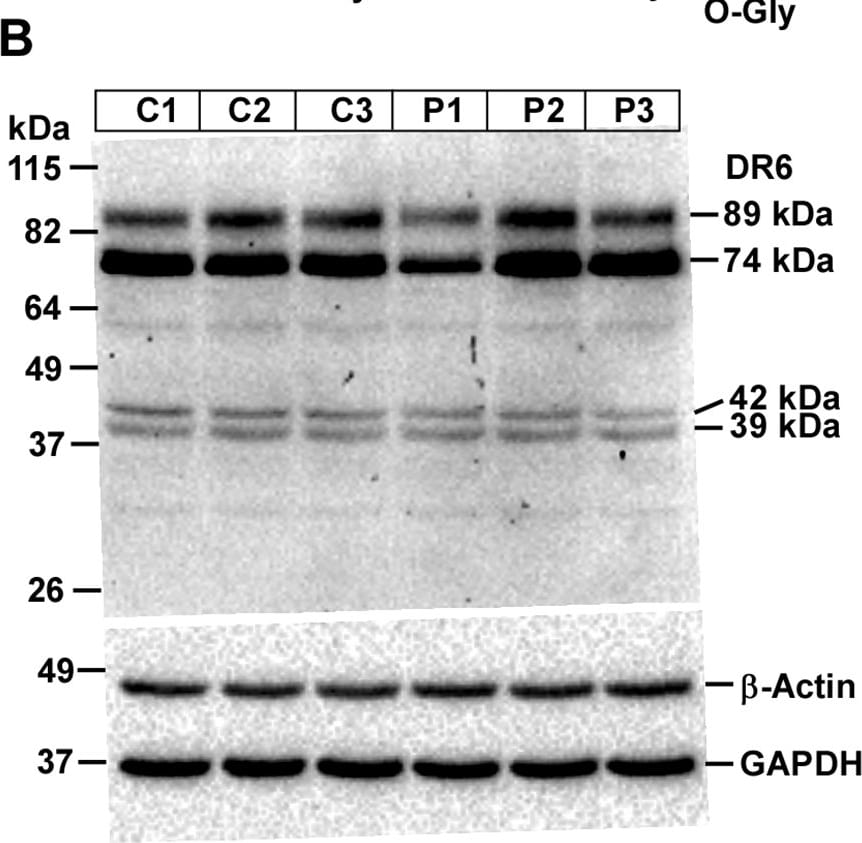
Expression pattern of DR6 and p75NTR protein isoforms varies between control (C) and prion-diseased (P) C57BL/6J mice brains. A Schema of a 68-kDa DR6 protein structure shows various domains, glycosylation sites and epitope of anti-DR6 antibody. B Western blot analysis of DR6 on lysates showing similar expression pattern of all DR5 isoforms between control and prion-diseased mice brains. D The densitometry of 89-kDa, D 74-kDa DR6 isoforms show similar levels of expression within prion-diseased and control mice brains. E Schematic structure of p75NTR protein exhibiting various domains, glycosylation sites and epitope of anti-p75NTR antibody. E Western blot analysis of p75NTR shows a dominant 75 kDa band with increased p75NTR level in prion-diseased mice brains than controls. F The densitometry of 75 kDa band show significant increased intensity in prion diseased mice brain lysates than controls. GAPDH and beta -actin were used as loading controls. Each histogram represents mean ± SD of a set of three brain lysates from control or prion-diseased mice. Two-tailed unpaired t-test was performed to compare the significance of difference between two groups. P ≤ 0.05 is considered statistically significant Image collected and cropped by CiteAb from the following open publication (https://pubmed.ncbi.nlm.nih.gov/38802941), licensed under a CC-BY license. Not internally tested by R&D Systems.Applications for Human DR6/TNFRSF21 Antibody
Western Blot
Sample: Recombinant Human DR6/TNFRSF21 Fc Chimera (Catalog # 144-DR)
Human DR6/TNFRSF21 Sandwich Immunoassay
Formulation, Preparation, and Storage
Purification
Reconstitution
Reconstitute at 0.2 mg/mL in sterile PBS. For liquid material, refer to CoA for concentration.
Formulation
Shipping
Stability & Storage
- 12 months from date of receipt, -20 to -70 °C as supplied.
- 1 month, 2 to 8 °C under sterile conditions after reconstitution.
- 6 months, -20 to -70 °C under sterile conditions after reconstitution.
Calculators
Background: DR6/TNFRSF21
References
- Benschop, R. et al. (2009) Adv. Exp. Med. Biol. 647:186.
- Pan, G. et al. (1998) FEBS Lett. 431:351.
- Klima, M. et al. (2009) Biochim. Biophys. Acta 1793:1579.
- Kasof, G.M. et al. (2001) Oncogene 20:7965.
- Matesanz-Isabel, J. et al. (2011) Immunol. Lett. 134:104.
- Schmidt, C.S. et al. (2003) J. Exp. Med. 197:51.
- Liu, J. et al. (2001) Immunity 15:23.
- Zhao, H. et al. (2001) J. Exp. Med. 194:1441.
- Venkataraman, C. et al. (2006) Immunol. Lett. 106:42.
- Schmidt, C.S. et al. (2005) J. Immunol. 175:2286.
- Liu, J. et al. (2002) J. Immunol. 169:3993.
- Nikolaev, A. et al. (2009) Nature 457:981.
- Kuester, M. et al. (2011) J. Mol. Biol. 409:189.
Long Name
Alternate Names
Gene Symbol
UniProt
Additional DR6/TNFRSF21 Products
Product Documents for Human DR6/TNFRSF21 Antibody
Certificate of Analysis
To download a Certificate of Analysis, please enter a lot or batch number in the search box below.
Note: Certificate of Analysis not available for kit components.
Product Specific Notices for Human DR6/TNFRSF21 Antibody
For research use only
Related Research Areas
Citations for Human DR6/TNFRSF21 Antibody
 Powered by Bioz
Customer Reviews for Human DR6/TNFRSF21 Antibody
There are currently no reviews for this product. Be the first to review Human DR6/TNFRSF21 Antibody and earn rewards!
Have you used Human DR6/TNFRSF21 Antibody?
Submit a review and receive an Amazon gift card!
$25/€18/£15/$25CAN/¥2500 for a review with an image
$10/€7/£6/$10CAN/¥1110 for a review without an image
Submit a review
Protocols
Find general support by application which include: protocols, troubleshooting, illustrated assays, videos and webinars.
- Cellular Response to Hypoxia Protocols
- R&D Systems Quality Control Western Blot Protocol
- Troubleshooting Guide: Western Blot Figures
- Western Blot Conditions
- Western Blot Protocol
- Western Blot Protocol for Cell Lysates
- Western Blot Troubleshooting
- Western Blot Troubleshooting Guide
- View all Protocols, Troubleshooting, Illustrated assays and Webinars
Associated Pathways
